Carbon-carbon double-bond shift in the biosynthesis of the
antibiotic corallopyronin A
CorJ DH*: a shift domain
Dissertation
zur
Erlangung des Doktorgrades (Dr. rer. nat.)
der
Mathematisch-Naturwissenschaftlichen Fakultät
der
Rheinischen Friedrich-Wilhelms-Universität Bonn
vorgelegt von
Diplom-Pharmazeutin (Dipl.-Pharm.)
Friederike Lohr
aus
Haan
Bonn 2014
II
Angefertigt mit Genehmigung der Mathematisch-Naturwissenschaftlichen
Fakultät der Rheinischen Friedrich-Wilhelms-Universität Bonn
1. Referentin : Prof. Dr. G. M. König
2. Referent : Prof. Dr. M. Gütschow
Tag der Promotion : 16. Dezember 2015
Erscheinungsdatum : 2015

III
Für meine Eltern und Maxim.

IV
V
Publications
Ö. Erol, T. F. Schäberle, A. Schmitz, S. Rachid, C. Gurgui, M. El Omari, F. Lohr, S.
Kehraus, J. Piel, R. Müller, G. M. König; Biosynthesis of the myxobacterial antibiotic
corallopyronin A. Chem Bio Chem 2010, 11, 1253–1265
M. Frizler, F. Lohr, N. Furtmann, J. Kläs, M. Gütschow; Structural optimization of
azadipeptide nitriles strongly increases association rates and allows the
development of selective cathepsin inhibitors. J Med Chem 2011, 54, 396–400
M. Frizler, F. Lohr, M. Lüllsdorf, M. Gütschow; Facing the gem-dialkyl effect in
enzyme inhibitor design: preparation of homocycloleucine-based azadipeptide
nitriles. Chemistry 2011, 17, 11419–11423
A. Schmitz, S. Felder, T. Höver, S. Kehraus, F. Lohr, G. M. König, T. F. Schäberle;
Antibiotics from gliding bacteria. Phytochem. Rev. 2013, 12, 507–516
F. Lohr, I. Jenniches, M. Frizler, M. J. Meehan, M. Sylvester, A. Schmitz, M. Gütschow,
P. C. Dorrestein, G. M. König, T. F. Schäberle; alpha, beta –> beta, gamma double-
bond migration in corallopyronin A biosynthesis. Chem Sci 2013, 4, 4175–4180
T. F. Schäberle, M. Mir Mohseni, F. Lohr, A. Schmitz, G. M. König; Function of the
loading module in CorI and of the O-methyltransferase CorH in vinyl carbamate
biosynthesis of the antibiotic corallopyronin A. Antimicrob. Agents Chemother. 2014,
58, 950–956
T. F. Schäberle, F. Lohr, A. Schmitz, G. M. König; Antibiotics from Myxobacteria.
DOI:10.1039/C4NP00011K.
VI
Conferences
International VAAM (Vereinigung für angewandte und allgemeine Mikrobiologie)-
Workshop 2010 „Biology of bacteria producing natural products“; 26–28 september
2010 in Tübingen, Germany. Poster presentation: “Biosynthesis of the myxobacterial
antibiotic corallopyronin A”. Abstract published in program & abstract book
International VAAM-Workshop 2010 “Biology of bacteria producing natural
products”.
International VAAM (Vereinigung für angewandte und allgemeine Mikrobiologie)-
Workshop 2011 „Biology of bacteria producing natural products“; 28–30 september
2011 in Bonn, Germany.
XIV International symposium on marine natural products (MaNaPro) 2013, 8 ECMNP
(European conference on marine natural products); 15–20 september 2013 in La Toja
Island, Spain. Poster presentation: “Investigation of the double-bond shift in
corallopyronin A biosynthesis”. Abstract published in program & abstract book XIV
International symposium on marine natural products.
Internationale DPhG-Doktorandentagung 2014; 10–12 march 2014 in Wuppertal,
Germany. Oral presentation: “Investigation of the double-bond shift in corallopyronin
A biosynthesis”. Abstract published in program & abstract book Internationale DPhG-
Doktorandentagung 2014.
Workshop
International workshop for young researchers 2013; 10–12 july 2013 in Lille-
Villeneuve d’Ascq, France. Bioinformatics tools for NRPS discovery: from genomic
data to the products.

VII
Further professional education
Fortbildungsveranstaltung zur Ausbildung von Projektleitern und Beauftragten für
biologische Sicherheit nach §15 (2) und (4) GenTSV 2012; 25–27 september 2012 at
the University of Cologne, Germany.
VIII
Danke!
Ich möchte meinen besonderen Dank meiner Doktormutter Frau Professorin König
aussprechen, dafür dass sie mich in ihre Arbeitsgruppe aufgenommen hat und mir die
Möglichkeit gegeben hat, auf dem vielseitigen Feld der bakteriellen Biosynthese zu
arbeiten. Ich möchte ihr sehr dafür danken, dass sie mir während meiner praktischen
Arbeit und während des Schreibens der Dissertation immer zur Seite stand,
wissenschaftlich und persönlich.
Herrn Professor Gütschow möchte ich zum einen für die erfolgreiche Kooperation im
Bereich der hier beschriebenen Synthese und zum anderen für die Bereitschaft zur
Übernahme des Coreferats dieser Arbeit danken.
Frau Professorin Wägele und Herrn Professor Wagner danke ich für ihre Bereitschaft
Teil der Prüfungskommission zu sein.
Meinem Freund Dr. Maxim Frizler möchte ich an dieser Stelle herzlich für seinen
Einsatz bei der Synthese des in dieser Arbeit verwendeten Substrats danken, welche
im Rahmen der Kooperation mit dem Arbeitskreis von Professor Gütschow
durchgeführt wurde. Ich bin ihm dankbar für etliche wissenschaftliche Diskussionen
und für seine liebevolle Unterstützung zu jeder Zeit. Danke.
Bei Herrn Dr. Marc Sylvester (Institut für Biochemie und Molekularbiologie der Uni
Bonn) möchte ich mich für die massenspektrometrischen Messungen im Rahmen des
„ppant ejection assays“ bedanken.
Herrn Dr. Till Schäberle danke ich für die Betreuung während meiner Promotion, für
viele konstruktive Diskussionen und für das Korrekturlesen einiger Teile meiner
Arbeit.
IX
Herrn Dr. Stefan Kehraus möchte ich für die Unterstützung bei allen HPLC- und NMR-
Fragen und für die Aufnahme von NMR Spektren in der Pharmazeutischen Chemie
danken. Vielen Dank auch für das Korrekturlesen einiger Teile meiner Arbeit.
Bei Frau Edith Neu möchte ich mich für die Einführung in das Arbeiten mit
Myxobakterien, besonders deren Isolierung und Kultivierung, bedanken. Frau
Ekatarina Eguereva danke ich für LC/MS Messungen und Frau Mila Goralski für Tips
und Hilfestellungen im S1 Labor. Allen dreien danke ich von Herzen für eine richtig
schöne Zeit.
Ich danke Herrn Thomas Kögler für seine Hilfe bei Problemen aller Art mit dem
Computer und anderen technischen Geräten.
Den Damen Kirsten Knapp und Annika Orland danke ich für die voranbringende
Zusammenarbeit während der Promotion und noch viel mehr für ihre Freundschaft.
Den Herren Alexander Bogdanov, Stephan Felder, Henrik Harms und Peter Hufendiek
möchte Danke sagen für die Unterstützung bei HPLC und NMR Fragen, für manches
gemeinsam getrunkene Bier und für eine tolle Zeit.
Allen Kollegen und Kolleginnen des Arbeitskreises der Pharmazeutischen Biologie in
Bonn danke ich herzlich für die gute Zusammenarbeit, für vielseitige Gespräche und
für die ausgewogene Mischung aus lustiger und konzentrierter Arbeitsatmosphäre.
Meiner Mutter danke ich für das Korrekturlesen der Arbeit.
Meinen Eltern und Maxim danke ich für ihren Glauben an mich, der mir immer sicher
ist und der alles leicht macht.

X
Contents
XI
1 Introduction ....................................................................................... 1
1.1 Myxobacterial antibiotics that target bacterial RNA polymerase ............................... 1
1.1.1 Corallopyronins and myxopyronins ......................................................................... 2
1.1.2 Ripostatins .............................................................................................................. 8
1.1.3 Sorangicins ............................................................................................................ 10
1.1.4 Etnangien .............................................................................................................. 12
1.2 Myxobacterial antibiotics targeting bacterial protein biosynthesis.......................... 14
1.2.1 Althiomycin ........................................................................................................... 14
1.2.2 Angiolam A............................................................................................................ 17
1.2.3 Myxovalargins ...................................................................................................... 18
1.3 Myxobacterial antibiotics targeting the respiratory chain ................................ 20
1.3.1 Aurachins .............................................................................................................. 20
1.3.2 Thuggacins ............................................................................................................ 23
1.4 Myxobacterial antibiotics influencing biofilm formation ................................... 27
1.4.1 Carolacton............................................................................................................. 27
1.5 Myxobacterial antibiotics targeting the type II signal peptidase LspA ..................... 29
1.5.1 Myxovirescins ....................................................................................................... 29
1.6 Myxobacterial antibiotics with an unknown mode of action ............................ 32
1.6.1 Chondrochlorens ................................................................................................. 32
1.6.2 Indiacens A and B ................................................................................................. 34
1.6.3 Maracin A and Maracen A .................................................................................. 34
1.6.4 Nannochelins ........................................................................................................ 35
1.6.5 Roimatacene......................................................................................................... 36
1.6.6 Sorangiadenosine ................................................................................................ 37
1.6.7 Sulfangolids and Kulkenon ................................................................................. 38
1.7 Antibiotics from marine myxobacteria ................................................................. 39
2 Scope of the study ............................................................................. 41
3 Material and Methods ....................................................................... 43
3.1 Solvents and Reagents ............................................................................................... 43
3.2 Enzymes ..................................................................................................................... 43
3.3 Molecular biological kits ............................................................................................ 43
Contents
XII
3.4 Media, stock solutions and buffers ........................................................................... 43
3.5 Bacterial strains ......................................................................................................... 46
3.6 Vectors ....................................................................................................................... 47
3.7 DNA constructs in this study ..................................................................................... 47
3.8 Primers ....................................................................................................................... 48
3.9 Software and databases ............................................................................................ 48
3.10 General molecular biological methods...................................................................... 49
3.10.1 Sterilization ......................................................................................................... 49
3.10.2 Cultivation, storage and disposal of organisms ................................................. 49
3.10.3 Antibiotic selectivity test .................................................................................... 50
3.11 Molecular biological methods concerning bacterial organisms................................ 50
3.11.1 Transformation of bacteria ................................................................................ 50
3.12 Molecular biological methods concerning nucleic acids ........................................... 52
3.12.1 Isolation of DNA ................................................................................................. 52
3.12.2 PCR...................................................................................................................... 52
3.12.3 Restriction digestion........................................................................................... 54
3.12.4 Dephosphorylation of linear DNA ...................................................................... 54
3.12.5 Agarose gel electrophoresis and DNA recovery................................................. 55
3.12.6 Ligation of DNA into a vector ............................................................................. 55
3.12.7 Sequencing of DNA constructs and PCR fragments ........................................... 55
3.13 Molecular biological methods concerning proteins .................................................. 56
3.13.1 Heterologous expression of the proteins........................................................... 56
3.13.2 Cell lysis by sonication ........................................................................................ 56
3.13.3 Purification of the recombinant protein by Ni-NTA affinity chromatography .. 57
3.13.4 SDS-Polyacrylamind gel electrophoresis (SDS-PAGE) and Coomassie staining . 57
3.13.5 Concentration of the proteins and buffer exchange ......................................... 59
3.13.6 Determination of the protein concentration ..................................................... 59
3.14 Chromatography ........................................................................................................ 60
3.15 NMR spectroscopy ..................................................................................................... 60
3.16 Mass spectrometry .................................................................................................... 61
3.17 In vitro assays to prove the functional role of the DH* ............................................ 61
3.17.1 Phosphopantethein (Ppant) ejection assay ....................................................... 61
3.17.2 NMR based assay ............................................................................................... 62
Contents
XIII
3.18 Chemical syntheses of compounds 1, 5–8, 10–13 .................................................... 63
3.18.1 (E)-4-Methoxycarbonylaminobut 2-enoic acid (6) ............................................. 63
3.18.2 (E)-S-2-Acetamidoethyl 4-(methoxycarbonylamino)but-3-enethioate (7) ........ 65
3.18.3 S-2-Acetamidoethyl 2-bromoethanethioate (10) .............................................. 66
3.18.4 S-2-Acetamidoethyl 2-(diethoxyphosphoryl)ethanethioate (11) ...................... 67
3.18.5 (E)-S-2-Acetamidoethyl 4-(tert-butyloxycarbonylamino)but-2-enethioate (12)68
3.18.6 (E)-S-2-Acetamidoethyl 4-(methoxycarbonylamino)but-2-enethioate (1) ........ 69
4 Results .............................................................................................. 71
4.1 Corallopyronin A and its biosynthesis: An overview ................................................. 71
4.2 Attemps to prove the putative biosynthetic gene cluster of coralloyronin A .......... 75
4.3 Double-bond migration in corallopyronin A biosynthesis: investigation of the
protein CorJ DH* ........................................................................................................ 78
4.4 In vitro assays envisaged to investigate the functional role of CorJ DH* ................. 83
4.5 Heterologous expression of CorJ DH*-ACP, CorJ DH* and CorJ ACP ........................ 84
4.5.1 Single amino acid exchange in the shift domain CorJ DH* .................................... 88
4.6 Syntheses of the N-acetylcysteamine (SNAC) activated substrate for the shift
domain CorJ DH* ....................................................................................................... 91
4.6.1 Synthesis of compound 7 ....................................................................................... 92
4.6.2 Synthesis of compound 8 ....................................................................................... 94
4.6.3 Abruzov and Horner-Wittig-Emmons reaction resulted in compound 1 .............. 98
4.7 In vitro assays to prove the functional role of CorJ DH* ......................................... 100
4.7.1 Phosphopantetheine (ppant) ejection assays ..................................................... 100
4.7.1.1 Proof of the successful linkage of substrate 1 to the phosphopantethein
(ppant) arm of CorJ holo-ACP ........................................................................... 103
4.7.1.2 Investigation of potential unspecific hydrogen/deuterium exchanges under
assay conditions ............................................................................................... 106
4.7.1.3 Proof of the β,γ double-bond migration using the ppant ejection assay ........ 108
4.7.1.4 PPant ejection assay with the substrate-loaded active site mutant
CorJ DH*H47A holo-ACP................................................................................... 113
4.7.1.5 PPant ejection assay with the substrate-loaded active site mutant
CorJ DH*D211N holo-ACP ................................................................................ 114
4.7.2 Investigating CorJ DH* in an NMR based assay ................................................... 115
5 Conclusion ....................................................................................... 117
5.1 Drug discovery from natural products .................................................................... 117
Contents
XIV
5.2 Biosynthesis of myxobacterial natural products focussing on corallopyronin A .... 118
5.3 Outlook .................................................................................................................... 121
6 Summary........................................................................................ 123
7 References ...................................................................................... 127
8 Appendix ......................................................................................... 143
8.1 Primer sequences .................................................................................................... 143
8.2 Protein sequences ................................................................................................... 145
8.2.1 Protein sequence of CorJ ACP .............................................................................. 145
8.2.2 Protein sequence of CorJ DH* ............................................................................. 145
8.2.3 Protein sequence of CorJ DH*ACP ....................................................................... 146
8.2.4 Protein sequence of CorJ DH*H47A ACP ............................................................. 146
8.2.5 Protein sequence of CorJ DH*D211N ACP ........................................................... 146
8.3 Analytical data of compounds 1, 6, 7, 8, 10, 11 and 12 .......................................... 147
Abbreviations
XV
Abbreviations
A Alanine
A domain Adenylation domain
AT Acyltransferase
ACP Acyl-carrier protein
°C Degree Celsius
1D One-dimensional
2D Two-dimensional
∂ NMR chemical shift (ppm)
ε Extinction coefficient (UV/VIS spectroscopy)
λ Wavelength (nm)
µ Micro (10-6)
br Broad
c Concentration
CID Collision-induced dissociation
CoA Coenzyme A
COSY Correlated spectroscopy
CP Carrier protein
CYP Cytochrome
d Doublet (in connection with NMR data)
D Aspartat
Da Dalton
DH Dehydrogenase domain
DAD Diode array detector
DCM Dichloromethane
DEPT Distortionless enhancement by polarisation transfer
DMAP 4-Dimethylaminopyridine
DMSO Dimetyhlsulfoxide
DNA Deoxyribonucleic acid
EDTA Ethylenediamine-tetraacetic acid
Abbreviations
XVI
e.g. „Exempli gratia“ (Latin); for example
EE Ethylacetat
ER Enoylreductase v
EtOH Ethanol
ESI Electro spray ionisation
et al. „Et alii“ (Latin); and others
H Histidine
h Hour
H/D Proton/deuterium
HMBC Heteronuclear multiple-bond correlation
HMG Hydroxymethylglutaryl
HPLC High performance liquid chromatography
HSQC Heteronuclear single quantum correlation
Hz Hertz
IC50 Inhibitory concentration - concentration of a drug that is required for
50 % inhibition of viral replication, protein binding etc.
i.e. „Id est“ (Latin); that is
J Spin-spin coupling constant [Hz]
KR Ketoreductase domain
KS Ketosynthase domain
kbp Kilo base pairs
L Leucine
L Litre
log Decadic logarithm
m Multiplet (in connection with NMR data)
M Molar (mol/L)
Mr Molecular mass
max Maximum
min Minute
MeOH Methanol
Abbreviations
XVII
MHz Megahertz
MIC Minimal inhibitory concentration
MRSA Methicilline resistant Staphylococcus aureus
MS Mass spectrometry
mult. Multiplicity
m/z Mass-to-charge ratio
N Asparagine
n.d. Not determined
n.i. Not investigated
NMR Nuclear magnetic resonance
No. Number
NRPS Nonribosomal peptide synthetase
P Proline
ppm Parts per million
PCR Polymerase chain reaction
PE Petroleum ether
pH Potentia hydrogenii
PKS Polyketide synthase
ppant Phosphopantethein
pant Pantethein
q Quartet (in connection with NMR data)
R Residue (in combination with chemical structures)
rpm Rounds per minute
RNA Ribonucleic acid
RNAP RNA Polymerase
RP Reversed phase
rt Room temperature
Rt Retention time
s Singlet (in connection with NMR data)
SAM S-Adenosyl-L-methionine
Abbreviations
XVIII
SDS Sodium dodecyl sulfate
SNAC N-acetylcysteamine
t Triplet (in connection with NMR data)
Taq Thermostable polymerase from the thermophilic bacterium Thermus aquaticus
TEMED Tetramethylethylendiamin
TFA Trifluoroacetic acid
Tris Tris(hydroxymethyl)-aminomethan
UV Ultraviolet
VIS Visible
Introduction
1 |
1 Introduction1
The increased emergence of bacteria resistant to antibiotics is a serious threat to
modern medicine (Schäberle and Hack, 2014). The successful treatment of bacterial
infections is in danger, since ever more multi-, and even pan-resistant bacteria evolve.
This development is aggravated by the fact that, since the golden age of antibiotics in
the 70ies, the number of new antibiotically active drugs introduced into therapy is
dramatically dwindling. Therefore, research to identify new putative antibiotics has to
be pursued and intensified. Natural products, especially microbe-derived compounds,
proved themselves as a good source for antibiotics. Besides the well-known
proliferative producer organisms like the streptomycetes and bacilli, currently
myxobacteria move into the focus. This group of bacteria synthesises structurally
diverse secondary metabolites, distinct from the classes known so far from traditional
antibiotic producers. An example for a myxobacterial metabolite successfully
introduced into therapy, albeit in another therapeutic area, is the anti-cancer drug
ixabepilon, a derivative of the myxobacterial metabolite epothilone, which was
launched in 2007 (Thompson, 2007). Interestingly, many myxobacterial compounds
showing promising antibacterial activities were identified to date, however none of
these was further developed as a drug.
In this review all myxobacterial compounds with antibiotic activity, which could serve
as lead structures for future developments are discussed, according to their mode of
action.
1.1 Myxobacterial antibiotics that target bacterial RNA polymerase
Bacterial RNA polymerase (RNAP) is an established target for antibiotics (Chopra,
2007; Ho et al., 2009; Mariani and Maffioli, 2009; Villain-Guillot et al., 2007). It is an
essential enzyme and well suited for the attack of antibiotics, since the bacterial
subunits are highly conserved, but differ from the eukaryotic ones. This way, such
antibiotics are highly selective, have a broad-spectrum activity and low toxicity.
RNAP-inhibitors in clinical use are the rifamycins, natural products and their
derivatives originating from actinomycetes, which are of particular importance in the
1 The introduction is published in Schäberle et al., 2014; Antibiotics from myxobacteria.
Introduction
2 |
treatment of tuberculosis. Other infections are also amenable to therapy with
rifamycins, e.g. in infections with Bacillus anthracis (inhalation anthrax) a combination
therapy using a rifamycin together with ciprofloxacin or doxycycline proved successful
in the 2001 anthrax attacks (Srivastava et al., 2011). Fidoxamicin, another RNA
synthesis inhibitor was only recently approved for Clostridium difficile infections
(Artsimovitch et al., 2012).
Up to date four antibiotics and their corresponding derivatives are known from
myxobacteria, which inhibit bacterial RNAP, namely corallopyronin A, myxopyronin A,
ripostatin A, and sorangicin A.
1.1.1 Corallopyronins and myxopyronins
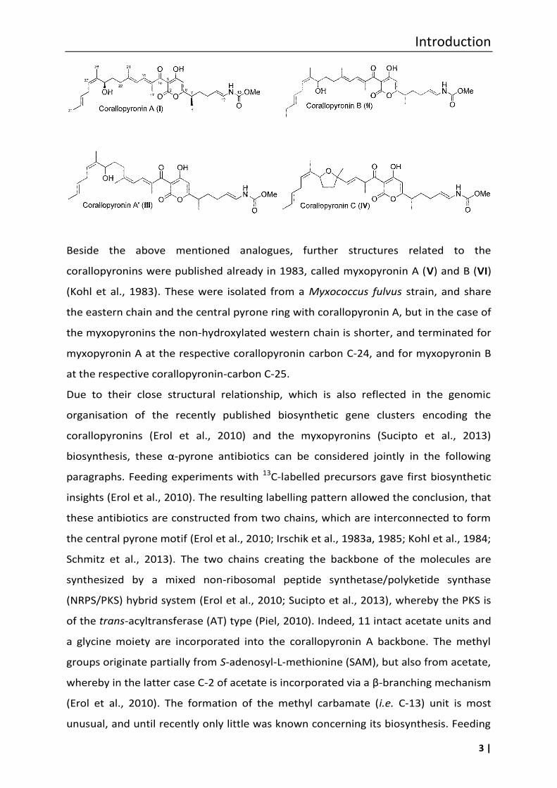
Corallopyronins were first isolated in 1985 from a Corallococcus coralloides strain
from Tunisia (Jansen et al., 1985). Corallopyronin A (I) has several interesting
structural features. A pyrone ring forms the central rigid core of the molecule, to
which two conformationally more flexible chains are attached, i.e. the lipophilic
western chain with three methyl groups, a hydroxyl group, and a diene element, and
the eastern chain with one methyl group, an enamide function, and a methyl
carbamate moiety. Carbamates are a rarely found structural moiety in secondary
metabolites from bacteria.
Three analogues are known, i.e. corallopyronin A´ (III), corallopyronin B (II) and
corallopyronin C (IV). The double-bond Δ19,20 is Z-configurated in corallopyronin A’,
whereas in the main metabolite corallopyronin A the configuration of this double-
bond is E. Corallopyronin A´ may be an artefact formed during isolation and storage of
corallopyronin A. Corallopyronin B differs from A in the western chain by an
additional methylene group, assumed to be derived from the respectively
incorporated starter unit, i.e. a propionyl instead of an acetyl moiety, during the
biosynthesis of this chain (Erol et al., 2010). Corallopyronin C is characterized by a
tetrahydrofuran ring in the western chain. However also in this case, it cannot be
excluded that corallopyronin C is an artefact of the isolation process, and might be
formed through a reaction of the C-24 hydroxyl group of corallopyronin A with the
diene motif.
Introduction
3 |
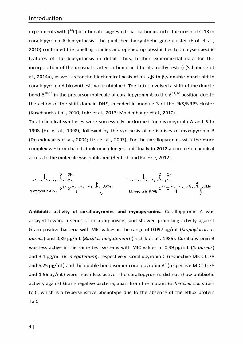
Beside the above mentioned analogues, further structures related to the
corallopyronins were published already in 1983, called myxopyronin A (V) and B (VI)
(Kohl et al., 1983). These were isolated from a Myxococcus fulvus strain, and share
the eastern chain and the central pyrone ring with corallopyronin A, but in the case of
the myxopyronins the non-hydroxylated western chain is shorter, and terminated for
myxopyronin A at the respective corallopyronin carbon C-24, and for myxopyronin B
at the respective corallopyronin-carbon C-25.
Due to their close structural relationship, which is also reflected in the genomic
organisation of the recently published biosynthetic gene clusters encoding the
corallopyronins (Erol et al., 2010) and the myxopyronins (Sucipto et al., 2013)
biosynthesis, these α-pyrone antibiotics can be considered jointly in the following
paragraphs. Feeding experiments with 13C-labelled precursors gave first biosynthetic
insights (Erol et al., 2010). The resulting labelling pattern allowed the conclusion, that
these antibiotics are constructed from two chains, which are interconnected to form
the central pyrone motif (Erol et al., 2010; Irschik et al., 1983a, 1985; Kohl et al., 1984;
Schmitz et al., 2013). The two chains creating the backbone of the molecules are
synthesized by a mixed non-ribosomal peptide synthetase/polyketide synthase
(NRPS/PKS) hybrid system (Erol et al., 2010; Sucipto et al., 2013), whereby the PKS is
of the trans-acyltransferase (AT) type (Piel, 2010). Indeed, 11 intact acetate units and
a glycine moiety are incorporated into the corallopyronin A backbone. The methyl
groups originate partially from S-adenosyl-L-methionine (SAM), but also from acetate,
whereby in the latter case C-2 of acetate is incorporated via a β-branching mechanism
(Erol et al., 2010). The formation of the methyl carbamate (i.e. C-13) unit is most
unusual, and until recently only little was known concerning its biosynthesis. Feeding
Introduction
4 |
experiments with [13C]bicarbonate suggested that carbonic acid is the origin of C-13 in
corallopyronin A biosynthesis. The published biosynthetic gene cluster (Erol et al.,
2010) confirmed the labelling studies and opened up possibilities to analyse specific
features of the biosynthesis in detail. Thus, further experimental data for the
incorporation of the unusual starter carbonic acid (or its methyl ester) (Schäberle et
al., 2014a), as well as for the biochemical basis of an , to β,γ double-bond shift in
corallopyronin A biosynthesis were obtained. The latter involved a shift of the double
bond Δ10,11 in the precursor molecule of corallopyronin A to the Δ11,12 position due to
the action of the shift domain DH*, encoded in module 3 of the PKS/NRPS cluster
(Kusebauch et al., 2010; Lohr et al., 2013; Moldenhauer et al., 2010).
Total chemical syntheses were successfully performed for myxopyronin A and B in
1998 (Hu et al., 1998), followed by the synthesis of derivatives of myxopyronin B
(Doundoulakis et al., 2004; Lira et al., 2007). For the corallopyronins with the more
complex western chain it took much longer, but finally in 2012 a complete chemical
access to the molecule was published (Rentsch and Kalesse, 2012).
Antibiotic activity of corallopyronins and myxopyronins. Corallopyronin A was
assayed toward a series of microorganisms, and showed promising activity against
Gram-positive bacteria with MIC values in the range of 0.097 µg/mL (Staphylococcus
aureus) and 0.39 µg/mL (Bacillus megaterium) (Irschik et al., 1985). Corallopyronin B
was less active in the same test systems with MIC values of 0.39 µg/mL (S. aureus)
and 3.1 µg/mL (B. megaterium), respectively. Corallopyronin C (respective MICs 0.78
and 6.25 µg/mL) and the double bond isomer corallopyronin A´ (respective MICs 0.78
and 1.56 µg/mL) were much less active. The corallopyronins did not show antibiotic
activity against Gram-negative bacteria, apart from the mutant Escherichia coli strain
tolC, which is a hypersensitive phenotype due to the absence of the efflux protein
TolC.
Introduction
5 |
The antibiotic profile of the myxopyronins was comparable, whereby the activity was
not as high as that of corallopyronin A. Myxopyronin B performed better than
myxopyronin A (MIC MyxA 1.0 and 6.0 µg/mL; MIC MyxB 0.3 and 0.8 µg/mL against
S. aureus and B. megaterium, respectively) (Irschik et al., 1983a). In the initial activity
assessments in the 80ies neither activity of the myxopyronins, nor of the
corallopyronins was observed against Mycobacterium phlei (Irschik et al., 1983a,
1985). Our recent evaluation of corallopyronin A required 64 μg/mL in Müller-Hinton
medium and 128 μg/mL of corallopyronin A in Lysogeny Broth medium to inhibit
Mycobacterium smegmatis. An MIC of 16 μg/mL of corallopyronin A was determined
for the sensitive strain Mycobacterium bovis Bacillus Calmette-Guérin (BCG), the
latter causing animal tuberculosis with only subordinate relevance for human
tuberculosis (Ayele et al., 2004; Schiefer et al., 2012). Furthermore, we observed an
MIC value of 0.25 µg/mL toward a methicillin resistant (MRSA) strain of S. aureus SG
511 (Institute collection of IMMIP, University of Bonn, Germany) (Schmitz, 2013). It
should be noted, that the recently determined MICs toward S. aureus are much
higher as the ones described in 1985, but nevertheless in a very promising range. In
our experiments the MIC against Micrococcus luteus H78S 1–3 was found to be
0.5 µg/mL while toward Bacillus subtilis 168 instead, an MIC of 32 µg/mL was
determined (Rentsch and Kalesse, 2012). The low sensitivity of B. subtilis towards
pyrone antibiotics was also noted in another study, in this case using racemic
myxopyronin B, which produced only slight inhibition zones in disk diffusion assays at
a concentration of 30 µg/ml (Yakushiji et al., 2013).
The activity of corallopyronin A was further determined against Wolbachia species,
intracellular bacteria of nematodes (Schiefer et al., 2012). These Gram-negative
proteobacteria of the order Rickettsiales are obligate endosymbionts of nematodes,
and considered as a novel target for controlling filarial infections like lymphatic
filariasis or onchocerciasis (Taylor et al., 2010). As one of a multitude of screened
substances, corallopyronin A proved itself to be in vivo active. In the model applied,
mice were infected with the filarial nematode Litomosoides sigmodontis. Beginning
the day after the infection, mice were untreated or given intraperitoneal injections
containing corallopyronin A (35 mg/kg/day) for 28 days. Five weeks post infection,
worms were recovered from the pleural cavity and depletion of Wolbachia was
Introduction
6 |
monitored by qPCR. More than 99 % of Wolbachia were depleted from L. sigmodontis
worms after corallopyronin A treatment (P < 0.0001 compared with untreated)
(Schiefer et al., 2012). This treatment does finally also kill the nematodes, since they
are dependent on their bacterial symbionts. It should be emphasised that, the
antibiotic is in vivo effective against intracellular Wolbachia despite the many
boundaries, and membranes the drug has to penetrate, like blood vessels, pleura,
worm cuticle, worm cells, vesicles, Wolbachia inner and outer membranes (Schäberle
et al., 2014b; Schiefer et al., 2012). Toxicity in mice was not detected up to the
maximum tested of 100 mg/kg (Irschik et al., 1983a).
The low activity against mycobacteria may here be regarded as an advantage of
corallopyronin A, since it opens up the possibility to develop a drug for filariasis
elimination without concern for cross-resistance development in tuberculosis
(Schäberle et al., 2014b; Schiefer et al., 2012).
A report in 2009 stated that no activity was observed for corallopyronin A in a
S. aureus sepsis model in mice after parenteral dosage, but no experimental details
for the respective experiments were given. The authors assumed that the lack of in
vivo activity was due to high serum protein binding (Haebich and von Nussbaum,
2009). Indeed, in a later study Moy et al. described that the MIC of myxopyronin B
toward S. aureus increased > 128-fold in the presence of human serum albumin (Moy
et al., 2011). In the light of the above discussed in vivo experiments, however,
corallopyronin A has to be judged as very promising for further development at least
as an antinematodal agent targeting intracellular Wolbachia.
Mode of action. The mode of action of these natural products was determined by
studying, protein, RNA and DNA synthesis in antibiotic treated S. aureus cells by
adding the radioactive precursors [U-14C]leucine, or [2-14C]uracil, or [U-14C]thymidine.
The result of these incorporation experiments showed that thymidine incorporation
was not affected, while leucine and uracil incorporation decreased. The reduction of
leucine incorporation was clearly delayed with respect to the immediate inhibiting
effect on uracil-incorporation. Thus, inhibition of RNA synthesis was suggested as
primary target. Consequently, the influence of myxopyronin A directly on the enzyme
RNAP of Thermus thermophilus was determined in in vitro experiments. It was found
that myxopyronin A acts specifically on bacterial RNAP, while the corresponding
Introduction
7 |
eukaryotic enzyme was not affected even at the highest concentration tested, i.e. up
to 200 µg/mL myxopyronin A and 40 µg/mL corallopyronin A, respectively (Irschik et
al., 1983a, 1985). Interestingly, corallopyronin A inhibited the growth of rifampin-
resistant S. aureus (O’Neill et al., 2000). Therefore, it was concluded that
corallopyronin A must address a new binding pocket on RNAP and thus represented a
novel mode of action. Subsequent X-ray analysis and biochemical data on
T. thermophilus RNAP complexed with myxopyronin A, and independently of a
desmethyl derivative of myxopyronin B, revealed the mode of action of these
antibiotics on the molecular level (Belogurov et al., 2008; Mukhopadhyay et al.,
2008).
Mukhopadhyay et al. showed that myxopyronin A interacts with the RNAP ‘‘switch
region’’, i.e. the hinge that mediates opening and closing of the RNAP active centre
cleft (Mukhopadhyay et al., 2008). By this binding the correct interaction of RNAP
with the template promoter DNA is prevented. It was further suggested that
myxopyronin A acts by inhibiting transcription initiation, since inhibition requires
myxopyronin-RNAP-interaction prior to interaction with promoter DNA. Thus, it was
proposed that myxopyronin A interferes with the opening and closing of the RNAP
clamp by jamming the hinge. Belogurov et al. also found desmethyl myxopyronin B
binding to the same pocket deep inside the RNA polymerase clamp head domain
(Belogurov et al., 2008). Through this binding the interaction with the DNA template
in the transcription bubble is disturbed and might compromise binding to, or directly
clash with, the melted template DNA strand (Belogurov et al., 2008). Footprinting
data showed that promoter DNA is indeed melted, but that its propagation towards
the active site is blocked.
The X-ray structures pictured that adjacent to the myxopyronin A binding pocket an
additional hydrophobic pocket is situated (Belogurov et al., 2008). This organization
may provide an explanation for the decrease of antibiotic activity going along with a
decrease in length of the western chain. It seems that a complete jamming of the
binding pocket infers more efficient with the hinge region, and consequently results
in a better antibiotic activity. Modelling corallopyronin A into Wolbachia RNAP
indicated that the binding pockets analysed by Mukhopadhyay et al., and by
Belogurov et al., for myxopyronins, were completely occupied, explaining the
Introduction
8 |
superior activity of the molecule (Schiefer et al., 2012). Correspondingly, the weaker
activity of corallopyronin B, possessing a longer western chain may result in partial
repulsion, since this molecule seems already too large.
The detailed knowledge on the binding mode of myxopyronin on RNAP was taken by
several studies as a starting point for a structure-based ligand design of novel RNAP
inhibitors. Described are either hybrid compounds, which include structural features
of the myxopyronins (Sahner et al., 2013; Yakushiji et al., 2013) or molecules with a
pyridyl-benzamide skeleton (McPhillie et al., 2011) or so-called squaramide
derivatives (Buurman et al., 2012) which are structurally completely different to the
respective natural product. All synthesized compounds that were found based on this
approach are considerably less active than the natural products.
Besides the RNAP-inhibiting effect, the α-pyrone-containing antibiotics might also
possess an additional mechanism of action, since inhibitory effects on fatty acid
synthesis were shown for antibiotic agents with an α-pyrone moiety (Giddens et al.,
2008). Further, 1 slightly induced the fabHB biosensor that is responsive to inhibition
of fatty acid biosynthesis (Mariner et al., 2011).
Resistance development. Resistance development is well described for the RNAP-
targeting rifamycins (Wehrli and Staehelo, 1971). Likewise resistance can develop
against the above described inhibitors of the RNAP switch region by mutations of the
RNAP resulting in a change of the respective binding pocket (Mariner et al., 2011;
Moy et al., 2011; Srivastava et al., 2011). Despite this observation, it would be
worthwhile considering whether the corallopyronin-type antibiotics could be useful in
combination therapy, as well known for the rifamycins. In this context it is also of
interest – as mentioned above – that there is no concern about cross-resistance in
tuberculosis-causing pathogens. Corallopyronin A is now in the focus of a translational
project to be developed as a drug for filariasis elimination (Annual report 2012 of the
German Centre for Infection Research).
1.1.2 Ripostatins
Ripostatin A–C were isolated from Sorangium cellulosum So ce377 (VII–IX)
(Augustiniak et al., 1996; Irschik et al., 1995). Ripostatin A (VII) and B (VIII) are 14-
membered macrolides with three 2,5,8-positioned double-bonds, whereas ripostatin
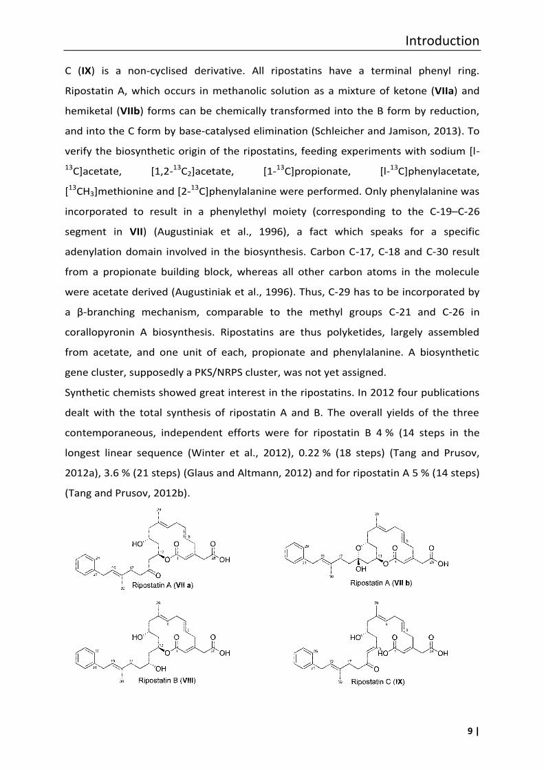
Introduction
9 |
C (IX) is a non-cyclised derivative. All ripostatins have a terminal phenyl ring.
Ripostatin A, which occurs in methanolic solution as a mixture of ketone (VIIa) and
hemiketal (VIIb) forms can be chemically transformed into the B form by reduction,
and into the C form by base-catalysed elimination (Schleicher and Jamison, 2013). To
verify the biosynthetic origin of the ripostatins, feeding experiments with sodium [l-
13C]acetate, [1,2-13C2]acetate, [1-13C]propionate, [l-13C]phenylacetate,
[13CH3]methionine and [2-13C]phenylalanine were performed. Only phenylalanine was
incorporated to result in a phenylethyl moiety (corresponding to the C-19–C-26
segment in VII) (Augustiniak et al., 1996), a fact which speaks for a specific
adenylation domain involved in the biosynthesis. Carbon C-17, C-18 and C-30 result
from a propionate building block, whereas all other carbon atoms in the molecule
were acetate derived (Augustiniak et al., 1996). Thus, C-29 has to be incorporated by
a β-branching mechanism, comparable to the methyl groups C-21 and C-26 in
corallopyronin A biosynthesis. Ripostatins are thus polyketides, largely assembled
from acetate, and one unit of each, propionate and phenylalanine. A biosynthetic
gene cluster, supposedly a PKS/NRPS cluster, was not yet assigned.
Synthetic chemists showed great interest in the ripostatins. In 2012 four publications
dealt with the total synthesis of ripostatin A and B. The overall yields of the three
contemporaneous, independent efforts were for ripostatin B 4 % (14 steps in the
longest linear sequence (Winter et al., 2012), 0.22 % (18 steps) (Tang and Prusov,
2012a), 3.6 % (21 steps) (Glaus and Altmann, 2012) and for ripostatin A 5 % (14 steps)
(Tang and Prusov, 2012b).
Introduction
10 |
Antibiotic activity. The two compounds VII and VIII showed nearly the same
antimicrobial activity against certain Gram-positive bacteria, mainly S. aureus strains,
and toward E. coli tolC with MICs in the range of about 1 µg/mL. Ripostatin B
displayed additionally minor activity against several yeasts and fungi (MIC 20 µg/mL
against Nadsonia fulvescens and 80 µg/mL against Debaryomyces hansenii,
respectively) (Irschik et al., 1995). The acyclic ripostatin C is biologically inactive
(Augustiniak et al., 1996). Furthermore, it was found that no cross-resistance occurs
between ripostatins and rifampin or sorangicin (Irschik et al., 1995). Indeed, ripostatin
A was effective against rifampin-resistant bacteria harbouring point mutations in the
rpoB gene sequence coding for their RNAP (Moy et al., 2011). Ripostatin A showed no
inhibitory effect on wheat germ RNAP II at a concentration of 20 µg/mL. However,
when applied to mouse fibroblasts L929 cells (10 µg/disc) an inhibition zone of 74 mm
indicated a toxic effect (Irschik et al., 1995). No other toxicity data are known.
Ripostatin A and B, even though being RNAP inhibitors such as the rifamycins, seem
to have no activity towards mycobacteria (Irschik et al., 1995).
Mode of action. In S. aureus cultures treated with ripostatin A (VII) RNA synthesis was
completely blocked (Irschik et al., 1995). The antibiotic also inhibited isolated E. coli
RNAP with an IC50 of 0.1 µg/mL (complete inhibition at 50 µg/mL). The earlier
assumption that the ripostatin binding site differs from the one of the rifamycins was
confirmed by analysing the cross-resistance patterns of mutagenized E. coli RNAP
with myxopyronin A, corallopyronin A, ripostatin A, and rifampin, respectively. Thus,
based on the Thermus thermophilus RNAP-myxopyronin A X-ray structure, it was
concluded that despite lack of structural similarity between the ripostatins and the α-
pyrone antibiotics, both target the RNAP switch region – a binding site different to
that of the rifamycin antibacterial agents (Mukhopadhyay et al., 2008).
Overall, there may be a risk of toxicity concerning the ripostatins. Since the published
data are not extensive, a detailed in vitro evalution would be valuable though.
1.1.3 Sorangicins
The sorangicin antibiotics, as the ripostatins, also originate from a myxobacterial
strain of the genus Sorangium. Fermentation of S. cellulosum So ce12 yielded, by
activity based screening, sorangicin A (X), the desoxygenated variant sorangicin B (XI),
Introduction
11 |
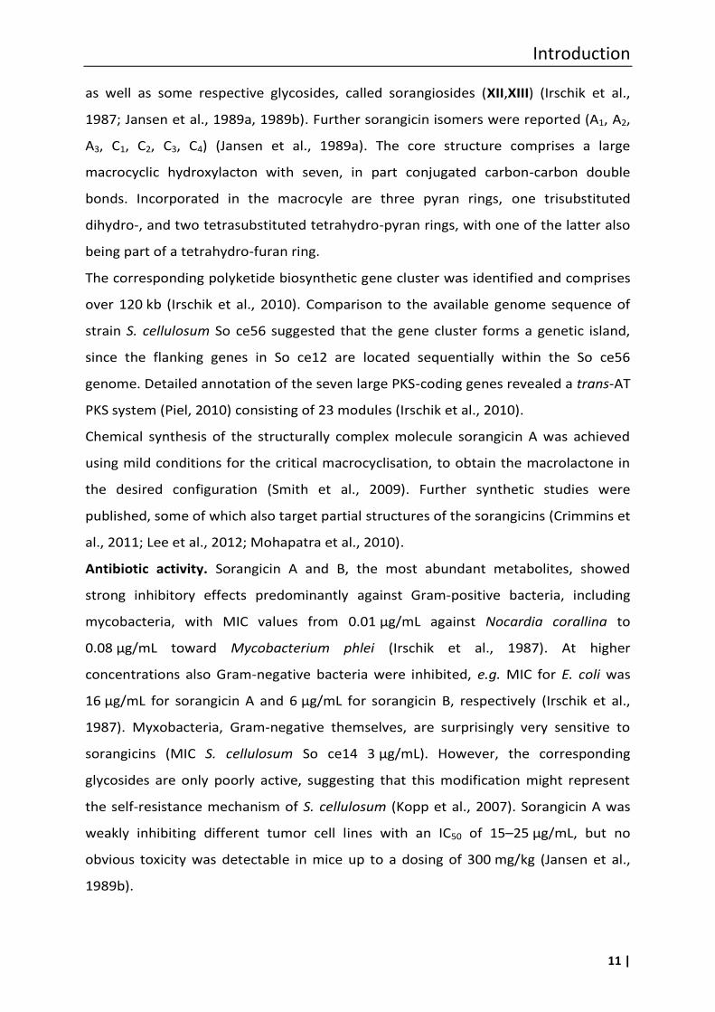
as well as some respective glycosides, called sorangiosides (XII,XIII) (Irschik et al.,
1987; Jansen et al., 1989a, 1989b). Further sorangicin isomers were reported (A1, A2,
A3, C1, C2, C3, C4) (Jansen et al., 1989a). The core structure comprises a large
macrocyclic hydroxylacton with seven, in part conjugated carbon-carbon double
bonds. Incorporated in the macrocyle are three pyran rings, one trisubstituted
dihydro-, and two tetrasubstituted tetrahydro-pyran rings, with one of the latter also
being part of a tetrahydro-furan ring.
The corresponding polyketide biosynthetic gene cluster was identified and comprises
over 120 kb (Irschik et al., 2010). Comparison to the available genome sequence of
strain S. cellulosum So ce56 suggested that the gene cluster forms a genetic island,
since the flanking genes in So ce12 are located sequentially within the So ce56
genome. Detailed annotation of the seven large PKS-coding genes revealed a trans-AT
PKS system (Piel, 2010) consisting of 23 modules (Irschik et al., 2010).
Chemical synthesis of the structurally complex molecule sorangicin A was achieved
using mild conditions for the critical macrocyclisation, to obtain the macrolactone in
the desired configuration (Smith et al., 2009). Further synthetic studies were
published, some of which also target partial structures of the sorangicins (Crimmins et
al., 2011; Lee et al., 2012; Mohapatra et al., 2010).
Antibiotic activity. Sorangicin A and B, the most abundant metabolites, showed
strong inhibitory effects predominantly against Gram-positive bacteria, including
mycobacteria, with MIC values from 0.01 µg/mL against Nocardia corallina to
0.08 µg/mL toward Mycobacterium phlei (Irschik et al., 1987). At higher
concentrations also Gram-negative bacteria were inhibited, e.g. MIC for E. coli was
16 µg/mL for sorangicin A and 6 µg/mL for sorangicin B, respectively (Irschik et al.,
1987). Myxobacteria, Gram-negative themselves, are surprisingly very sensitive to
sorangicins (MIC S. cellulosum So ce14 3 µg/mL). However, the corresponding
glycosides are only poorly active, suggesting that this modification might represent
the self-resistance mechanism of S. cellulosum (Kopp et al., 2007). Sorangicin A was
weakly inhibiting different tumor cell lines with an IC50 of 15–25 µg/mL, but no
obvious toxicity was detectable in mice up to a dosing of 300 mg/kg (Jansen et al.,
1989b).
Introduction
12 |
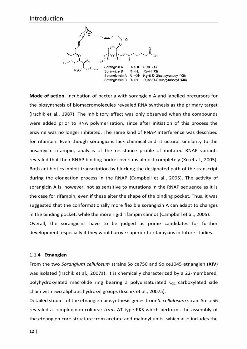
Mode of action. Incubation of bacteria with sorangicin A and labelled precursors for
the biosynthesis of biomacromolecules revealed RNA synthesis as the primary target
(Irschik et al., 1987). The inhibitory effect was only observed when the compounds
were added prior to RNA polymerisation, since after initiation of this process the
enzyme was no longer inhibited. The same kind of RNAP interference was described
for rifampin. Even though sorangicins lack chemical and structural similarity to the
ansamycin rifampin, analysis of the resistance profile of mutated RNAP variants
revealed that their RNAP binding pocket overlaps almost completely (Xu et al., 2005).
Both antibiotics inhibit transcription by blocking the designated path of the transcript
during the elongation process in the RNAP (Campbell et al., 2005). The activity of
sorangicin A is, however, not as sensitive to mutations in the RNAP sequence as it is
the case for rifampin, even if these alter the shape of the binding pocket. Thus, it was
suggested that the conformationally more flexible sorangicin A can adapt to changes
in the binding pocket, while the more rigid rifampin cannot (Campbell et al., 2005).
Overall, the sorangicins have to be judged as prime candidates for further
development, especially if they would prove superior to rifamycins in future studies.
1.1.4 Etnangien
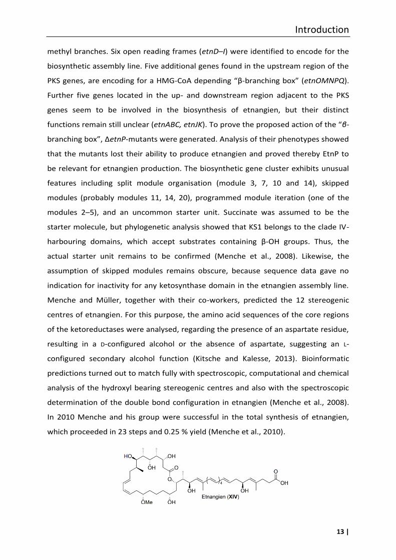
From the two Sorangium cellulosum strains So ce750 and So ce1045 etnangien (XIV)
was isolated (Irschik et al., 2007a). It is chemically characterized by a 22-membered,
polyhydroxylated macrolide ring bearing a polyunsaturated C21 carboxylated side
chain with two aliphatic hydroxyl groups (Irschik et al., 2007a).
Detailed studies of the etnangien biosynthesis genes from S. cellulosum strain So ce56
revealed a complex non-colinear trans-AT type PKS which performs the assembly of
the etnangien core structure from acetate and malonyl units, which also includes the
Introduction
13 |
methyl branches. Six open reading frames (etnD–I) were identified to encode for the
biosynthetic assembly line. Five additional genes found in the upstream region of the
PKS genes, are encoding for a HMG-CoA depending “β-branching box” (etnOMNPQ).
Further five genes located in the up- and downstream region adjacent to the PKS
genes seem to be involved in the biosynthesis of etnangien, but their distinct
functions remain still unclear (etnABC, etnJK). To prove the proposed action of the “β-
branching box”, ΔetnP-mutants were generated. Analysis of their phenotypes showed
that the mutants lost their ability to produce etnangien and proved thereby EtnP to
be relevant for etnangien production. The biosynthetic gene cluster exhibits unusual
features including split module organisation (module 3, 7, 10 and 14), skipped
modules (probably modules 11, 14, 20), programmed module iteration (one of the
modules 2–5), and an uncommon starter unit. Succinate was assumed to be the
starter molecule, but phylogenetic analysis showed that KS1 belongs to the clade IV-
harbouring domains, which accept substrates containing β-OH groups. Thus, the
actual starter unit remains to be confirmed (Menche et al., 2008). Likewise, the
assumption of skipped modules remains obscure, because sequence data gave no
indication for inactivity for any ketosynthase domain in the etnangien assembly line.
Menche and Müller, together with their co-workers, predicted the 12 stereogenic
centres of etnangien. For this purpose, the amino acid sequences of the core regions
of the ketoreductases were analysed, regarding the presence of an aspartate residue,
resulting in a D-configured alcohol or the absence of aspartate, suggesting an L-
configured secondary alcohol function (Kitsche and Kalesse, 2013). Bioinformatic
predictions turned out to match fully with spectroscopic, computational and chemical
analysis of the hydroxyl bearing stereogenic centres and also with the spectroscopic
determination of the double bond configuration in etnangien (Menche et al., 2008).
In 2010 Menche and his group were successful in the total synthesis of etnangien,
which proceeded in 23 steps and 0.25 % yield (Menche et al., 2010).
Introduction
14 |
Antibiotic activity and mode of action. Etnangien is effective against a broad panel of
Gram-positive bacteria, some belonging to the Corynebacteria like Nocardia corallia
and mycobacteria. Of special note is its antibiotic activity against rifampin-resistant
S. aureus (MIC 0.62 µg/mL) (Irschik et al., 2007a). Investigations of the DNA, RNA and
protein synthesis of etnangien-treated Micrococcus luteus cells revealed an inhibitory
effect on the formation of all of these macromolecules. Inhibition assays using
purified RNA (EcRNAP) and DNA polymerase (EcDNAP) and reverse transcriptase
(HIVRT) showed comparable dose-effect curves, with a maximal inhibition reached at
60 µg/mL etnangien. The reverse transcriptase of Moloney murine leukemia virus
(MuLVRT) was the most sensitive virus with a nearly complete inhibition at 5 µg/mL
etnangien. Although, eukaryotic DNA polymerase is a sensitive target for etnangien,
only a low toxicity against mammalian cells (IC50 of 74 µg/mL against mouse
fibroblasts cells L929) was observed (Irschik et al., 2007a). Analogs of entnangien with
an absent or a shortened polyene side chain, or a contracted macrocycle lost their
antibiotic activity, whereas the activity of the carboxy-methylester analogue was
comparable with that of the natural product (Menche et al., 2010). Derivatives with
modifications in the highly labile polyene portion of the side chain had no or merely
marginal activity (Altendorfer et al., 2012, 2013). These synthetic studies showed that
the macrocycle as well as the side chain are essential parts of the pharmacophore.
1.2 Myxobacterial antibiotics targeting bacterial protein biosynthesis
Ribosomes play a key role in all living organisms including microbes, and due to
distinct differences in their molecular structure represent an important target for
antibacterial agents. A large number of clinically useful antibiotics, e.g.
aminoglycosides and tetracyclines, target this complex machinery responsible for
protein synthesis. A few myxobacterial metabolites were identified, which interfere
with this ribonucleoprotein machinery.
1.2.1 Althiomycin
The sulphur-containing antibiotic althiomycin (XV) was first isolated in 1957 from a
Streptomyces althioticus strain (Yamaguchi et al., 1957). However, also members of
Introduction
15 |
the myxobacterial genera Cystobacter and Myxococcus are producers of this
compound (Kunze et al., 1982), as well as the insect pathogen Serratia marcescens
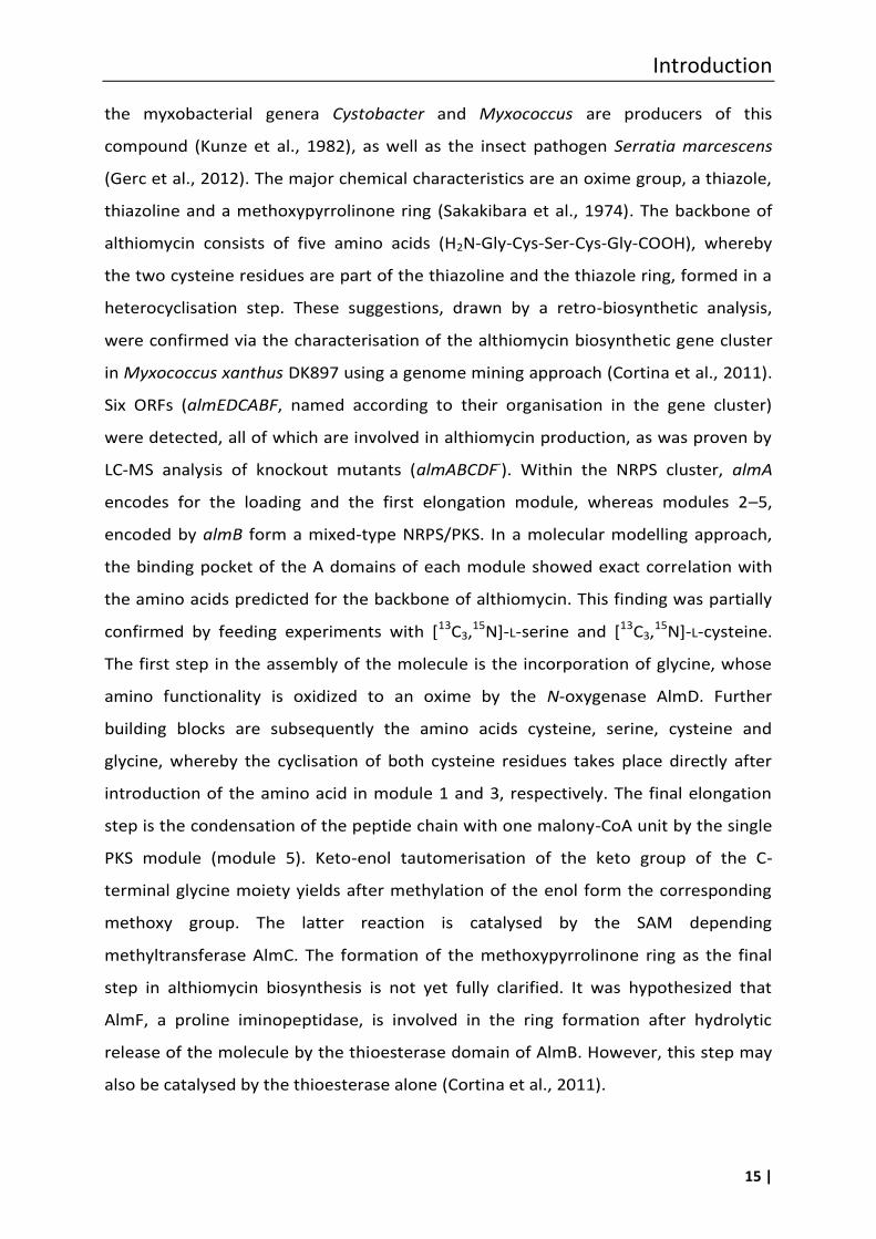
(Gerc et al., 2012). The major chemical characteristics are an oxime group, a thiazole,
thiazoline and a methoxypyrrolinone ring (Sakakibara et al., 1974). The backbone of
althiomycin consists of five amino acids (H2N-Gly-Cys-Ser-Cys-Gly-COOH), whereby
the two cysteine residues are part of the thiazoline and the thiazole ring, formed in a
heterocyclisation step. These suggestions, drawn by a retro-biosynthetic analysis,
were confirmed via the characterisation of the althiomycin biosynthetic gene cluster
in Myxococcus xanthus DK897 using a genome mining approach (Cortina et al., 2011).
Six ORFs (almEDCABF, named according to their organisation in the gene cluster)
were detected, all of which are involved in althiomycin production, as was proven by
LC-MS analysis of knockout mutants (almABCDF-). Within the NRPS cluster, almA
encodes for the loading and the first elongation module, whereas modules 2–5,
encoded by almB form a mixed-type NRPS/PKS. In a molecular modelling approach,
the binding pocket of the A domains of each module showed exact correlation with
the amino acids predicted for the backbone of althiomycin. This finding was partially
confirmed by feeding experiments with [13C3,15N]-L-serine and [13C3,15N]-L-cysteine.
The first step in the assembly of the molecule is the incorporation of glycine, whose
amino functionality is oxidized to an oxime by the N-oxygenase AlmD. Further
building blocks are subsequently the amino acids cysteine, serine, cysteine and
glycine, whereby the cyclisation of both cysteine residues takes place directly after
introduction of the amino acid in module 1 and 3, respectively. The final elongation
step is the condensation of the peptide chain with one malony-CoA unit by the single
PKS module (module 5). Keto-enol tautomerisation of the keto group of the C-
terminal glycine moiety yields after methylation of the enol form the corresponding
methoxy group. The latter reaction is catalysed by the SAM depending
methyltransferase AlmC. The formation of the methoxypyrrolinone ring as the final
step in althiomycin biosynthesis is not yet fully clarified. It was hypothesized that
AlmF, a proline iminopeptidase, is involved in the ring formation after hydrolytic
release of the molecule by the thioesterase domain of AlmB. However, this step may
also be catalysed by the thioesterase alone (Cortina et al., 2011).
Introduction
16 |
Comparative analysis of the NRPS/PKS biosynthetic gene clusters from Serratia
marcescens and Myxococcus xanthus DK897 showed similarity in the range of 59–
72 % on the protein level. The predicted functions of the biosynthetic proteins are
comparable with each other, except for the proteins (Alb6 vs. AlmF), encoded by the
sixth gene (alb6 vs. almF), which differs completely. Alb6 is predicted to be a type II
thioesterase with a proofreading function in between the NRPS/PKS machinery (Gerc
et al., 2012), whereas AlmF is proposed to be a proline iminopeptidase and may affect
the methoxypyrrolinone formation.
Antibiotic activity and mode of action
Althiomycin showed antibiotic activity against several Gram-negative and -positive
bacteria, e.g. an MIC of 6.3 µg/mL against Klebsiella pneumoniae, of 1 µg/mL against
E. coli 1852E PM, of 16 µg/mL against S. aureus 853E, and of 0.8 µg/mL against
Corynebacterium diphteriae was observed (Inami and Shiba, 1986; Zarantonello et al.,
2002).
Studies regarding the mode of action of althiomycin were performed with E. coli cells.
Monitoring the effect of althiomycin on the synthesis of DNA, RNA and proteins
revealed that althiomycin primarily inhibits protein synthesis (Fujimoto et al., 1970).
This mechanism could be confirmed by a cell free inhibition assay of polypeptide
synthesis in a ribosome system, using native mRNA. Further studies suggested that
althiomycin effects the peptide bond formation by interfering with the amino acid
bound to the A site of the ribosome. However, althiomycin did not inhibit aminoacyl-
tRNA synthesis or binding of the aminoacyl-tRNA to ribosomes. No significant
inhibition effect of althiomycin on the protein synthesis was observed in rabbit
reticulocytes. Thus, a low cytotoxicity and a good selectivity towards prokaryotic cells
may be concluded (Fujimoto et al., 1970; Inami and Shiba, 1986).
To evaluate the pharmacophore, several analogues of althiomycin have been
synthesised. In bioactivity assays only one of the synthetic althiomycin derivatives, i.e.
dehydroxymethyl-althiomycin, a molecule without the C-7 hydroxymethylene
Introduction
17 |
function, retained weak antibiotic activity. The MICs for this compound were
determined to be 32 µg/mL against S. aureus and 16 µg/mL against E. coli 1852E PM
(Zarantonello et al., 2002). From the synthetic studies it could be deduced that the
following chemical features have major impact on the antibiotic activity: (i) the
configuration of the C-10 chiral centre of the thiazoline ring, (ii) the
methoxypyrrolinone ring, (iii) the oxime moiety, and (iv) the hydroxymethyl group
(Inami and Shiba, 1986; Zarantonello et al., 2002).
It was reported that the pharmaceutical industry had some interest in the antibiotic
althiomycin (Kirst et al., 1975; Zarantonello et al., 2002), mainly because of its
antibiotic effects against Gram-negative bacteria, and despite the fact that its potency
toward several clinically relevant Gram-positives is low. Althiomycin is water-insoluble
and all efforts to modify the structure resulted in strongly decreased activity. There
seems to be no current interest in the molecule, it may however, be worthwhile to
explore SAR more extensively to exploit the lead structure offered by this natural
product.
1.2.2 Angiolam A
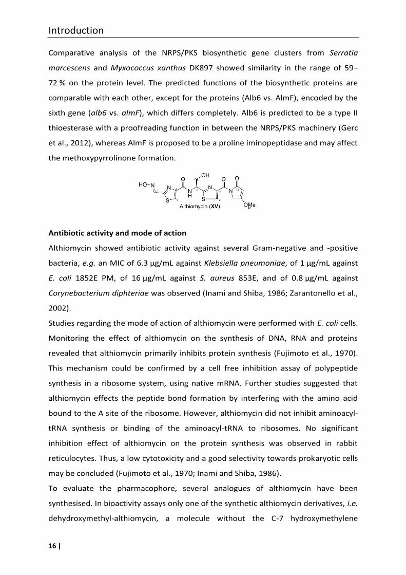
Angiolam A (XVI) is a lactam-lactone antibiotic from Angiococcus disciformis An d30
(Kohl et al., 1985). Very recently the total synthesis of angiolam A was accomplished.
The material synthesized by this 18 step procedure enabled the revision of the
absolute configuration and confirmed the C2–C3 double-bond of XVI to be E-
configured (Gieseler and Kalesse, 2014). The 19-membered macrocycle is decorated
with methyl, carbonyl and hydroxyl groups and contains a single carbon-carbon
double-bond. The side chain is monohydroxylated with three double-bonds including
a terminal diene system. To date, no data on the biosynthesis of this molecule are
available.
Introduction
18 |
Antibiotic activity and mode of action. The antibiotic activity profile was found to be
very narrow, in that only some members of the Gram-positive Bacillaceae, including
anaerobic Clostridium perfringens, were sensitive (MIC of the latter 0.78 µg/mL) (Kohl
et al., 1985). Gram-negative bacteria were in general resistant, except of E. coli
mutants with increased permeability (MIC of 2.5 µg/mL against E. coli tolC) (Kohl et
al., 1985).
The antibiotic effect was bacteriostatic. This was tested by adding up to 10 µg/mL of
angiolam A to growing Bacillus cells; the latter were subsequently still able to form
colonies. The effect on macromolecule biosynthesis revealed that protein
biosynthesis stopped completely 5 minutes after addition of angiolam A. In terms of
toxicity to mice, no acute toxicity was observed up to a dosing of 300 mg/kg
subcutaneously (s.c.) (Kunze et al., 1985).
In general it seems that the antibiotic activity of angiolam A towards only a very few
bacteria does not speak for the development of the natural product itself, unless a
narrow spectrum of activity is aimed for. It would be worthwhile though, to analyse
the activity of analogues for a potentially better profile.
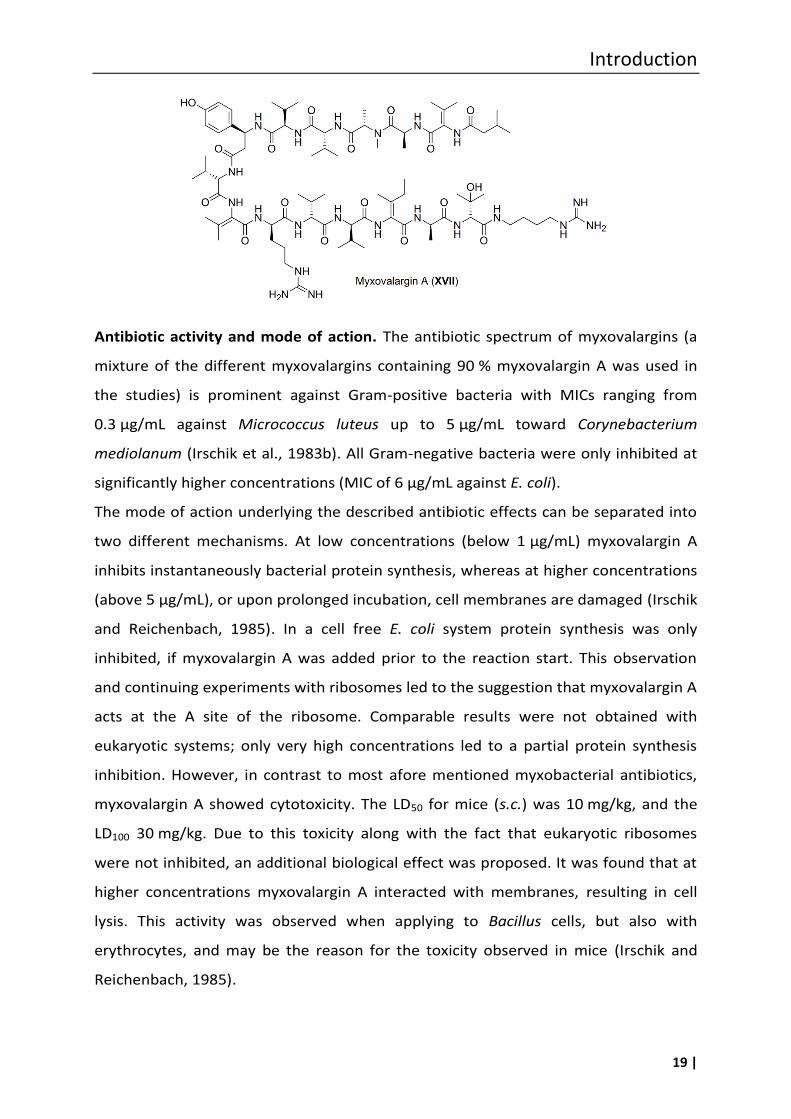
1.2.3 Myxovalargins
Myxovalargins A (XVII) and the derivatives myxovalargin B and C were obtained from
Myxococcus fulvus strain Mx f65 (Irschik et al., 1983b). These compounds are linear
peptides consisting of 14 amino acids, and hydrolysis proved that many of these are
non-proteinogenic. Among others, N-methylalanine, β-hydroxyvaline, agmatine, 3-
methylbutyric acid, α,β-dehydrovaline, α,β-dehydroleucine, and (S)-β-Tyr are
incorporated into myxovalargins. The conversion of L-Tyr into (S)-β-Tyr by the
catalytic action of the M. fulvus Mx f65 derived tyrosine aminomutase was proven
(Krug and Müller, 2009), providing this essential precursor for incorporation into the
nascent myxovalargin peptide chain. A corresponding gene cluster is not published
yet.
Introduction
19 |
Antibiotic activity and mode of action. The antibiotic spectrum of myxovalargins (a
mixture of the different myxovalargins containing 90 % myxovalargin A was used in
the studies) is prominent against Gram-positive bacteria with MICs ranging from
0.3 µg/mL against Micrococcus luteus up to 5 µg/mL toward Corynebacterium
mediolanum (Irschik et al., 1983b). All Gram-negative bacteria were only inhibited at
significantly higher concentrations (MIC of 6 µg/mL against E. coli).
The mode of action underlying the described antibiotic effects can be separated into
two different mechanisms. At low concentrations (below 1 µg/mL) myxovalargin A
inhibits instantaneously bacterial protein synthesis, whereas at higher concentrations
(above 5 µg/mL), or upon prolonged incubation, cell membranes are damaged (Irschik
and Reichenbach, 1985). In a cell free E. coli system protein synthesis was only
inhibited, if myxovalargin A was added prior to the reaction start. This observation
and continuing experiments with ribosomes led to the suggestion that myxovalargin A
acts at the A site of the ribosome. Comparable results were not obtained with
eukaryotic systems; only very high concentrations led to a partial protein synthesis
inhibition. However, in contrast to most afore mentioned myxobacterial antibiotics,
myxovalargin A showed cytotoxicity. The LD50 for mice (s.c.) was 10 mg/kg, and the
LD100 30 mg/kg. Due to this toxicity along with the fact that eukaryotic ribosomes
were not inhibited, an additional biological effect was proposed. It was found that at
higher concentrations myxovalargin A interacted with membranes, resulting in cell
lysis. This activity was observed when applying to Bacillus cells, but also with
erythrocytes, and may be the reason for the toxicity observed in mice (Irschik and
Reichenbach, 1985).
Introduction
20 |
Overall, based on the results obtained for myxovalarin A, these compounds seem to
be too toxic for an application as an antibiotic. However, it cannot be excluded today
that the derivatives B–D or other derivatives will show only minor toxicity.
1.3 Myxobacterial antibiotics targeting the respiratory chain
Two antibiotically active myxobacterial metabolites were found that target the
respiratory chain, i.e. aurachins and thuggacins. Enzymes of the respiratory chain do
not represent a classical target in antibiotic therapy, since these proteins are highly
conserved in all organisms. Therefore, the chance of toxicity is high. However, there
might be the chance of finding specific inhibitors within the variants described below.
1.3.1 Aurachins
A range of isoprenoid quinoline alkaloids were isolated from Stigmatella aurantiaca
strain SG a15, and the three main metabolites were named aurachin A (XVIII), B (XIX)
and C (XX), while D (XXI) and E (XXII) are minor products (Kunze et al., 1987). All of
these compounds share the quinoline nucleus, in some cases with the nitrogen being
present as N-oxide, and are substituted with a sesquiterpene unit. In addition to
various Stigmatella strains also Rhodococcus species were now identified as
producers of aurachins (Kitagawa et al., 2013; Nachtigall et al., 2010).
Concerning the biosynthesis, first insights were gained by feeding studies with
assumed precursors like 13C- and 18O-labelled anthranilic acid, C-1 and C-2 13C-
enriched acetate and 18O-labelled molecular oxygen ( e and un e, 2008). It was
proven that anthranilic acid is a building block of the aurachins, presenting a
biosynthetic bottle neck, since medium supplementation with anthranilic acid
increased the yield of aurachins. Unexpectedly, the farnesyl residue was constructed
in parallel via different pathways, i.e. isoprenoid biosynthesis by the mevalonate and
non-mevalonate (methyl-erythritol phosphate/deoxy-xylulose phosphate,
MEP/DOXP) pathway, as well as leucine degradation ( e and un e, 2008).
Concerning the decoration of the quinoline alkaloid moiety with an isoprenoid side
chain, biochemical investigations showed AuaA to be the responsible enzyme, in that
it catalyses the prenylation of 2-methyl-4-hydroxyquinoline in the presence of
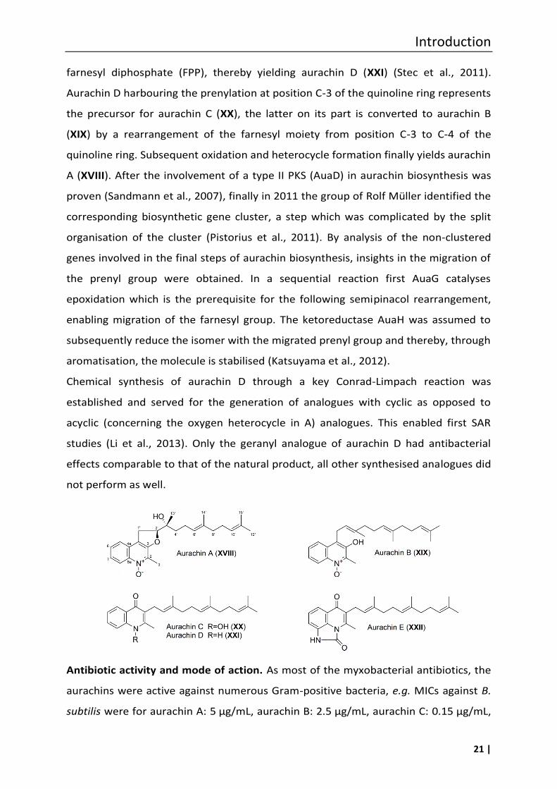
Introduction
21 |
farnesyl diphosphate (FPP), thereby yielding aurachin D (XXI) (Stec et al., 2011).
Aurachin D harbouring the prenylation at position C-3 of the quinoline ring represents
the precursor for aurachin C (XX), the latter on its part is converted to aurachin B
(XIX) by a rearrangement of the farnesyl moiety from position C-3 to C-4 of the
quinoline ring. Subsequent oxidation and heterocycle formation finally yields aurachin
A (XVIII). After the involvement of a type II PKS (AuaD) in aurachin biosynthesis was
proven (Sandmann et al., 2007), finally in 2011 the group of Rolf Müller identified the
corresponding biosynthetic gene cluster, a step which was complicated by the split
organisation of the cluster (Pistorius et al., 2011). By analysis of the non-clustered
genes involved in the final steps of aurachin biosynthesis, insights in the migration of
the prenyl group were obtained. In a sequential reaction first AuaG catalyses
epoxidation which is the prerequisite for the following semipinacol rearrangement,
enabling migration of the farnesyl group. The ketoreductase AuaH was assumed to
subsequently reduce the isomer with the migrated prenyl group and thereby, through
aromatisation, the molecule is stabilised (Katsuyama et al., 2012).
Chemical synthesis of aurachin D through a key Conrad-Limpach reaction was
established and served for the generation of analogues with cyclic as opposed to
acyclic (concerning the oxygen heterocycle in A) analogues. This enabled first SAR
studies (Li et al., 2013). Only the geranyl analogue of aurachin D had antibacterial
effects comparable to that of the natural product, all other synthesised analogues did
not perform as well.
Antibiotic activity and mode of action. As most of the myxobacterial antibiotics, the
aurachins were active against numerous Gram-positive bacteria, e.g. MICs against B.
subtilis were for aurachin A: 5 µg/mL, aurachin B: 2.5 µg/mL, aurachin C: 0.15 µg/mL,
Introduction
22 |
and aurachin D: 0.15 µg/mL. Against Gram-negative E. coli no activity of was observed
at all (Kunze et al., 1987). Additionally, a weak but incomplete inhibition of fungi was
found, e.g. MIC of aurachin A was 50 µg/mL against Debaryomyces hansenii and
Saccharoymyces cerevisiae, whereby a turbidity of up to 25 % remained. In general
the aurachins C and D were more active than A and B.
The effects of the aurachins on the NADH oxidation were tested on beef heart sub-
mitochondrial particles, due to their structural similarity to the respiratory chain
inhibitor 2-heptyl-4-hydroxyquinoline-N-oxide (HQNO). The required concentration to
reach 50 %inhibition was about ten-times lower than for HQNO (Kunze et al., 1987).
This potent inhibitory effect on the bacterial and eukaryotic respiratory chains was
the focus of subsequent biochemical studies. Thus, it was found that the cytochromes
bo and bd, both terminal oxidases of E. coli, were inhibited by aurachin C, whereas
aurachin D and its analogues showed selectivity for inhibition of cytochrome bd
(Meunier et al., 1995). Using a chemically synthesized derivative, i.e. decyl-aurachin D,
it was shown that this molecule acts on the donor side of haem b-558, thereby
preventing electron flow from the quinol substrate (Jünemann et al., 1997). In the
following, the aurachins became useful tools for probing of the ubiquinol-binding site
in cytochromes, due to their strong inhibitory effect on the respiratory chains (Mogi
et al., 2006).
From early on, the aurachins were suspected to have an antimalarial activity, due to
their structural similarity with antiplasmodial drugs. This was proven by a first in vitro
screening against Plasmodium falciparum provided by the WHO (Geneva). Indeed,
aurachins C (IC50 [ng/mL] 26/0.9) and E (13/0.4) showed good activity against P.
falciparum clones W-2 and D-6, respectively. These values are comparable to those of
chloroquine (35/1.2) and artemisinine (0.43/1.1) ( e et al., 2008). Further, it was
found that the derivative aurachin E (XXII), in contrast to the aurachins A–D, did not
show mitochondrial respiratory inhibition and had a low cytotoxicity. The IC50 against
mouse fibroblasts L929 was 25 µg/mL for aurachin E (XXII), compared to values
between 1.3 and 3.2 µg/mL for the derivatives A–D. The rare E variant can be
obtained in a semisynthetic approach by using a one-step reaction starting with the
better accessible aurachin C ( e et al., 2008). However, no in vivo activity was
Introduction
23 |
observed in a murine malaria model with Plasmodium berghei at 100 mg/kg, whereas
chloroquine showed an ED90 of 2.8 mg/kg (Milhous et al., 1985).
1.3.2 Thuggacins
Three thiazole-containing macrolides (XXIII-XXVIII) were isolated in 2007 from
Sorangium cellulosum strain So ce895 (Steinmetz et al., 2007). Due to their origin they
were named Soce-thuggacin A (XXIII), B (XXIV) and C (XXV) (sometimes, however only
named thuggacins). A further compound identified in S. cellulosum strain So ce895
was 13-methyl-thuggacin A (XXVI). Special features of Soce-thuggacin A are, besides
the thiazole ring a diene moiety (11E, 13Z), an α,β unsaturated lactone with an n-
hexyl side chain attached at C-2 and, additionally a side chain at C-16 containing three
hydroxyl and a diene functionality. In solution Soce-thuggacin A, a 17-membered
macrolide, rearranges under acyl migration to give Soce-thuggacin B, a 18-membered
macrolide, and Soce-thuggacin C, a 19-membered macrolide. For the determination
of the stereochemistry of Soce-thuggacins A–C a combination of chemical methods
was applied, e.g. chemical derivatisation, NMR studies, molecular modelling and
bioinformatic analysis of the ketoreductase domains of the biosynthetic genes (TugA,
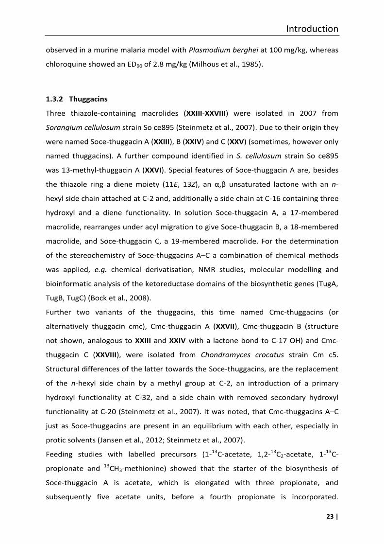
TugB, TugC) (Bock et al., 2008).
Further two variants of the thuggacins, this time named Cmc-thuggacins (or
alternatively thuggacin cmc), Cmc-thuggacin A (XXVII), Cmc-thuggacin B (structure
not shown, analogous to XXIII and XXIV with a lactone bond to C-17 OH) and Cmc-
thuggacin C (XXVIII), were isolated from Chondromyces crocatus strain Cm c5.
Structural differences of the latter towards the Soce-thuggacins, are the replacement
of the n-hexyl side chain by a methyl group at C-2, an introduction of a primary
hydroxyl functionality at C-32, and a side chain with removed secondary hydroxyl
functionality at C-20 (Steinmetz et al., 2007). It was noted, that Cmc-thuggacins A–C
just as Soce-thuggacins are present in an equilibrium with each other, especially in
protic solvents (Jansen et al., 2012; Steinmetz et al., 2007).
Feeding studies with labelled precursors (1-13C-acetate, 1,2-13C2-acetate, 1-13C-
propionate and 13CH3-methionine) showed that the starter of the biosynthesis of
Soce-thuggacin A is acetate, which is elongated with three propionate, and
subsequently five acetate units, before a fourth propionate is incorporated.
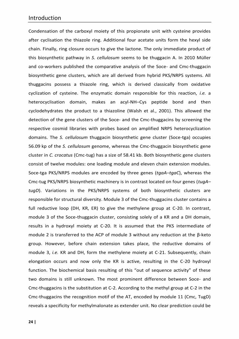
Introduction
24 |
Condensation of the carboxyl moiety of this propionate unit with cysteine provides
after cyclisation the thiazole ring. Additional four acetate units form the hexyl side
chain. Finally, ring closure occurs to give the lactone. The only immediate product of
this biosynthetic pathway in S. cellulosum seems to be thuggacin A. In 2010 Müller
and co-workers published the comparative analysis of the Soce- and Cmc-thuggacin
biosynthetic gene clusters, which are all derived from hybrid PKS/NRPS systems. All
thuggacins possess a thiazole ring, which is derived classically from oxidative
cyclization of cysteine. The enzymatic domain responsible for this reaction, i.e. a
heterocyclisation domain, makes an acyl-NH–Cys peptide bond and then
cyclodehydrates the product to a thiazoline (Walsh et al., 2001). This allowed the
detection of the gene clusters of the Soce- and the Cmc-thuggacins by screening the
respective cosmid libraries with probes based on amplified NRPS heterocyclization
domains. The S. cellulosum thuggacin biosynthetic gene cluster (Soce-tga) occupies
56.09 kp of the S. cellulosum genome, whereas the Cmc-thuggacin biosynthetic gene
cluster in C. crocatus (Cmc-tug) has a size of 58.41 kb. Both biosynthetic gene clusters
consist of twelve modules: one loading module and eleven chain extension modules.
Soce-tga PKS/NRPS modules are encoded by three genes (tgaA–tgaC), whereas the
Cmc-tug PKS/NRPS biosynthetic machinery is in contrast located on four genes (tugA–
tugD). Variations in the PKS/NRPS systems of both biosynthetic clusters are
responsible for structural diversity. Module 3 of the Cmc-thuggacins cluster contains a
full reductive loop (DH, KR, ER) to give the methylene group at C-20. In contrast,
module 3 of the Soce-thuggacin cluster, consisting solely of a KR and a DH domain,
results in a hydroxyl moiety at C-20. It is assumed that the PKS intermediate of
module 2 is transferred to the ACP of module 3 without any reduction at the β-keto
group. However, before chain extension takes place, the reductive domains of
module 3, i.e. KR and DH, form the methylene moiety at C-21. Subsequently, chain
elongation occurs and now only the KR is active, resulting in the C-20 hydroxyl
function. The biochemical basis resulting of this “out of sequence activity” of these
two domains is still unknown. The most prominent difference between Soce- and
Cmc-thuggacins is the substitution at C-2. According to the methyl group at C-2 in the
Cmc-thuggacins the recognition motif of the AT, encoded by module 11 (Cmc, TugD)
reveals a specificity for methylmalonate as extender unit. No clear prediction could be
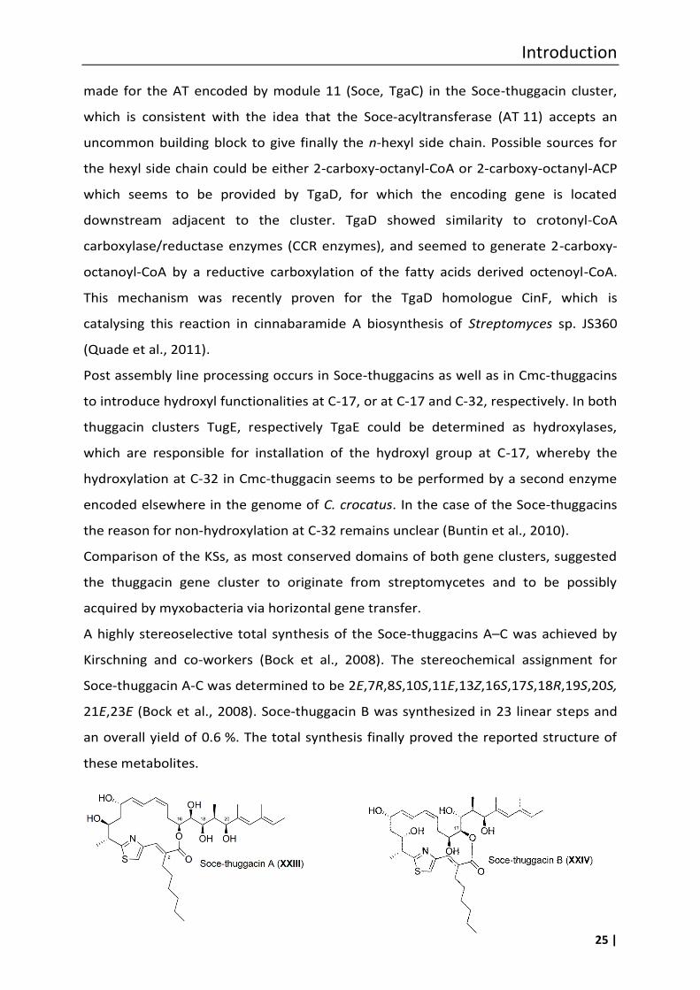
Introduction
25 |
made for the AT encoded by module 11 (Soce, TgaC) in the Soce-thuggacin cluster,
which is consistent with the idea that the Soce-acyltransferase (AT 11) accepts an
uncommon building block to give finally the n-hexyl side chain. Possible sources for
the hexyl side chain could be either 2-carboxy-octanyl-CoA or 2-carboxy-octanyl-ACP
which seems to be provided by TgaD, for which the encoding gene is located
downstream adjacent to the cluster. TgaD showed similarity to crotonyl-CoA
carboxylase/reductase enzymes (CCR enzymes), and seemed to generate 2-carboxy-
octanoyl-CoA by a reductive carboxylation of the fatty acids derived octenoyl-CoA.
This mechanism was recently proven for the TgaD homologue CinF, which is
catalysing this reaction in cinnabaramide A biosynthesis of Streptomyces sp. JS360
(Quade et al., 2011).
Post assembly line processing occurs in Soce-thuggacins as well as in Cmc-thuggacins
to introduce hydroxyl functionalities at C-17, or at C-17 and C-32, respectively. In both
thuggacin clusters TugE, respectively TgaE could be determined as hydroxylases,
which are responsible for installation of the hydroxyl group at C-17, whereby the
hydroxylation at C-32 in Cmc-thuggacin seems to be performed by a second enzyme
encoded elsewhere in the genome of C. crocatus. In the case of the Soce-thuggacins
the reason for non-hydroxylation at C-32 remains unclear (Buntin et al., 2010).
Comparison of the KSs, as most conserved domains of both gene clusters, suggested
the thuggacin gene cluster to originate from streptomycetes and to be possibly
acquired by myxobacteria via horizontal gene transfer.
A highly stereoselective total synthesis of the Soce-thuggacins A–C was achieved by
Kirschning and co-workers (Bock et al., 2008). The stereochemical assignment for
Soce-thuggacin A-C was determined to be 2E,7R,8S,10S,11E,13Z,16S,17S,18R,19S,20S,
21E,23E (Bock et al., 2008). Soce-thuggacin B was synthesized in 23 linear steps and
an overall yield of 0.6 %. The total synthesis finally proved the reported structure of
these metabolites.
Introduction
26 |
Antibiotic activity and mode of action
Soce-thuggacin A showed inhibitory activity against Gram-positive bacteria like
Micrococcus luteus (MIC 3 ng/mL). Of special interest is its activity toward several
mycobacteria, i.e. MIC against Mycobacterium phlei was found to be 0.03 µg/mL, M.
chitae 0.60 µg/mL and M. tuberculosis 8.0 µg/mL. The activities of Soce-thuggacin A
and B were similar to each other, but Soce-thuggacin C was much less active (Irschik
et al., 2007b). Antibacterial effects, most importantly also against Mycobacteria, was
demonstrated as well for the cmc-thuggacins (Walsh et al., 2001).
Mode of action studies on Soce-thuggacin A were performed using M. luteus. After
addition of Soce-thuggacin A to the bacterium, the first observation using radioactive
precursors was the immediate stop of macromolecule synthesis like that of DNA, RNA
and proteins. Further, Irschik and co-workers could show, due to the observation of
the oxygen consumption of M. luteus, that a total inhibition of respiration was
reached with 2.5 ng/mL Soce-thuggacin A (Irschik et al., 2007b). Experiments with
cytoplasmatic membranes gave evidence for the inhibition of the NADH oxidase (90 %
inhibition at 10 ng/mL Soce-thuggacin A). Additionally, the inhibition of the reduction
of cytochromes a, b and c by NADH in membranes was detected. Summing up, Soce-
thuggacin A seems to inhibit late stages of the respiratory chain which results in a
disruption of the energy supply for the cells. The toxicity test against mouse fibroblast
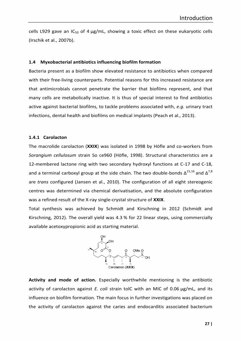
Introduction
27 |
cells L929 gave an IC50 of 4 µg/mL, showing a toxic effect on these eukaryotic cells
(Irschik et al., 2007b).
1.4 Myxobacterial antibiotics influencing biofilm formation
Bacteria present as a biofilm show elevated resistance to antibiotics when compared
with their free-living counterparts. Potential reasons for this increased resistance are
that antimicrobials cannot penetrate the barrier that biofilms represent, and that
many cells are metabolically inactive. It is thus of special interest to find antibiotics
active against bacterial biofilms, to tackle problems associated with, e.g. urinary tract
infections, dental health and biofilms on medical implants (Peach et al., 2013).
1.4.1 Carolacton
The macrolide carolacton (XXIX) was isolated in 1998 by Höfle and co-workers from
Sorangium cellulosum strain So ce960 (Höfle, 1998). Structural characteristics are a
12-membered lactone ring with two secondary hydroxyl functions at C-17 and C-18,
and a terminal carboxyl group at the side chain. The two double-bonds Δ15,16 and Δ7,8
are trans configured (Jansen et al., 2010). The configuration of all eight stereogenic
centres was determined via chemical derivatisation, and the absolute configuration
was a refined result of the X-ray single-crystal structure of XXIX.
Total synthesis was achieved by Schmidt and Kirschning in 2012 (Schmidt and
Kirschning, 2012). The overall yield was 4.3 % for 22 linear steps, using commercially
available acetoxypropionic acid as starting material.
Activity and mode of action. Especially worthwhile mentioning is the antibiotic
activity of carolacton against E. coli strain tolC with an MIC of 0.06 µg/mL, and its
influence on biofilm formation. The main focus in further investigations was placed on
the activity of carolacton against the caries and endocarditis associated bacterium
Introduction
28 |
Streptococcus mutans. The latter is able to form biofilms and proved to be sensitive
towards carolacton. At a concentration of 5 ng/mL, 33 % of the cells in the biofilm
died, whereas a concentration of 25 ng/mL resulted in 66 % dead cells (Jansen et al.,
2010). Inhibition of biofilms in nanomolar concentrations implied that carolacton
addresses a primary target, present only in a few copies per cell (Reck et al., 2011).
Wagner-Döbler and co-workers suggested carolacton to target quorum sensing
systems of S. mutans (Kunze et al., 2010). With the help of LIVE/DEAD BacLight
bacterial viability staining, they gained a dose-response-relationship curve which
showed a sigmoidal shape with a low threshold of 10 nM. No substantial increase of
activity above this concentration was observed, which indicated that carolacton
targets a signalling pathway. Confocal laser scanning microscopic images with
LIVE/DEAD stained carolacton treated S. mutans cells visualized changes in cell
morphology. Cells were elongated, bulged, and the number of bacterial cells stringed
together in one chain increased (Kunze et al., 2010). Further, the applied method
indicated that carlocaton induces membrane damages, which was confirmed by the
detection of cytoplasmatic proteins and external DNA in an analysis (SDS-page and
quantitative PCR) of the supernatant of carolacton treated biofilms. A time related
profile of the transcriptional response of S. mutans to carolacton treatment indicated
the regulation of genes with an impact on biofilm formation, autolysis, cell shape, cell
division and pyrimidine and histidine metabolism. The investigation of correlated
two-component signal transduction systems (TCS) (e.g. VicKRX, SMU.1037c/1038c,
SMU.659/660 and ComDE) revealed that carolacton mainly interacts with the
serine/threonine protein kinase (STPK) PknB (Banu et al., 2010). The latter conclusion
could be confirmed by the insensitivity of a pknB deletion mutant S. mutans EA 72 to
carolacton treatment (Banu et al., 2010). These conclusions were further supported
by the close similarity of the transcriptome of the pknB deletion mutant with that of
the carolacton treated biofilm.
Summing up, recent insights into the mode of action of carolacton indicated that this
compound interferes with the STPK PknB and hence with PknB-mediated signalling.
This in turn influences pyrimidine biosynthesis, cell wall and biofilm formation, as well
as the ComDE mediated bacteriocin production. The alterations in cell wall
composition result in weakened cell walls, leading to loss of integrity at low pH and
Introduction
29 |
leakage of cytoplasmatic proteins and DNA, and finally cause cell death (Reck et al.,
2011). In 2013 the biofilm reducing activity of carolacton in comparison to
chlorhexidine and triclosan was reported (Apel et al., 2013). The incorporation of
XXIX at a concentration of 0.002 %in dental filling material gave promising results. No
adverse effect on the mechanical properties of the latter, and a significant effect on
biofilm-formation were observed.
Antifungal activity against Aspergillus niger, Pythium debaryanum and Sclerotina
sclerotiorum in the range of 16–20 µg/mL was also reported. In 2002 the scaffold of
carolacton was protected by a Japanese patent (Ishihara et al., 2002).
1.5 Myxobacterial antibiotics targeting the type II signal peptidase LspA
The novel target LspA is part of the lipoprotein processing system, which is essential
in all Gram-negative bacteria, and can be conditionally essential in Gram-positives.
Lipoproteins are ubiquitous in bacteria, play an important role in viability, and are
also key factors in pathogenesis, since these outer membrane proteins represent
virulence factors (Kovacs-Simon et al., 2011). In eukaryotic cells instead, LspA is
absent, making it an attractive target for antibiotics (Hutchings et al., 2009; Tjalsma,
1999; Xiao et al., 2012). Recently, the activity of a myxobacterial compound against
LspA was described (Xiao et al., 2012).
1.5.1 Myxovirescins
The myxovirescin family is represented by closely related antibiotics produced by
many myxobacterial strains, whereby the first isolation and structure elucidation was
performed using Myxococcus virescens strain Mx v48 (Gerth et al., 1982). The basic
structure of the molecules is a 28-membered ring, with a lactone and a lactam
functionality. Several reports on these compounds can be found in the literature, in
which myxovirescin A1 (XXX) is also named as antibiotic TA (from strain Tel Aviv),
megovalicin, or M-230B (Onishi et al., 1984; Takayama et al., 1988).
The biosynthetic gene cluster coding for the enzymes responsible for the assembly of
this mixed PKS/NRPS-derived product (XXX) was identified in the genome-sequenced
strain Myxococcus xanthus DK1622. The cluster has a size of approximately 83 kb and
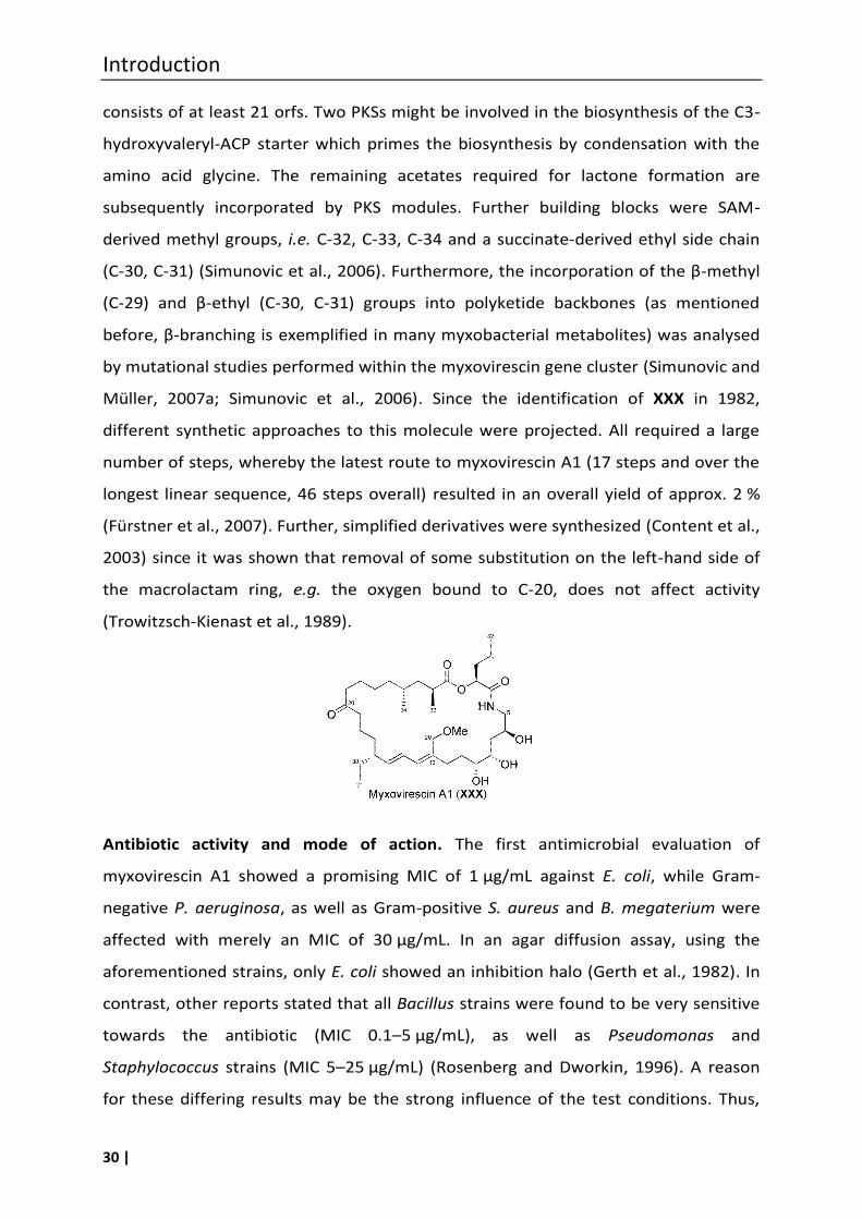
Introduction
30 |
consists of at least 21 orfs. Two PKSs might be involved in the biosynthesis of the C3-
hydroxyvaleryl-ACP starter which primes the biosynthesis by condensation with the
amino acid glycine. The remaining acetates required for lactone formation are
subsequently incorporated by PKS modules. Further building blocks were SAM-
derived methyl groups, i.e. C-32, C-33, C-34 and a succinate-derived ethyl side chain
(C-30, C-31) (Simunovic et al., 2006). Furthermore, the incorporation of the β-methyl
(C-29) and β-ethyl (C-30, C-31) groups into polyketide backbones (as mentioned
before, β-branching is exemplified in many myxobacterial metabolites) was analysed
by mutational studies performed within the myxovirescin gene cluster (Simunovic and
Müller, 2007a; Simunovic et al., 2006). Since the identification of XXX in 1982,
different synthetic approaches to this molecule were projected. All required a large
number of steps, whereby the latest route to myxovirescin A1 (17 steps and over the
longest linear sequence, 46 steps overall) resulted in an overall yield of approx. 2 %
(Fürstner et al., 2007). Further, simplified derivatives were synthesized (Content et al.,
2003) since it was shown that removal of some substitution on the left-hand side of
the macrolactam ring, e.g. the oxygen bound to C-20, does not affect activity
(Trowitzsch-Kienast et al., 1989).
Antibiotic activity and mode of action. The first antimicrobial evaluation of
myxovirescin A1 showed a promising MIC of 1 µg/mL against E. coli, while Gram-
negative P. aeruginosa, as well as Gram-positive S. aureus and B. megaterium were
affected with merely an MIC of 30 µg/mL. In an agar diffusion assay, using the
aforementioned strains, only E. coli showed an inhibition halo (Gerth et al., 1982). In
contrast, other reports stated that all Bacillus strains were found to be very sensitive
towards the antibiotic (MIC 0.1–5 µg/mL), as well as Pseudomonas and
Staphylococcus strains (MIC 5–25 µg/mL) (Rosenberg and Dworkin, 1996). A reason
for these differing results may be the strong influence of the test conditions. Thus,
Introduction
31 |
sensitive E. coli could be protected by the addition of calcium or bovine serum. In in
vivo assays XXX did not protect mice infected with lethal doses of (not further
specified) pathogenic bacteria (Rosenberg and Dworkin, 1996). This was due to the
strong tissue binding properties of the compound (Rosenberg et al., 1984). Thus,
myxovirescin A1 was not distributed in the organism as such, however retained its
activity when bound to membranes or surfaces (Rosenberg and Dworkin, 1996). Due
to its high adhesive properties, the compound was tested with 8 human volunteers
suffering from gingivitis. They were treated with 4 applications of 0.1 mg
myxovirescin A1. In this experiment the 3 indices, i.e. plaque, gingival and bleeding,
showed a rapid decrease (Manor et al., 1989). Concerning toxicity it was reported
that “relatively large doses of myxovirescin A were not toxic to rats” (Rosenberg and
Dworkin, 1996). The bactericidal activity of myxovirescin A1 needs ongoing protein
synthesis and thus cell metabolism, which indicated that the compound kills bacteria
in a target-specific manner. Analysis of the resistance mechanism of several E. coli
mutants led to the conclusion that the type II signal peptidase (LspA) is the target of
myxovirescin A1, since overexpression of LspA, and also inactivation of lpp (coding for
the outer membrane “Braun’s” lipoprotein), both specifically conferred resistance to
E. coli. (Xiao et al., 2012). Thus, two consequences of the LspA interaction could be
responsible for the bactericidal effect: (i) a toxic build-up of Lpp inside the cell leading
to lethal cross-linking of the cell wall and the inner membrane, and (ii) the inhibition
of the proper localization of essential lipoproteins to the outer membrane (Xiao et al.,
2012). This conclusion is also supported by the fact that the biosynthetic gene cluster
of myxovirescin encodes two lspA paralogs that might play a role in self-resistance of
the producer strain. The identification of the molecular target, i.e. LspA, for
myxovirescin A1 opened up new opportunities for lead optimization. LspA represents
a novel antibiotic target and therefore the highly active myxovirescin A1 can be seen
as a promising lead structure for further studies.
Introduction
32 |
1.6 Myxobacterial antibiotics with an unknown mode of action
1.6.1 Chondrochlorens
Chondromyces crocatus strain Cm c5, reported as the producer of thuggacins (see
above), was also found to produce the chondrochlorens. 2003 the group of Höfle
reported two chloro-hydroxy-styryl amides, the characteristic styrene moiety of
which is linked by an amide bond with a 14-membered aliphatic side chain (Jansen et
al., 2003). Chondrochloren A (XXXI) and B (XXXII) differ merely concerning the
methoxy or ethoxy functionality at C-2, respectively.
Sequencing efforts of a cosmid library of the genome of C. crocatus strain Cm c5 led
to the elucidation of the biosynthetic gene cluster of the chondrochlorens, which
spans over a contiguous stretch of ~68 kb on the C. crocatus chromosome (Rachid et
al., 2009). The biosynthetic mixed type PKS/NRPS gene cluster consists of 10 core
genes (cndA–cndF) and several post PKS/NRPS processing genes (cndI, encoding for a
SAM depended methyltransferase; cndH, encoding for a tyrosine halogenase; cndG,
encoding for an oxidative decarboxylase). The genetic architecture and the scaffold of
the natural products are highly colinear. The biosynthetic process uses butyrate as
the starter unit, succeeded by the condensation with three methylmalonate units,
and subsequently with two methoxymalonyl units. Finally, NRPS related proteins
incorporate tyrosine or 3-chloro-L-tyrosine into the PKS chain, and then the molecule
is released from the biosynthetic enzymes by a thioesterase. The FAD-linked oxidative
carboxylase CndG is responsible for tyrosine decarboxylation from pre-
chondrochlorens A and B, which yields the styryl moiety in chondrochlorens (Rachid
et al., 2010).
Investigation of the chlorination reaction performed by CndH implied that this occurs
before release of the intermediate from the assembly line. This was supported by the
fact that a cndH-deficient mutant did neither produce chondrochloren A nor B,
whereas the biosynthesis of chondrochlorens could be restored to a good extent (i.e.
approx. to 50 %) by the addition of 3-chloro-L-tyrosin. Nevertheless, investigation of
the corresponding A-domain (encoded by cndF) in an ATP-PPi exchange assay
exhibited an insignificant preference of CndF for 3-chloro-L-tyrosine as compared with
L-tyrosine (Rachid et al., 2009). CndH belongs to the FAD-dependent halogenases
which are closely related to the FAD-dependent aromatic hydroxylases
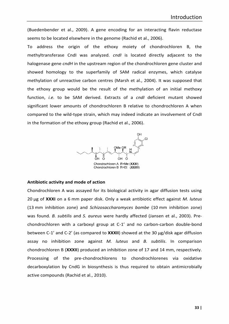
Introduction
33 |
(Buedenbender et al., 2009). A gene encoding for an interacting flavin reductase
seems to be located elsewhere in the genome (Rachid et al., 2006).
To address the origin of the ethoxy moiety of chondrochloren B, the
methyltransferase CndI was analyzed. cndI is located directly adjacent to the
halogenase gene cndH in the upstream region of the chondrochloren gene cluster and
showed homology to the superfamily of SAM radical enzymes, which catalyse
methylation of unreactive carbon centres (Marsh et al., 2004). It was supposed that
the ethoxy group would be the result of the methylation of an initial methoxy
function, i.e. to be SAM derived. Extracts of a cndI deficient mutant showed
significant lower amounts of chondrochloren B relative to chondrochloren A when
compared to the wild-type strain, which may indeed indicate an involvement of CndI
in the formation of the ethoxy group (Rachid et al., 2006).
Antibiotic activity and mode of action
Chondrochloren A was assayed for its biological activity in agar diffusion tests using
20 µg of XXXI on a 6 mm paper disk. Only a weak antibiotic effect against M. luteus
(13 mm inhibition zone) and Schizosaccharomyces bombe (10 mm inhibition zone)
was found. B. subtilis and S. aureus were hardly affected (Jansen et al., 2003). Pre-
chondrochloren with a carboxyl group at C-1’ and no carbon-carbon double-bond
between C-1’ and C-2’ (as compared to XXXII) showed at the 30 µg/disk agar diffusion
assay no inhibition zone against M. luteus and B. subtilis. In comparison
chondrochloren B (XXXII) produced an inhibition zone of 17 and 14 mm, respectively.
Processing of the pre-chondrochlorens to chondrochlorenes via oxidative
decarboxylation by CndG in biosynthesis is thus required to obtain antimicrobially
active compounds (Rachid et al., 2010).
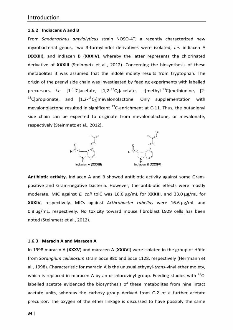
Introduction
34 |
1.6.2 Indiacens A and B
From Sandaracinus amylolyticus strain NOSO-4T, a recently characterized new
myxobacterial genus, two 3-formylindol derivatives were isolated, i.e. indiacen A
(XXXIII), and indiacen B (XXXIV), whereby the latter represents the chlorinated
derivative of XXXIII (Steinmetz et al., 2012). Concerning the biosynthesis of these
metabolites it was assumed that the indole moiety results from tryptophan. The
origin of the prenyl side chain was investigated by feeding experiments with labelled
precursors, i.e. [1-13C]acetate, [1,2-13C2]acetate, L-[methyl-13C]methionine, [2-
13C]propionate, and [1,2-13C2]mevalonolactone. Only supplementation with
mevalonolactone resulted in significant 13C-enrichment at C-11. Thus, the butadienyl
side chain can be expected to originate from mevalonolactone, or mevalonate,
respectively (Steinmetz et al., 2012).
Antibiotic activity. Indiacen A and B showed antibiotic activity against some Gram-
positive and Gram-negative bacteria. However, the antibiotic effects were mostly
moderate. MIC against E. coli tolC was 16.6 µg/mL for XXXIII, and 33.0 µg/mL for
XXXIV, respectively. MICs against Arthrobacter rubellus were 16.6 µg/mL and
0.8 µg/mL, respectively. No toxicity toward mouse fibroblast L929 cells has been
noted (Steinmetz et al., 2012).
1.6.3 Maracin A and Maracen A
In 1998 maracin A (XXXV) and maracen A (XXXVI) were isolated in the group of Höfle
from Sorangium cellulosum strain Soce 880 and Soce 1128, respectively (Herrmann et
al., 1998). Characteristic for maracin A is the unusual ethynyl-trans-vinyl ether moiety,
which is replaced in maracen A by an α-chlorovinyl group. Feeding studies with 13C-
labelled acetate evidenced the biosynthesis of these metabolites from nine intact
acetate units, whereas the carboxy group derived from C-2 of a further acetate
precursor. The oxygen of the ether linkage is discussed to have possibly the same
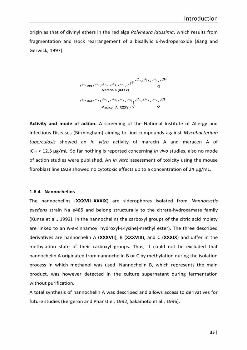
Introduction
35 |
origin as that of divinyl ethers in the red alga Polyneura latissima, which results from
fragmentation and Hock rearrangement of a bisallylic 6-hydroperoxide (Jiang and
Gerwick, 1997).
Activity and mode of action. A screening of the National Institute of Allergy and
Infectious Diseases (Birmingham) aiming to find compounds against Mycobacterium
tuberculosis showed an in vitro activity of maracin A and maracen A of
IC99 < 12.5 µg/mL. So far nothing is reported concerning in vivo studies, also no mode
of action studies were published. An in vitro assessment of toxicity using the mouse
fibroblast line L929 showed no cytotoxic effects up to a concentration of 24 µg/mL.
1.6.4 Nannochelins
The nannochelins (XXXVII–XXXIX) are siderophores isolated from Nannocystis
exedens strain Na e485 and belong structurally to the citrate-hydroxamate family
(Kunze et al., 1992). In the nannochelins the carboxyl groups of the citric acid moiety
are linked to an N-ε-cinnamoyl hydroxyl-L-lysine(-methyl ester). The three described
derivatives are nannochelin A (XXXVII), B (XXXVIII), and C (XXXIX) and differ in the
methylation state of their carboxyl groups. Thus, it could not be excluded that
nannochelin A originated from nannochelin B or C by methylation during the isolation
process in which methanol was used. Nannochelin B, which represents the main
product, was however detected in the culture supernatant during fermentation
without purification.
A total synthesis of nannochelin A was described and allows access to derivatives for
future studies (Bergeron and Phanstiel, 1992; Sakamoto et al., 1996).
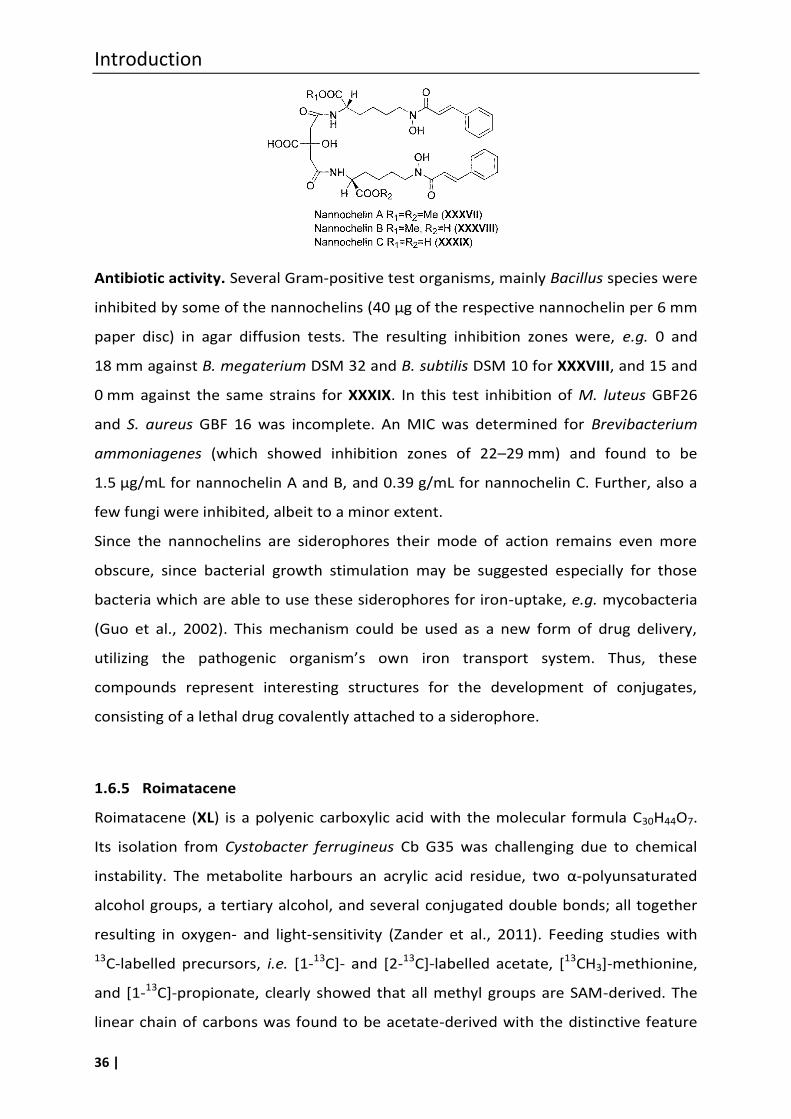
Introduction
36 |
Antibiotic activity. Several Gram-positive test organisms, mainly Bacillus species were
inhibited by some of the nannochelins (40 µg of the respective nannochelin per 6 mm
paper disc) in agar diffusion tests. The resulting inhibition zones were, e.g. 0 and
18 mm against B. megaterium DSM 32 and B. subtilis DSM 10 for XXXVIII, and 15 and
0 mm against the same strains for XXXIX. In this test inhibition of M. luteus GBF26
and S. aureus GBF 16 was incomplete. An MIC was determined for Brevibacterium
ammoniagenes (which showed inhibition zones of 22–29 mm) and found to be
1.5 µg/mL for nannochelin A and B, and 0.39 g/mL for nannochelin C. Further, also a
few fungi were inhibited, albeit to a minor extent.
Since the nannochelins are siderophores their mode of action remains even more
obscure, since bacterial growth stimulation may be suggested especially for those
bacteria which are able to use these siderophores for iron-uptake, e.g. mycobacteria
(Guo et al., 2002). This mechanism could be used as a new form of drug delivery,
utilizing the pathogenic organism’s own iron transport system. Thus, these
compounds represent interesting structures for the development of conjugates,
consisting of a lethal drug covalently attached to a siderophore.
1.6.5 Roimatacene
Roimatacene (XL) is a polyenic carboxylic acid with the molecular formula C30H44O7.
Its isolation from Cystobacter ferrugineus Cb G35 was challenging due to chemical
instability. The metabolite harbours an acrylic acid residue, two α-polyunsaturated
alcohol groups, a tertiary alcohol, and several conjugated double bonds; all together
resulting in oxygen- and light-sensitivity (Zander et al., 2011). Feeding studies with
13C-labelled precursors, i.e. [1-13C]- and [2-13C]-labelled acetate, [13CH3]-methionine,
and [1-13C]-propionate, clearly showed that all methyl groups are SAM-derived. The
linear chain of carbons was found to be acetate-derived with the distinctive feature
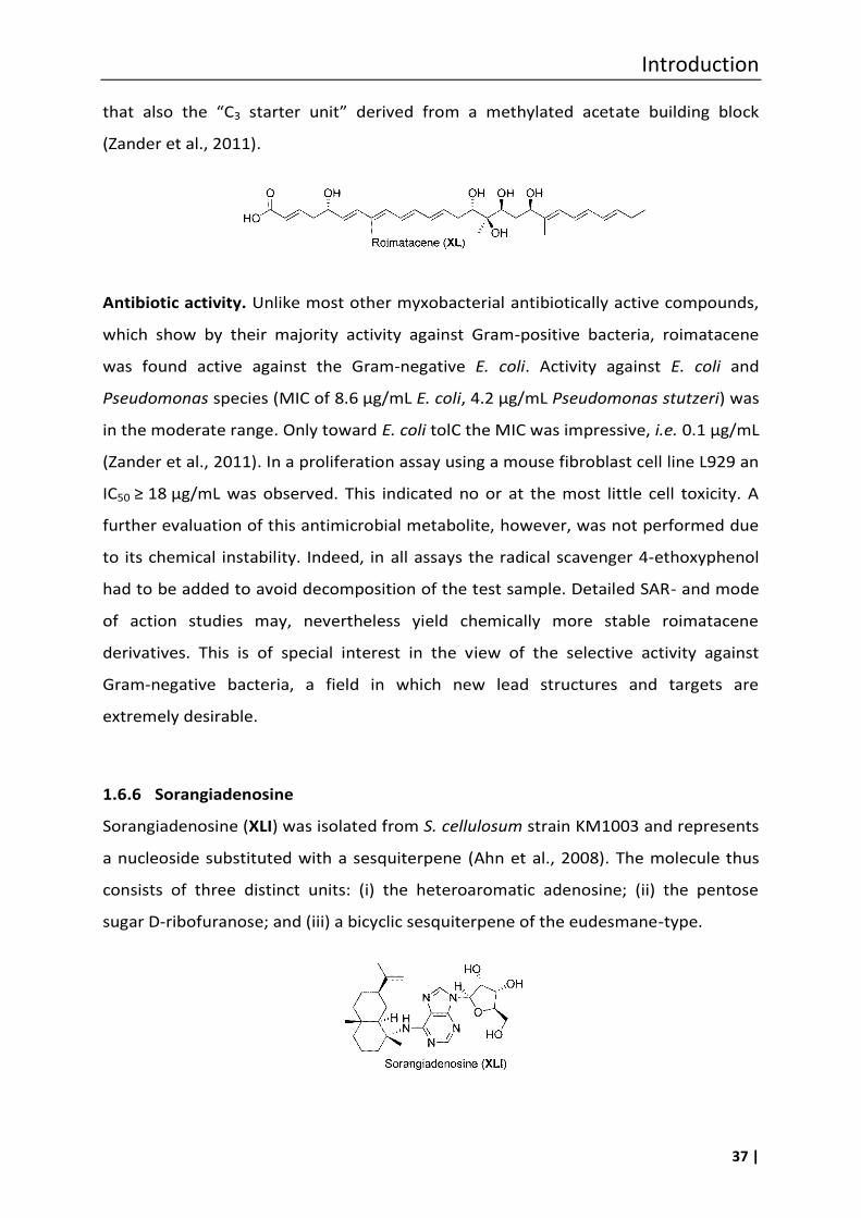
Introduction
37 |
that also the “C3 starter unit” derived from a methylated acetate building block
(Zander et al., 2011).
Antibiotic activity. Unlike most other myxobacterial antibiotically active compounds,
which show by their majority activity against Gram-positive bacteria, roimatacene
was found active against the Gram-negative E. coli. Activity against E. coli and
Pseudomonas species (MIC of 8.6 µg/mL E. coli, 4.2 µg/mL Pseudomonas stutzeri) was
in the moderate range. Only toward E. coli tolC the MIC was impressive, i.e. 0.1 µg/mL
(Zander et al., 2011). In a proliferation assay using a mouse fibroblast cell line L929 an
IC50 ≥ 18 µg/mL was observed. This indicated no or at the most little cell toxicity. A
further evaluation of this antimicrobial metabolite, however, was not performed due
to its chemical instability. Indeed, in all assays the radical scavenger 4-ethoxyphenol
had to be added to avoid decomposition of the test sample. Detailed SAR- and mode
of action studies may, nevertheless yield chemically more stable roimatacene
derivatives. This is of special interest in the view of the selective activity against
Gram-negative bacteria, a field in which new lead structures and targets are
extremely desirable.
1.6.6 Sorangiadenosine
Sorangiadenosine (XLI) was isolated from S. cellulosum strain KM1003 and represents
a nucleoside substituted with a sesquiterpene (Ahn et al., 2008). The molecule thus
consists of three distinct units: (i) the heteroaromatic adenosine; (ii) the pentose
sugar D-ribofuranose; and (iii) a bicyclic sesquiterpene of the eudesmane-type.
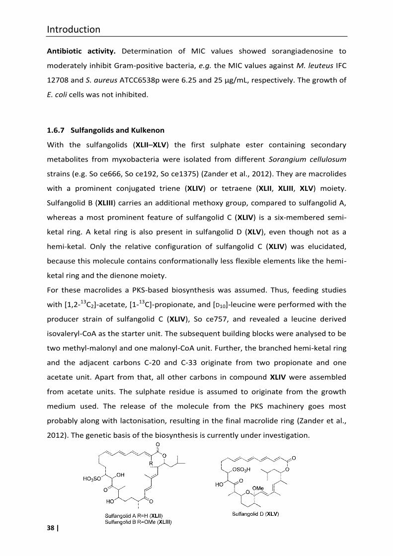
Introduction
38 |
Antibiotic activity. Determination of MIC values showed sorangiadenosine to
moderately inhibit Gram-positive bacteria, e.g. the MIC values against M. leuteus IFC
12708 and S. aureus ATCC6538p were 6.25 and 25 µg/mL, respectively. The growth of
E. coli cells was not inhibited.
1.6.7 Sulfangolids and Kulkenon
With the sulfangolids (XLII–XLV) the first sulphate ester containing secondary
metabolites from myxobacteria were isolated from different Sorangium cellulosum
strains (e.g. So ce666, So ce192, So ce1375) (Zander et al., 2012). They are macrolides
with a prominent conjugated triene (XLIV) or tetraene (XLII, XLIII, XLV) moiety.
Sulfangolid B (XLIII) carries an additional methoxy group, compared to sulfangolid A,
whereas a most prominent feature of sulfangolid C (XLIV) is a six-membered semi-
ketal ring. A ketal ring is also present in sulfangolid D (XLV), even though not as a
hemi-ketal. Only the relative configuration of sulfangolid C (XLIV) was elucidated,
because this molecule contains conformationally less flexible elements like the hemi-
ketal ring and the dienone moiety.
For these macrolides a PKS-based biosynthesis was assumed. Thus, feeding studies
with [1,2-13C2]-acetate, [1-13C]-propionate, and [D10]-leucine were performed with the
producer strain of sulfangolid C (XLIV), So ce757, and revealed a leucine derived
isovaleryl-CoA as the starter unit. The subsequent building blocks were analysed to be
two methyl-malonyl and one malonyl-CoA unit. Further, the branched hemi-ketal ring
and the adjacent carbons C-20 and C-33 originate from two propionate and one
acetate unit. Apart from that, all other carbons in compound XLIV were assembled
from acetate units. The sulphate residue is assumed to originate from the growth
medium used. The release of the molecule from the PKS machinery goes most
probably along with lactonisation, resulting in the final macrolide ring (Zander et al.,
2012). The genetic basis of the biosynthesis is currently under investigation.
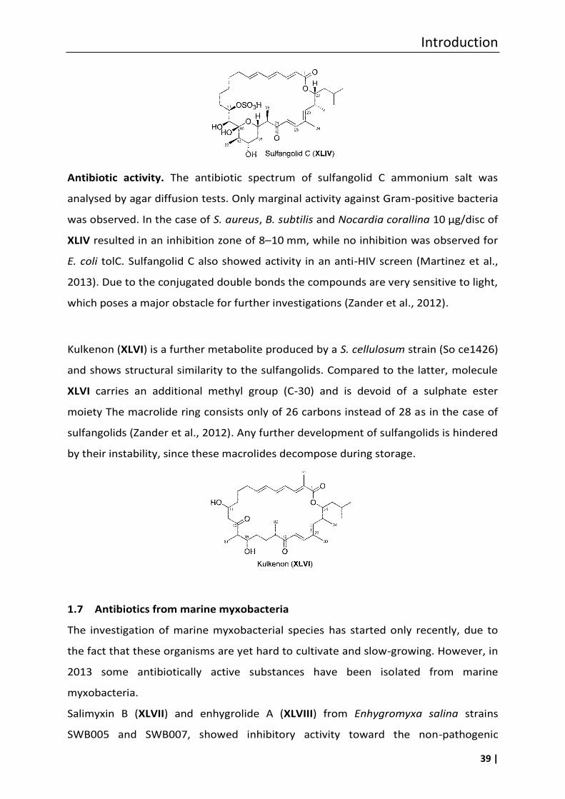
Introduction
39 |
Antibiotic activity. The antibiotic spectrum of sulfangolid C ammonium salt was
analysed by agar diffusion tests. Only marginal activity against Gram-positive bacteria
was observed. In the case of S. aureus, B. subtilis and Nocardia corallina 10 µg/disc of
XLIV resulted in an inhibition zone of 8–10 mm, while no inhibition was observed for
E. coli tolC. Sulfangolid C also showed activity in an anti-HIV screen (Martinez et al.,
2013). Due to the conjugated double bonds the compounds are very sensitive to light,
which poses a major obstacle for further investigations (Zander et al., 2012).
Kulkenon (XLVI) is a further metabolite produced by a S. cellulosum strain (So ce1426)
and shows structural similarity to the sulfangolids. Compared to the latter, molecule
XLVI carries an additional methyl group (C-30) and is devoid of a sulphate ester
moiety The macrolide ring consists only of 26 carbons instead of 28 as in the case of
sulfangolids (Zander et al., 2012). Any further development of sulfangolids is hindered
by their instability, since these macrolides decompose during storage.
1.7 Antibiotics from marine myxobacteria
The investigation of marine myxobacterial species has started only recently, due to
the fact that these organisms are yet hard to cultivate and slow-growing. However, in
2013 some antibiotically active substances have been isolated from marine
myxobacteria.
Salimyxin B (XLVII) and enhygrolide A (XLVIII) from Enhygromyxa salina strains
SWB005 and SWB007, showed inhibitory activity toward the non-pathogenic
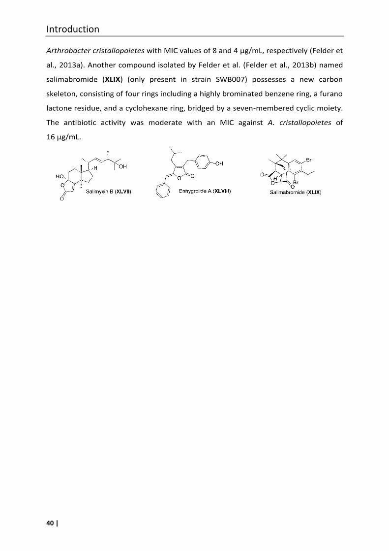
Introduction
40 |
Arthrobacter cristallopoietes with MIC values of 8 and 4 µg/mL, respectively (Felder et
al., 2013a). Another compound isolated by Felder et al. (Felder et al., 2013b) named
salimabromide (XLIX) (only present in strain SWB007) possesses a new carbon
skeleton, consisting of four rings including a highly brominated benzene ring, a furano
lactone residue, and a cyclohexane ring, bridged by a seven-membered cyclic moiety.
The antibiotic activity was moderate with an MIC against A. cristallopoietes of
16 µg/mL.
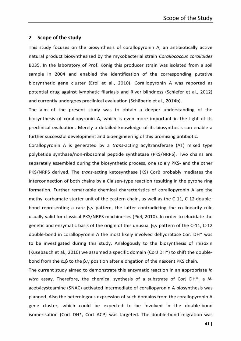
Scope of the Study
41 |
2 Scope of the study
This study focuses on the biosynthesis of corallopyronin A, an antibiotically active
natural product biosynthesized by the myxobacterial strain Corallococcus coralloides
B035. In the laboratory of Prof. König this producer strain was isolated from a soil
sample in 2004 and enabled the identification of the corresponding putative
biosynthetic gene cluster (Erol et al., 2010). Corallopyronin A was reported as
potential drug against lymphatic filariasis and River blindness (Schiefer et al., 2012)
and currently undergoes preclinical evaluation (Schäberle et al., 2014b).
The aim of the present study was to obtain a deeper understanding of the
biosynthesis of corallopyronin A, which is even more important in the light of its
preclinical evaluation. Merely a detailed knowledge of its biosynthesis can enable a
further successful development and bioengineering of this promising antibiotic.
Corallopyronin A is generated by a trans-acting acyltransferase (AT) mixed type
polyketide synthase/non-ribosomal peptide synthetase (PKS/NRPS). Two chains are
separately assembled during the biosynthetic process, one solely PKS- and the other
PKS/NRPS derived. The trans-acting ketosynthase (KS) CorB probably mediates the
interconnection of both chains by a Claisen-type reaction resulting in the pyrone ring
formation. Further remarkable chemical characteristics of corallopyronin A are the
methyl carbamate starter unit of the eastern chain, as well as the C-11, C-12 double-
bond representing a rare β,γ pattern, the latter contradicting the co-linearity rule
usually valid for classical PKS/NRPS machineries (Piel, 2010). In order to elucidate the
genetic and enzymatic basis of the origin of this unusual β,γ pattern of the C-11, C-12
double-bond in corallopyronin A the most likely involved dehydratase CorJ DH* was
to be investigated during this study. Analogously to the biosynthesis of rhizoxin
(Kusebauch et al., 2010) we assumed a specific domain (CorJ DH*) to shift the double-
bond from the α,β to the β,γ position after elongation of the nascent PKS chain.
The current study aimed to demonstrate this enzymatic reaction in an appropriate in
vitro assay. Therefore, the chemical synthesis of a substrate of CorJ DH*, a N-
acetylcysteamine (SNAC) activated intermediate of corallopyronin A biosynthesis was
planned. Also the heterologous expression of such domains from the corallopyronin A
gene cluster, which could be expected to be involved in the double-bond
isomerisation (CorJ DH*, CorJ ACP) was targeted. The double-bond migration was
Scope oft the Study
42 |
envisaged to be detected by applying high-resolution MS measurements using the
phosphopantetheinyl (ppant) ejection assay (Meluzzi et al., 2008) and by NMR
analysis. To obtain evidence for the essential amino acids involved in the double-bond
migration process, mutated enzyme variants were planned to be constructed and
expressed.
The in vitro analysis of the heterologously expressed shift domain (CorJ DH*) and its
mutated variants should provide a basic understanding of carbon-carbon double-
bond shift reactions in polyketide biosynthesis beyond that of corallopyronin A.
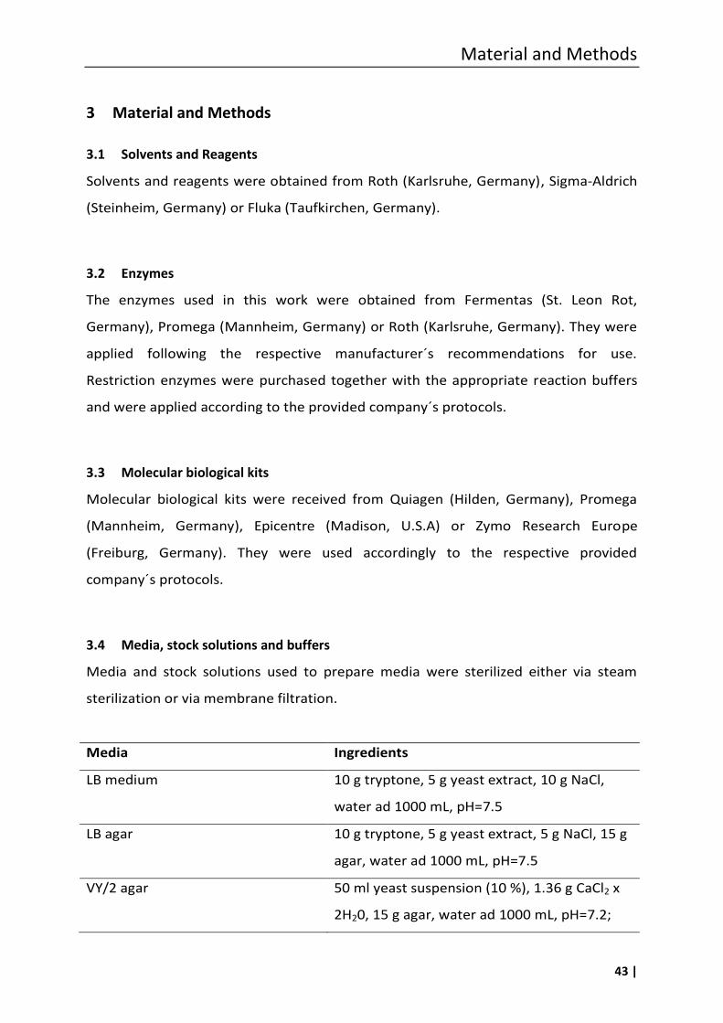
Material and Methods
43 |
3 Material and Methods
3.1 Solvents and Reagents
Solvents and reagents were obtained from Roth (Karlsruhe, Germany), Sigma-Aldrich
(Steinheim, Germany) or Fluka (Taufkirchen, Germany).
3.2 Enzymes
The enzymes used in this work were obtained from Fermentas (St. Leon Rot,
Germany), Promega (Mannheim, Germany) or Roth (Karlsruhe, Germany). They were
applied following the respective manufacturer´s recommendations for use.
Restriction enzymes were purchased together with the appropriate reaction buffers
and were applied according to the provided company´s protocols.
3.3 Molecular biological kits
Molecular biological kits were received from Quiagen (Hilden, Germany), Promega
(Mannheim, Germany), Epicentre (Madison, U.S.A) or Zymo Research Europe
(Freiburg, Germany). They were used accordingly to the respective provided
company´s protocols.
3.4 Media, stock solutions and buffers
Media and stock solutions used to prepare media were sterilized either via steam
sterilization or via membrane filtration.
Media Ingredients
LB medium 10 g tryptone, 5 g yeast extract, 10 g NaCl,
water ad 1000 mL, pH=7.5
LB agar 10 g tryptone, 5 g yeast extract, 5 g NaCl, 15 g
agar, water ad 1000 mL, pH=7.5
VY/2 agar 50 ml yeast suspension (10 %), 1.36 g CaCl2 x
2H20, 15 g agar, water ad 1000 mL, pH=7.2;
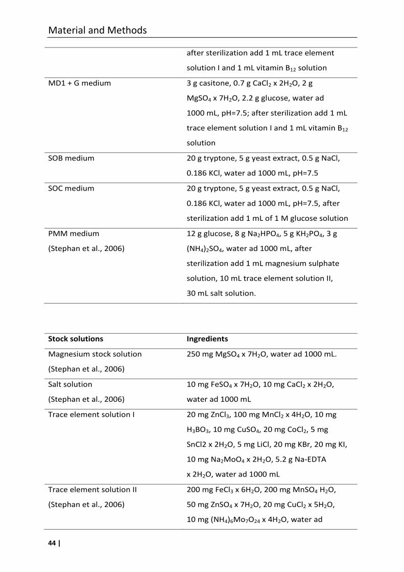
Material and Methods
44 |
after sterilization add 1 mL trace element
solution I and 1 mL vitamin B12 solution
MD1 + G medium 3 g casitone, 0.7 g CaCl2 x 2H2O, 2 g
MgSO4 x 7H2O, 2.2 g glucose, water ad
1000 mL, pH=7.5; after sterilization add 1 mL
trace element solution I and 1 mL vitamin B12
solution
SOB medium 20 g tryptone, 5 g yeast extract, 0.5 g NaCl,
0.186 KCl, water ad 1000 mL, pH=7.5
SOC medium 20 g tryptone, 5 g yeast extract, 0.5 g NaCl,
0.186 KCl, water ad 1000 mL, pH=7.5, after
sterilization add 1 mL of 1 M glucose solution
PMM medium
(Stephan et al., 2006)
12 g glucose, 8 g Na2HPO4, 5 g KH2PO4, 3 g
(NH4)2SO4, water ad 1000 mL, after
sterilization add 1 mL magnesium sulphate
solution, 10 mL trace element solution II,
30 mL salt solution.
Stock solutions Ingredients
Magnesium stock solution
(Stephan et al., 2006)
250 mg MgSO4 x 7H2O, water ad 1000 mL.
Salt solution
(Stephan et al., 2006)
10 mg FeSO4 x 7H2O, 10 mg CaCl2 x 2H2O,
water ad 1000 mL
Trace element solution I 20 mg ZnCl3, 100 mg MnCl2 x 4H2O, 10 mg
H3BO3, 10 mg CuSO4, 20 mg CoCl2, 5 mg
SnCl2 x 2H2O, 5 mg LiCl, 20 mg KBr, 20 mg KI,
10 mg Na2MoO4 x 2H2O, 5.2 g Na-EDTA
x 2H2O, water ad 1000 mL
Trace element solution II
(Stephan et al., 2006)
200 mg FeCl3 x 6H2O, 200 mg MnSO4 H2O,
50 mg ZnSO4 x 7H2O, 20 mg CuCl2 x 5H2O,
10 mg (NH4)6Mo7O24 x 4H2O, water ad
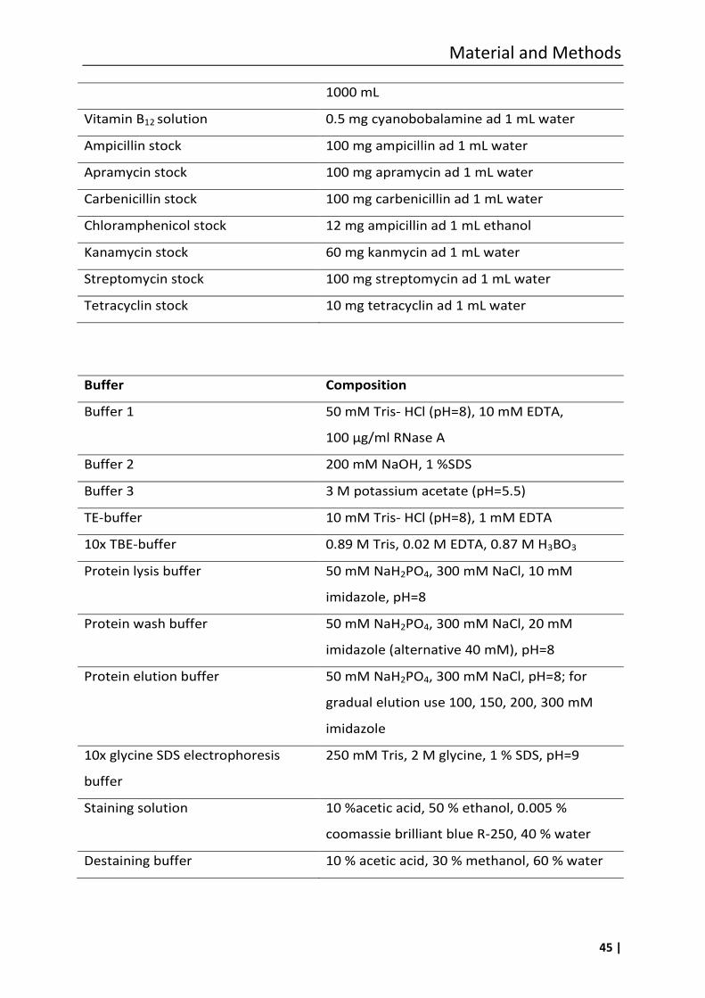
Material and Methods
45 |
1000 mL
Vitamin B12 solution 0.5 mg cyanobobalamine ad 1 mL water
Ampicillin stock 100 mg ampicillin ad 1 mL water
Apramycin stock 100 mg apramycin ad 1 mL water
Carbenicillin stock 100 mg carbenicillin ad 1 mL water
Chloramphenicol stock 12 mg ampicillin ad 1 mL ethanol
Kanamycin stock 60 mg kanmycin ad 1 mL water
Streptomycin stock 100 mg streptomycin ad 1 mL water
Tetracyclin stock 10 mg tetracyclin ad 1 mL water
Buffer Composition
Buffer 1 50 mM Tris- HCl (pH=8), 10 mM EDTA,
100 µg/ml RNase A
Buffer 2 200 mM NaOH, 1 %SDS
Buffer 3 3 M potassium acetate (pH=5.5)
TE-buffer 10 mM Tris- HCl (pH=8), 1 mM EDTA
10x TBE-buffer 0.89 M Tris, 0.02 M EDTA, 0.87 M H3BO3
Protein lysis buffer 50 mM NaH2PO4, 300 mM NaCl, 10 mM
imidazole, pH=8
Protein wash buffer 50 mM NaH2PO4, 300 mM NaCl, 20 mM
imidazole (alternative 40 mM), pH=8
Protein elution buffer 50 mM NaH2PO4, 300 mM NaCl, pH=8; for
gradual elution use 100, 150, 200, 300 mM
imidazole
10x glycine SDS electrophoresis
buffer
250 mM Tris, 2 M glycine, 1 % SDS, pH=9
Staining solution 10 %acetic acid, 50 % ethanol, 0.005 %
coomassie brilliant blue R-250, 40 % water
Destaining buffer 10 % acetic acid, 30 % methanol, 60 % water
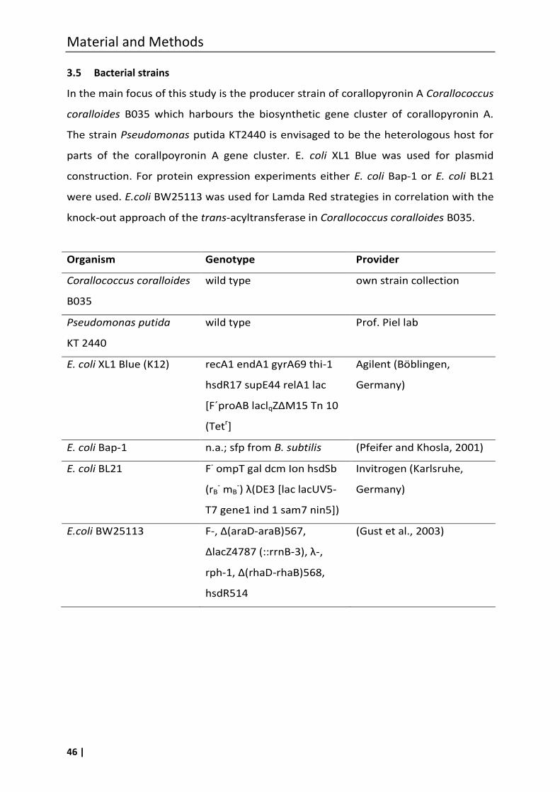
Material and Methods
46 |
3.5 Bacterial strains
In the main focus of this study is the producer strain of corallopyronin A Corallococcus
coralloides B035 which harbours the biosynthetic gene cluster of corallopyronin A.
The strain Pseudomonas putida KT2440 is envisaged to be the heterologous host for
parts of the corallpoyronin A gene cluster. E. coli XL1 Blue was used for plasmid
construction. For protein expression experiments either E. coli Bap-1 or E. coli BL21
were used. E.coli BW25113 was used for Lamda Red strategies in correlation with the
knock-out approach of the trans-acyltransferase in Corallococcus coralloides B035.
Organism Genotype Provider
Corallococcus coralloides
B035
wild type own strain collection
Pseudomonas putida
KT 2440
wild type Prof. Piel lab
E. coli XL1 Blue (K12) recA1 endA1 gyrA69 thi-1
hsdR17 supE44 relA1 lac
[F´proAB laclqZΔM15 Tn 10
(Tetr]
Agilent (Böblingen,
Germany)
E. coli Bap-1 n.a.; sfp from B. subtilis (Pfeifer and Khosla, 2001)
E. coli BL21 F- ompT gal dcm Ion hsdSb
(rB- mB
-) λ(DE3 [lac lacUV5-
T7 gene1 ind 1 sam7 nin5])
Invitrogen (Karlsruhe,
Germany)
E.coli BW25113 F-, Δ(araD-araB)567,
ΔlacZ4787 (::rrnB-3), λ-,
rph-1, Δ(rhaD-rhaB)568,
hsdR514
(Gust et al., 2003)
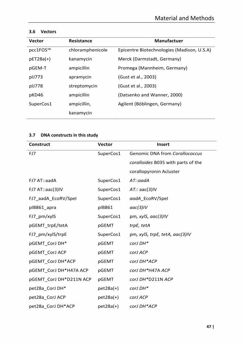
Material and Methods
47 |
3.6 Vectors
Vector Resistance Manufactuer
pcc1FOS™ chloramphenicole Epicentre Biotechnologies (Madison, U.S.A)
pET28a(+) kanamycin Merck (Darmstadt, Germany)
pGEM-T ampicillin Promega (Mannheim, Germany)
pIJ773 apramycin (Gust et al., 2003)
pIJ778 streptomycin (Gust et al., 2003)
pKD46 ampicillin (Datsenko and Wanner, 2000)
SuperCos1 ampicillin,
kanamycin
Agilent (Böblingen, Germany)
3.7 DNA constructs in this study
Construct Vector Insert
FJ7 SuperCos1 Genomic DNA from Corallococcus
coralloides B035 with parts of the
corallopyronin Acluster
FJ7 AT::aadA SuperCos1 AT::aadA
FJ7 AT::aac(3)IV SuperCos1 AT:: aac(3)IV
FJ7_aadA_EcoRV/SpeI SuperCos1 aadA_EcoRV/SpeI
pIB861_apra pIB861 aac(3)IV
FJ7_pm/xylS SuperCos1 pm, xylS, aac(3)IV
pGEMT_trpE/tetA pGEMT trpE, tetA
FJ7_pm/xylS/trpE SuperCos1 pm, xylS, trpE, tetA, aac(3)IV
pGEMT_CorJ DH* pGEMT corJ DH*
pGEMT_CorJ ACP pGEMT corJ ACP
pGEMT_CorJ DH*ACP pGEMT corJ DH*ACP
pGEMT_CorJ DH*H47A ACP pGEMT corJ DH*H47A ACP
pGEMT_CorJ DH*D211N ACP pGEMT corJ DH*D211N ACP
pet28a_CorJ DH* pet28a(+) corJ DH*
pet28a_CorJ ACP pet28a(+) corJ ACP
pet28a_CorJ DH*ACP pet28a(+) corJ DH*ACP
Material and Methods
48 |
pet28a_CorJ DH*H47A ACP pet28a(+) corJ DH*H47A ACP
pet28a_CorJ DH*D211N ACP pet28a(+) corJ DH*D211N ACP
3.8 Primers
Primers used in this work were designed from multi sequence alignment and
purchased from Eurofins MWG Operin (Ebersberg, Germany). Oligonucleotides were
reconstituted in sterile water and adjusted to a concentration of 100 pmol/µL. They
were stored at -20 °C for longer periods and at -4 °C for a short time. A list of the
sequences of primers out of this work is given in the appendix (8.1).
3.9 Software and databases
Basis Local Alignment Search Tool; Blast [www.blast.ncbi.nlm.nih.gov] provided by
the National Centre for Biotechnology Information (NCBI) is used for multiple
sequence alignment for protein primary sequences and for nucleotide sequences.
Blastx translates nucleotide sequences in its corresponding amino acid sequence
which subsequently are compared to the amino acid database. Blastp uses a protein
query for comparison with the protein database. Blastn was used to compare a
nucleotide sequence with the nucleotide database of NCBI.
ClustalW [www.ebi.ac.uk/Tools/msa/clustalw2] provides by the European
Bioinformatics Institute (EBI), part of the European Molecular Biology Lab (EMBL),
was applied to from multiple sequence alignments of nucleotide or amino acid
sequences. Given reference sequences are NCBI derived.
Clone Manager is a purchased program (Sci-Ed Software) and was used due to its set
of tools for primer design and for planning of cloning experiments as well as for
graphic map drawing.
NEBcutter V2 (www.tools.neb.com/NEBcutter2; New England Biolabs) was applied to
carry out restriction side analysis.
Artemis Genome Browser and Annotation Tool is a freely available program
provided by the Sanger Institute. It was utilized to visualize bioinformatic features and
to annotate open reading frames in sequences plasmid, cosmid or genome derived as
well as to analyze the GC-content of a sequence.
Material and Methods
49 |
ProtPram is a free bioinformatic tool provide by the Swiss Institute for Bioinformatics.
It was applied to calculate the molar extinction factor ε, necessary for determination
of the concentration of purified heterologously expressed proteins.
3.10 General molecular biological methods
3.10.1 Sterilization
Solutions, media and all working tools used to apply on microbial organisms were
sterilized by steam sterilization at 121 °C and 2 bar for 20 min in a Varioclav steam
sterilizer. Heat sensitive solutions were sterilized by filtration through an 0.22 µm
membrane, instead.
3.10.2 Cultivation, storage and disposal of organisms
All working-steps concerning cultivation of microorganisms were done on a lamina air
flow clean bench to provide sterile conditions. Instruments, solutions, media and
other working materials were beforehand sterilized via steam sterilization, membrane
filtration or by the heat of a Bunsen burner flame.
Corallococcus coralloides B035 was inoculated from a Petri dish and incubated in
300 mL flasks containing 100 mL MD1+G liquid medium on a horizontal shaker at
30 °C and 160 rpm or on VY/2 agar plates in an incubator at 30 °C. The incubation
time was between three and five days. Pseudomonas putida KT2440 was incubated
either in 100 mL liquid LB medium in 300 mL flasks on a horizontal shaker at 30 °C and
160 rpm, on LB agar plates or on PMM agar plates at 30 °C in an incubator. Incubation
times vary between 3 h and two days according to the experiment, respectively. Small
cultures of Escherichia coli cells were carried out in 10 mL flasks filled with 3 mL LB
liquid medium or in 2 mL tubes at 160 rpm at 30 °C or 37 °C, depending on the hosted
plasmid or cosmid. For cultivation on LB agar plates 250 µL of a Escherichia coli
suspension was spread on the agar plate. After the solvent was moved into the agar
the plate was incubated at 30 °C or 37 °C in an incubator. The incubation time was
16 h, if not stated otherwise.
For long terms of storage glycerin cultures were prepared. Therefore 500 µL of a fully
grown liquid culture was mixed with 500 µL of a sterile 87 % glycerin solution in a
Material and Methods
50 |
cryogenic vial. After thoroughly merging the vial was stored at -80 °C. For short period
storage glycerin cultures were frozen at -20 °C.
For waste disposal of microorganisms they were autoclaved for 20 min at 121 °C and
2 bar.
3.10.3 Antibiotic selectivity test
Corallococcus coralloides B035 (Erol et al., 2010) was tested toward natural resistance
against different antibiotics. VY/2 plates were incubated with 300 µL liquid culture at
30 °C for 5. Growth on the agar plates was determined and the inhibition capacity
was classified (Schmitz, PhD thesis, 2013).
3.11 Molecular biological methods concerning bacterial organisms
3.11.1 Transformation of bacteria
Preparation of competent cells for heat shock procedure. A 3 mL LB culture was
inoculated with a single bacteria colony and incubated over night at 37 °C and
160 rmp. The culture was further transferred into a 300 mL flask containing 70 mL
2xYT medium and incubated at 37 °C and 180 rpm to an OD600=0.3-0.4 and
subsequent harvested centrifugation for 10 min at 8000 rpm at 4 °C. To achieve
competence the cells were treated with 10 mL of ice cold CaCl2/MgCl2-solution
(70 mM CaCl2, 20 mM MgCl2) and incubated on ice for 30 min. After harvesting the
cells again by centrifugation they were suspended in 3.5 mL ice cold CaCl2/MgCl2-
solution and 875 µL glycerol was added. The chemically competed cells were finally
stored in 100 µL aliquots at -80 °C until usage.
Preparation of competed cells for electroporation procedure. Electro-competed cells
were always freshly prepared in order to use on the same day. Following procedure
was modified after Gust et al., 2003. During the whole procedure cells were strictly
kept ice cold. 100 mL SOB medium in a 500 mL flask were inoculated with 3 mL pre-
culture and incubated until OD600=0.5. The culture was splitted into two 50 mL falcon
tubes. After harvesting the cells via centrifugation, 6000 rpm for 5 min, the
supernatant was removed and each pellet was washed with 25 mL 10 % glycerol
Material and Methods
51 |
solution. This step was repeated and further washing steps were done with 20 mL,
10 mL and 5 mL 10 % glycerol solution. The pellets were combined, suspended in
300 µL 10 % glycerol solution and finally stored on ice in 100 µL aliquots.
Transformation of Escherichia coli cells by heat shock. For reasons of in vivo
amplification of plasmids or protein expression E. coli cells were transformed with
foreign DNA. Therefore a 100 µL aliquot of chemical competed cells were thawed on
ice, mixed with 5–10 µL DNA and incubated on ice for 30 min. Afterwards, the cells
were subjected to heat shock at 42 °C for 90 s and immediately replaced on ice for
2 min. For recovery purpose the cells were incubated in 1 mL LB medium at 37 °C and
1000 rpm for 1 h. 250 µL of the cell suspension were spread on agar plates containing
the suitable antibiotics for selection of positive transformants and incubated over
night at 30° or 37 °C. In the case of pGEM-T vector as introduced DNA the agar plates
contained x-gal (4 mg/mL) as additive in order to select via blue-white screening.
Positive clones were verify by whole-cell PCR (3.12.2).
Transformation of Escherichia coli cells by electroporation. Freshly prepared electro-
competed cells were mixed with 4–20 µL of the DNA to be introduced and filled into a
pre-chilled electroporation cuvette with a diameter of 2 mm. Following a voltage of
2.5 kV was applied in Biorad MicroPulser™. The cells were rapidly recovered with
1 mL ice cold SOC medium and incubated in a 10 mL tube at 30 °C at 160 rpm for 1–
2 h. Afterwards, 600 µL of the cell suspension were spread on agar plates containing
suitable antibiotics for selection and the plates were incubated at 30 °C over night.
Electroporation of Pseudomonas putida KT2440 cells. 2 mL of a pre-culture were
inoculated in 100 mL LB medium and incubated until a OD600=0.5 at 30 °C and
160 rpm. During the next steps it was important to keep the sample ice cold. Hence,
the cells were harvested via centrifugation at 6000 rpm for 5 min at 4 °C, the
supernatant was removed and the pellet was suspended in 25 mL ice cold 10 %
glycerol solution. Washing steps with 20 mL, 10 mL and 5 mL 10 % glycerol solution
followed. Finally the cell suspension was taken up in 300 µL 10 % glycerol solution and
allocated in 100 µL samples. The electro- competed cells were mixed with 4–20 µL of
Material and Methods
52 |
DNA to be transferred, and filled in a pre-chilled electroporation cuvette. To apply a
voltage of 2.5 kV a Biorad MicroPulser™ was used. The cells were recovered
immediately with 600 µL of ice cold LB medium and incubated for 2 h at 30 °C and
160 rpm. Positive transformants were selected on PMM solid agar containing suitable
antibiotics and verified via whole cell PCR (3.12.2).
3.12 Molecular biological methods concerning nucleic acids
3.12.1 Isolation of DNA
Isolation of vector DNA. Plasmids, fosmids or cosmids were isolated from the host
strain (usually E. coli). Normally 3 mL liquid media were inoculated with one single
colony and incubated over night. For larger demand of vector DNA midi or maxi
preparations were done. Thus means 10 mL or 100 mL medium were inoculated.
Vectors were purified with Promega´s PureYield Miniprep or with Qiagen Plasmid
Mini, Midi or Maxi Kit according to the manufactures instructions. Otherwise,
bacterial cells of a 3 mL culture were harvested via centrifugation and suspended in
350 µL buffer 1, treated with 350 µL buffer 2 and neutralized with 400 µL ice cold
buffer 3. Cell debris and proteins were pelleted by centrifugation and removed. The
supernatant containing the remaining DNA was again purified with 800 µL
phenol/chloroform in equal parts to remove protein remains. Solved DNA in the
aqueous phase was precipitated by isopropanol and centrifugation. The obtained
DNA pellet was finally washed with 70 % ethanol, dried and dissolved in 20–100 µL
sterile water.
Isolation of genomic DNA. For isolation purpose of Corallococcus coralloides B035
and Pseudomonas putida KT2440 derived genomic DNA the Promega Genomic Wizard
it was used according to the manufacturers’ instructions.
3.12.2 PCR
Polymerase chain reaction. PCR is a method exerted for amplification of DNA
sequences of interest inbetween two primer regions. Oligonucleotides appropriate to
the DNA sequence of interest were designed with the help of the program Clone
Material and Methods
53 |
Manager. First DNA was denaturated to single stranded DNA by heating what allows
the primers to anneal to the homolog DNA region. Then a thermostable DNA
polymerase elongates the DNA stand beginning at the primers 3’ end. Repetitive
cycles of different temperatures for denaturation, annealing and elongation make a
exponential amplification of determined DNA regions possible. In this study the
Thermus aquaticus (Taq) derived DNA- polymerase was used for all PCR reactions.
The composition of the PCR reaction mixture, appropriate in this study, is shown as
follows.
10x PCR buffer 4 µL
10x MgCl2-solution (25 mM) 1 µL
DMSO 1 µL
Primer 1 (100 µM) 0.5 µL
Primer 2 (100 µM) 0.5 µL
dNTPs (10 mM) 0.4 µL
Taq polymerase (5 U/µL) 0.16 µL
DNA template 1 µL
Water ad 20 µL
Whole cell PCR. For rapid testing on DNA introduced into bacteria, bacterial material
was directly stirred into the PCR mixture. The first denaturation step of repetitive
cycles of the PCR protocol was in this respect prolonged to ensure complete
denaturation of the double stranded DNA.
Sequential PCR amplification. In order to exchange one amino acid in a protein it was
necessary to gain an exchange of nucleotides in the protein encoding DNA sequence.
To achieve that side specific mutation a chimeric DNA molecule was created by
sequential PCR amplification. In the first round amplifications two PCR products were
gained containing a region of overlapping homology to each other. Both PCR products
were purified away from the primers, mixed in one reaction tube and annealed by
denaturation a renaturation. Taq DNA-polymerase was added and gained one PCR
fragment with the length to the sum of the two fragments. In the second round
Material and Methods
54 |
amplification complementary outside set primers were added to the mixture in order
to amplify the heteroduplex DNA species. Hereafter the applied PCR program is
shown.
1. Initial enaturation 95 °C 3.0 min
2. Denaturation 95 °C 30 s
3. Annealing 65 °C 30 s
4. Elongation 72 °C 1.0 min
5. Final elingation 72 °C 5.0 min
6. Cooling 4 °C hold
Steps 2–4 were repeated 10 times, without primers. In the following outside set
primers were added and the PCR program was run again. Then steps 2–4 were
repeated 20 times.
3.12.3 Restriction digestion
DNA restriction endonucleases belongs originally to bacterial defence mechanism
against foreign DNA. Their restriction sides are marked as palindromic DNA sequence
of 4–8 nucleotides. After cleavage of the phosphodiester-bonds within the DNA
backbone blunt or sticky ends remain corresponding to the restriction enzyme used,
respectively. The effectiveness of each restriction enzyme depends on temperature,
reaction medium and methylation pattern of the DNA to be restricted. Purchased
restriction en ymes in this work were applied according to the manufacturers’
instructions. Restriction digestion was performed to prepare PCR- fragments, vectors
and other DNA molecules for cloning purpose. Usually DNA was restricted by two
different restriction enzymes, e.g. for clear orientation of incorporated DNA
fragments in vectors. In the case of a digestion reaction with only one enzyme the
linearised vector was subsequently dephosphorylated to prevent self-ligation.
3.12.4 Dephosphorylation of linear DNA
Vectors cut with only one restriction enzyme had to be dephosphoylized to avoid self-
ligation when foreign linear DNA should be introduced. To prevent self-ligation the
Material and Methods
55 |
vector’s open ends were dephosphorylated by calf intestine alkaline phophatase
(CIAP) or by shrimp alkaline phosphatise (SAP) according to the manufacturers’
instructions. To remove the phophatase, which potentially disturb subsequent
ligation reactions, the DNA mixture was purified away from the enzyme using a
Qiagen MiniPrep Kit.
3.12.5 Agarose gel electrophoresis and DNA recovery
Mixtures of DNA fragments of different size were separated by gel electrophoresis,
e.g. restricted vectors, PCR fragments or genomic DNA. The areas of interests were
cut out of the gel and the DNA was purified with the help of a Qiagen gel extraction
kit following manufacturers’ instructions. Isolated DNA was dissolved in ultrapure,
sterile water.
3.12.6 Ligation of DNA into a vector
To introduce DNA fragments into a vector, both had to be subjected to a restriction
digestion (3.12.3). Subsequent ligation of the resulting linear DNA molecules were
performed by a T4 DNA- ligase, which is able to link the 5’-phopshodiester moiety and
the 3’-hydroxyl functionality of linear DNA molecules with each other. Thus, DNA
molecules with blunt or with compatible sticky ends could be stitched together. The
result was a circular double stranded DNA molecule consisting of a selected DNA
fragment and the vector scaffold. T4 DNA-ligase was used analogously to a standard
protocol and to the manufacturers’ instructions. For in vivo amplification of the
obtained vector construct and for save storage it was transferred into a bacterial host
like Escherichia coli XL1 blue (3.5 and 3.11.1).
3.12.7 Sequencing of DNA constructs and PCR fragments
To determine the sequence of PCR fragments and to exclude mutations in plasmids
their DNA sequence had to be analyzed. Sequencing was performed by GATC Biotech
AG (Konstanz, Germany) on an ABI3730xl after the Sanger dideoxy method (Sanger et
al., 1977). For sequencing reaction either specific primer pairs complementary to the

Material and Methods
56 |
PCR fragment, or general primer pairs complementary to the vector were used. The
obtained sequence information were processed with Clone Manager.
3.13 Molecular biological methods concerning proteins
3.13.1 Heterologous expression of the proteins
For the in vitro investigations of proteins a sufficient amount of the desired protein is
required. In order to obtain that usually over expression of the respective protein in a
heterologous host is applied. Therefore the DNA sequence of the target protein was
cloned into an expression vector (i.e. pET28a(+)). The pET28a(+) vector contains an
isopropyl-β-D-thiogalactopyranosid (IPTG) inducible promoter as well as an affinity tag
like the 6-his tag to facilitate the purification of the protein via affinity
chromatography.
In the first step the DNA sequence of the protein was amplified and the resulting PCR
fragment was ligated into the pGEMT vector. To exclude mutations in between the
amplified DNA sequence the construct was submitted to sequencing at GATC (3.12.7).
Second the DNA encoding for the protein was cut out of the pGEMT construct and
cloned into the multiple cloning side of the expression vector (pET28a(+) so that the
DNA sequence of the protein is in-frame with the 6-his tag within the vector. Finally E.
coli Bap-1 or BL21 cells were transformed with the generated plasmid, and positive
transformants were determined by whole-cell PCR (3.12.2).
A 3 mL pre-culture of the pET28a(+) construct was used to inoculate the main culture
of 1 L LB medium, containing kanamycin. The culture was grown at 37 °C to an OD600
of ~0.5 and then chilled to 16 °C. To induce protein expression IPTG was added to the
medium in a final concentration of 0.5 or 1 mM and the culture was further incubated
overnight at 16 °C.
3.13.2 Cell lysis by sonication
Cells used for protein expression were harvested by centrifugation at 4000 rpm for
30 min at 4 °C. Afterwards the pellet was resuspended in 2 mL lysis buffer and placed
on ice. Cells were lysed with the help of the Branson Sonifier 250, set to output level
4, 50 % duty cycle. The samples were sonified five times with ten pulses each. Cell
Material and Methods
57 |
debris and insoluble parts were pelleted by centrifugation at 8000 rpm for 15 min at
4 °C. The supernatant containing the soluble protein was collected for further
purification via affinity chromatography.
3.13.3 Purification of the recombinant protein by Ni-NTA affinity chromatography
Proteins containing a 6-his tag can easily be purified by affinity chromatography on a
Ni-NTA matrix. The twice positive charged Ni2+ ions interact with the histidine
residues of the 6-his tag and bind the protein to the matrix while other proteins elute.
With increasing concentrations of imidazole unspecific linked proteins can be eluted
due to the competition of imidazole with hisitine for the binding to Ni2+. The 6-his tag
ensure that the target protein elutes only at high imidazole concentrations.
The Ni-NTA gravity flow column consists of a Ni-NTA agarose matrix which is
equilibrated with lysis buffer. The sample, also dissolved in lysis buffer, was added to
the column and is allowed to pass the matrix. The resulting flow through was added
once again to the matrix to ensure a nearly complete binding of the protein to the Ni-
NTA matrix. Then the column was washed twice with 4 mL washing buffer.
Subsequent elution of the protein occurred in five elution steps with each 0.5 mL
elution buffer with increasing imidazole concentrations (100, 150, 200, 300, 300 mM
imidazole). All fractions, flow through fraction to last elution fraction, were collected
and stored on ice to avoid protein degradation.
3.13.4 SDS-Polyacrylamind gel electrophoresis (SDS-PAGE) and Coomassie staining
All collected fractions of the protein purification were subjected to SDS-PAGE to
record the purity and the average quantity of the target protein in each fraction. First
the fractions were boiled and treated with mercapto ethanol to reduce all di-sulfide
bonds to gain unfolded proteins. During electrophoresis in a SDS milieu the proteins
are charged completely negative and are able to be strictly separated due to their
molecule weight. The separating gel is gained by the polymerization of bis-acrylamid
to polyacrylamide which gives a molecular sieve. Depending on the molecular weight
of the proteins to be separated the concentration of polyacrylamide can be adapted.
To achieve focussed protein bands, discontinuous gels were used where the
Material and Methods
58 |
separating gel is covered with a stacking gel with a lower acrylamid concentration. In
a first step the separating gel was prepared, which was pipetted between two plastic
plates with a spacer distance of 1.5 mm directly after initiating the polymerisation by
APS (ammoniumperoxosulfate). To form a smooth surface the separating gel was
covered with isopropanol, which was removed before addition of the stacking gel.
The reaction mixture of both components of the discontinuous gels is as follows:
SDS stacking gel
Tris/HCl pH 6.8 (1 M) 375 µL
SDS (10 %) 30 µL
Bis-acrylamide (30 %) 510 µL
Water 2040 µL
APS (10 %) 30 µL
TEMED 3 µL
SDS separating gel
Tris/Hcl pH 6.8 (1 M) 2500 µL
SDS (10 %) 100 µL
Bis-acrylamide (30 %) 4000 µL
Water 3300 µL
APS (10 %) 100 µL
TEMED 4 µL
For each run the reservoir of the electrophoresis assembly were filled with fresh SDS
electrophoresis buffer. The protein samples were mixed with denaturing loading
buffer and boiled for 5 min at 90 °C, before loading them on the gel. Electrophoresis
was performed in a XCell SureLock® Mini-Cell. The voltage was 100 V until the
samples reached the separating gel, then it was increased to 130 V. As a reference a
molecule size marker was loaded on the gel as well.
Following directly after the electrophoresis the proteins were visualized by a
coomassie-staining. The gel was immersed in the staining solution and shortly heated
in a microwave and subsequent incubated several minutes on a horizontal shaker.
Material and Methods
59 |
Destaining of the background colour was performed by shaking with destaining buffer
for several hours at room temperature. Gels were documented with the INTAS
illuminator.
3.13.5 Concentration of the proteins and buffer exchange
Samples containing purified heterologously expressed proteins were concentrate
using spin filter column (Millipore, 10 kDa exclusion size). This method was also used
to remove imidazole from the elution buffer, which could disturb subsequent assays.
The elution buffer was replaced in several centrifugation steps with 50 mM Tris/HCl
(pH 8) to achieve a final volume of 250 to 500 µL protein solution. Proteins scheduled
for the MS based ejection assay (3.17.1) were buffered in 50 mM deuterated Tris/HCl
(pH 8), which was prepared beforehand: 50 mM aqueous Tris/HCl solution (1 mL each
sample) were dried in a SpeedVac completely, and were resolved in the same volume
of D2O.
3.13.6 Determination of the protein concentration
Proteins containing tyrosine and tryptophan residues and disulphide bonds will
absorb in the UV range of 280 nm making the correlation between absorbance and
protein-concentration for purified proteins possible. The calculation is based on the
Lambert-Beer equation (formula 3-1). It requires the knowledge of the molar
extinction factor (ε) which was calculated for a given protein sequence with the
program ProtPram provided by the Swiss Institute for Bioinformatics.
Formula 3-1:
dOD
L
molonconcentrai
OD280=optical density at λ=280 nm
d=dilution factor
ε=molar extinction factor [M-1cm-1]
Material and Methods
60 |
3.14 Chromatography
Affinity chromatography. See Molecular biological methods concerning proteins
(3.13.3).
Thin layer chromatography. For reaction control during synthesis of compounds 1, 5–
8, 10–13 thin layer chromatography was performed on Merck aluminum sheets, silica
gel 60 F254. After detection under UV light (254 nm) development took place using a
solution of ninhydrin in ethanol (0.2 % (m/V). As purification method column
chromatography was carried out on Merck silica gel 60 (70–230 mesh). As eluent a
mixture of ethyl acetate/petroleum ether or a mixture of dichloromethane/methanol
was used.
High performance liquid chromatography (HPLC). HPLC was performed on either a
Merck-Hitachi system equipped with an L-6200A pump, an L-4500A photodiode array
detector, a D-6000A interface with D-7000 HSM software and a Rheodyne 7725i
injection system, or a Waters system, controlled by a Waters millennium software,
consisting of a 717 puls autosampler, 600 pump with in-line degasser and a 996
photodiode array detector. Either a Waters Atlantis C18 column (5 µm, 4.6×250 mm),
or a Waters Symmetry 300 C4 column (5 μm, 4.6 mm×250 mm) were used for
preparative HPLC purification of synthesis products of enzyme assay reaction
products.
3.15 NMR spectroscopy
1H-NMR and 13C-NMR spectra were recorded on a Bruker Avance 300 DPX operating
at 300 MHz (1H) or 75 MHz (13C), on a Bruker Avance 500 DRX operating at 500 MHz
(1H) or 125 MHz (13C) or on a Brucker Avance 600 operating at 600 MHz (1H) or
300 MHz (13C) respectively. Processing of the NMR spectra was done using Bruker 1D
WIN-NMR, 2D WINNMR or XWIN-NMR Version 2.6 or 3.1. Chemical shifts were given
in ppm relating to the center of the solvent peak at reference: [D4]MeOH
3.35/49.3 ppm, [D6]DMSO 2.49/39.7 ppm. Multiplicity of carbon atoms was deduced
by DEPT experiments. Structural assignment were based on spectra resulting from
Material and Methods
61 |
one or more of the following NMR experiments: 1H, 13C, DEPT 135, 1H-1H COSY, 1H-13C
HSQC (direct correlation) and 1H-13C HMBC (long range correlation).
3.16 Mass spectrometry
HPLC-MS (ESI) measurements were performed by Frau M. Schneider (Pharmaceutical
Institute of the University of Bonn) or by Frau. E. Eguereva (Institute for
Pharmaceutical Biology of the University of Bonn) employing an Agilent 1100 Series
HPLC including DAD, with a RP 18 column (Macherey-Nagel Nucleodur 100,
125 mmx2 mm, 5 µm) coupled with an API 2000, Triple Quadrupole, LC/MS/MS,
applied Biosystems/MDS Sciex and ESI source. A gradient elution of (from 90 % H2O to
100 % MeOH in 10 min, then 100 % MeOH to 20 min, with added NH4Ac, 2 nM, DAD
220.0–400.0 nm) was chosen for compound characterization and purity
determination. Mass spectrometric analysis of proteins was done by M. Sylvester
(Institute for Biochemistry and Microbiology of the University of Bonn) using a
Thermo LTQ Orbitrap Velo coupled with an Advion TriVersaNanoMate enabling a
continues electron spray.
3.17 In vitro assays to prove the functional role of the DH*
3.17.1 Phosphopantethein (Ppant) ejection assay
The activity of the heterologously expressed shift domain CorJ DH* was determined in
a MS based in vitro assay: the ppant ejection assay, which enables the analysis of
carrier bound intermediates (Meluzzi et al., 2008). In the first reaction step the SNAC-
activated substrate should bind to the ppant arm of the holo-ACP protein. The second
reaction step should be the shift of the double-bond, performed by the “shift
domain§ CorJ DH*. Therefore the substrate 1 was loaded onto the
phosphopantethein arm of the CorJ ACP unit of the heterologously expressed
didomain CorJ DH*-ACP by co-incubation at room temperature. The shift reaction
started directly at this time (substrate addition was set as time point zero). The assay
volume was 100 µL containing 2 % DMSO: 10 µL protein solution, 2 µL substrate 1
(equal to 0.2 mM), 38 µL buffer (deuterated Tris 50 mM, pH 8.0). The reaction
mixture was incubated at room temperature for 1 h, 3 h and 20 h. To stop the
Material and Methods
62 |
reaction it was directly injected into the HPLC column (Waters Symmetry 300 C4;
solvent: linear gradient from 70 % water to 65 % acetonitrile over 30 min, flow:
1.0 mL/min) to obtain a desalted sample, which was immediately lyophilized with
liquid nitrogen.
The respective samples were dissolved in an adequate volume of electro spray
solution (49.5 % H2O, 49.5 % methanol, 1% formic acid). 10 µL samples were loaded
onto the 96 well plate of the NanoMate spray robot coupled to the LTQ Orbitrap
Velos. A spray chip with 5 µm nozzle diameter was used at a spray voltage of 1.6 kV
and 0.3 psi pressure setting. An environmental polysiloxane ion with m/z 445.12003
was used as lock mass for internal calibration. Typical mass deviation was <2 ppm.
Isolation and fragmentation were performed in the linear ion trap, detection of the
final product spectrum was done with the Orbitrap analyzer.
The same procedure and assay conditions were applied for investigating of CorJ holo-
ACP, CorJ DH*H47A holo-ACP and CorJ DH*D211N holo-ACP.
3.17.2 NMR based assay
The double bond shift mediated by the “shift domain” CorJ D * was additionally
monitored using a NMR based in vitro assay. To perform this assay the didomain CorJ
DH*-ACP was heterologously expressed in E. coli BL21, as an apo-enzyme, without a
ppant arm. The purified protein CorJ DH* apo-ACP (re-buffered in non-deuterated
Tris-buffer) was incubated with substrate 1 for 16 h. The assay volume was 500 µL
containing 1 % DMSO: 250 µL protein solution, 5 µL substrate 1 (equal to 0.5 mM),
245 µL buffer (Tris 50 mM, pH 8.0). The assay was stopped by adding an equal volume
of methanol. The protein was pelleted by centrifugation. The supernatant was
transferred to a new vial and dried in vacuo. Subsequently, the sample was prepared
for adjacent NMR experiments by dissolving it in deuterated methanol.
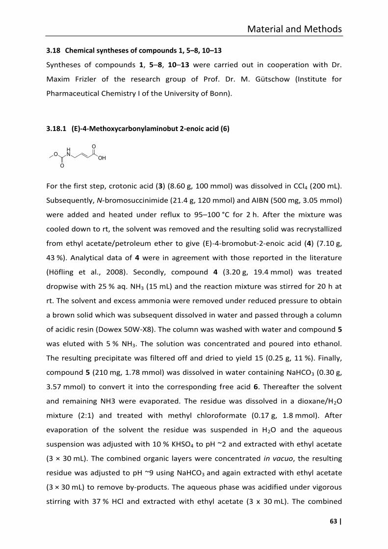
Material and Methods
63 |
3.18 Chemical syntheses of compounds 1, 5–8, 10–13
Syntheses of compounds 1, 5–8, 10–13 were carried out in cooperation with Dr.
Maxim Frizler of the research group of Prof. Dr. M. Gütschow (Institute for
Pharmaceutical Chemistry I of the University of Bonn).
3.18.1 (E)-4-Methoxycarbonylaminobut 2-enoic acid (6)
For the first step, crotonic acid (3) (8.60 g, 100 mmol) was dissolved in CCl4 (200 mL).
Subsequently, N-bromosuccinimide (21.4 g, 120 mmol) and AIBN (500 mg, 3.05 mmol)
were added and heated under reflux to 95–100 °C for 2 h. After the mixture was
cooled down to rt, the solvent was removed and the resulting solid was recrystallized
from ethyl acetate/petroleum ether to give (E)-4-bromobut-2-enoic acid (4) (7.10 g,
43 %). Analytical data of 4 were in agreement with those reported in the literature
(Höfling et al., 2008). Secondly, compound 4 (3.20 g, 19.4 mmol) was treated
dropwise with 25 % aq. NH3 (15 mL) and the reaction mixture was stirred for 20 h at
rt. The solvent and excess ammonia were removed under reduced pressure to obtain
a brown solid which was subsequent dissolved in water and passed through a column
of acidic resin (Dowex 50W-X8). The column was washed with water and compound 5
was eluted with 5 % NH3. The solution was concentrated and poured into ethanol.
The resulting precipitate was filtered off and dried to yield 15 (0.25 g, 11 %). Finally,
compound 5 (210 mg, 1.78 mmol) was dissolved in water containing NaHCO3 (0.30 g,
3.57 mmol) to convert it into the corresponding free acid 6. Thereafter the solvent
and remaining NH3 were evaporated. The residue was dissolved in a dioxane/H2O
mixture (2:1) and treated with methyl chloroformate (0.17 g, 1.8 mmol). After
evaporation of the solvent the residue was suspended in H2O and the aqueous
suspension was adjusted with 10 % KHSO4 to pH ~2 and extracted with ethyl acetate
(3 × 30 mL). The combined organic layers were concentrated in vacuo, the resulting
residue was adjusted to pH ~9 using NaHCO3 and again extracted with ethyl acetate
(3 × 30 mL) to remove by-products. The aqueous phase was acidified under vigorous
stirring with 37 % HCl and extracted with ethyl acetate (3 x 30 mL). The combined
O
O
HN
OH
O
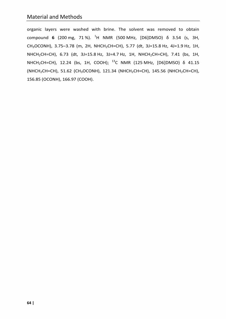
Material and Methods
64 |
organic layers were washed with brine. The solvent was removed to obtain
compound 6 (200 mg, 71 %). 1H NMR (500 M , [D6]DMSO) δ 3.54 (s, 3 ,
CH3OCONH), 3.75–3.78 (m, 2H, NHCH2CH=CH), 5.77 (dt, 3J=15.8 Hz, 4J=1.9 Hz, 1H,
NHCH2CH=CH), 6.73 (dt, 3J=15.8 Hz, 3J=4.7 Hz, 1H, NHCH2CH=CH), 7.41 (bs, 1H,
NHCH2CH=CH), 12.24 (bs, 1H, COOH); 13C NMR (125 M , [D6]DMSO) δ 41.15
(NHCH2CH=CH), 51.62 (CH3OCONH), 121.34 (NHCH2CH=CH), 145.56 (NHCH2CH=CH),
156.85 (OCONH), 166.97 (COOH).
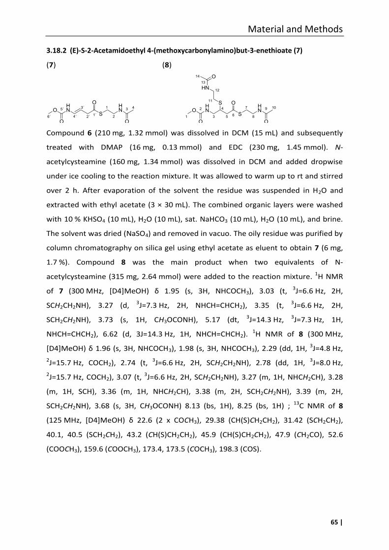
Material and Methods
65 |
3.18.2 (E)-S-2-Acetamidoethyl 4-(methoxycarbonylamino)but-3-enethioate (7)
(7) (8)
O
O
HN
S
OHN
O
S
HN
O
2
3 5 81
7 1094
6
11
12
13
14
Compound 6 (210 mg, 1.32 mmol) was dissolved in DCM (15 mL) and subsequently
treated with DMAP (16 mg, 0.13 mmol) and EDC (230 mg, 1.45 mmol). N-
acetylcysteamine (160 mg, 1.34 mmol) was dissolved in DCM and added dropwise
under ice cooling to the reaction mixture. It was allowed to warm up to rt and stirred
over 2 h. After evaporation of the solvent the residue was suspended in H2O and
extracted with ethyl acetate (3 × 30 mL). The combined organic layers were washed
with 10 % KHSO4 (10 mL), H2O (10 mL), sat. NaHCO3 (10 mL), H2O (10 mL), and brine.
The solvent was dried (NaSO4) and removed in vacuo. The oily residue was purified by
column chromatography on silica gel using ethyl acetate as eluent to obtain 7 (6 mg,
1.7 %). Compound 8 was the main product when two equivalents of N-
acetylcysteamine (315 mg, 2.64 mmol) were added to the reaction mixture. 1H NMR
of 7 (300 M , [D4]MeO ) δ 1.95 (s, 3 , N COC 3), 3.03 (t, 3J=6.6 Hz, 2H,
SCH2CH2NH), 3.27 (d, 3J=7.3 Hz, 2H, NHCH=CHCH2), 3.35 (t, 3J=6.6 Hz, 2H,
SCH2CH2NH), 3.73 (s, 1H, CH3OCONH), 5.17 (dt, 3J=14.3 Hz, 3J=7.3 Hz, 1H,
NHCH=CHCH2), 6.62 (d, 3J=14.3 Hz, 1H, NHCH=CHCH2). 1H NMR of 8 (300 MHz,
[D4]MeO ) δ 1.96 (s, 3 , N COC 3), 1.98 (s, 3H, NHCOCH3), 2.29 (dd, 1H, 3J=4.8 Hz,
2J=15.7 Hz, COCH2), 2.74 (t, 3J=6.6 Hz, 2H, SCH2CH2NH), 2.78 (dd, 1H, 3J=8.0 Hz,
2J=15.7 Hz, COCH2), 3.07 (t, 3J=6.6 Hz, 2H, SCH2CH2NH), 3.27 (m, 1H, NHCH2CH), 3.28
(m, 1H, SCH), 3.36 (m, 1H, NHCH2CH), 3.38 (m, 2H, SCH2CH2NH), 3.39 (m, 2H,
SCH2CH2NH), 3.68 (s, 3H, CH3OCONH) 8.13 (bs, 1H), 8.25 (bs, 1H) ; 13C NMR of 8
(125 M , [D4]MeO ) δ 22.6 (2 x COCH3), 29.38 (CH(S)CH2CH2), 31.42 (SCH2CH2),
40.1, 40.5 (SCH2CH2), 43.2 (CH(S)CH2CH2), 45.9 (CH(S)CH2CH2), 47.9 (CH2CO), 52.6
(COOCH3), 159.6 (COOCH3), 173.4, 173.5 (COCH3), 198.3 (COS).
O
O
HN
S
OHN
O2´
3´
4´
1
2
3 4
1´6´
5´
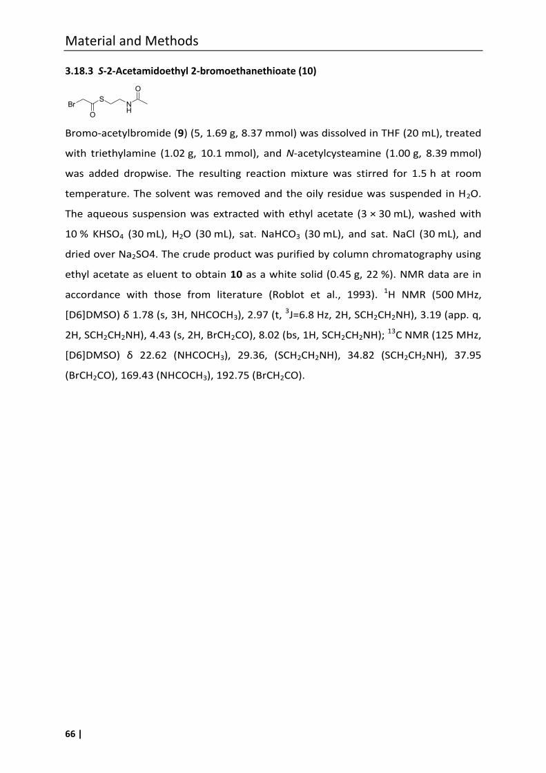
Material and Methods
66 |
3.18.3 S-2-Acetamidoethyl 2-bromoethanethioate (10)
Bromo-acetylbromide (9) (5, 1.69 g, 8.37 mmol) was dissolved in THF (20 mL), treated
with triethylamine (1.02 g, 10.1 mmol), and N-acetylcysteamine (1.00 g, 8.39 mmol)
was added dropwise. The resulting reaction mixture was stirred for 1.5 h at room
temperature. The solvent was removed and the oily residue was suspended in H2O.
The aqueous suspension was extracted with ethyl acetate (3 × 30 mL), washed with
10 % KHSO4 (30 mL), H2O (30 mL), sat. NaHCO3 (30 mL), and sat. NaCl (30 mL), and
dried over Na2SO4. The crude product was purified by column chromatography using
ethyl acetate as eluent to obtain 10 as a white solid (0.45 g, 22 %). NMR data are in
accordance with those from literature (Roblot et al., 1993). 1H NMR (500 MHz,
[D6]DMSO) δ 1.78 (s, 3 , N COC 3), 2.97 (t, 3J=6.8 Hz, 2H, SCH2CH2NH), 3.19 (app. q,
2H, SCH2CH2NH), 4.43 (s, 2H, BrCH2CO), 8.02 (bs, 1H, SCH2CH2NH); 13C NMR (125 MHz,
[D6]DMSO) δ 22.62 (N COC 3), 29.36, (SCH2CH2NH), 34.82 (SCH2CH2NH), 37.95
(BrCH2CO), 169.43 (NHCOCH3), 192.75 (BrCH2CO).
BrS
O
NH
O
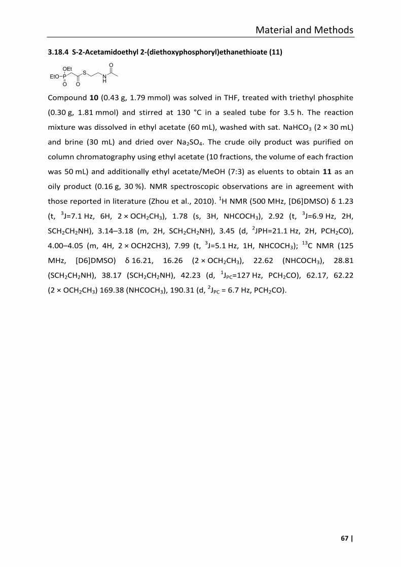
Material and Methods
67 |
3.18.4 S-2-Acetamidoethyl 2-(diethoxyphosphoryl)ethanethioate (11)
Compound 10 (0.43 g, 1.79 mmol) was solved in THF, treated with triethyl phosphite
(0.30 g, 1.81 mmol) and stirred at 130 °C in a sealed tube for 3.5 h. The reaction
mixture was dissolved in ethyl acetate (60 mL), washed with sat. NaHCO3 (2 × 30 mL)
and brine (30 mL) and dried over Na2SO4. The crude oily product was purified on
column chromatography using ethyl acetate (10 fractions, the volume of each fraction
was 50 mL) and additionally ethyl acetate/MeOH (7:3) as eluents to obtain 11 as an
oily product (0.16 g, 30 %). NMR spectroscopic observations are in agreement with
those reported in literature (Zhou et al., 2010). 1H NMR (500 M , [D6]DMSO) δ 1.23
(t, 3J=7.1 Hz, 6H, 2 × OCH2CH3), 1.78 (s, 3H, NHCOCH3), 2.92 (t, 3J=6.9 Hz, 2H,
SCH2CH2NH), 3.14–3.18 (m, 2H, SCH2CH2NH), 3.45 (d, 2JPH=21.1 Hz, 2H, PCH2CO),
4.00–4.05 (m, 4H, 2 × OCH2CH3), 7.99 (t, 3J=5.1 Hz, 1H, NHCOCH3); 13C NMR (125
M , [D6]DMSO) δ 16.21, 16.26 (2 × OCH2CH3), 22.62 (NHCOCH3), 28.81
(SCH2CH2NH), 38.17 (SCH2CH2NH), 42.23 (d, 1JPC=127 Hz, PCH2CO), 62.17, 62.22
(2 × OCH2CH3) 169.38 (NHCOCH3), 190.31 (d, 2JPC = 6.7 Hz, PCH2CO).
PS
O
NH
OOEt
O
EtO
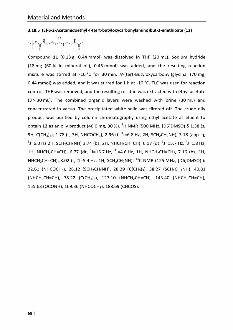
Material and Methods
68 |
3.18.5 (E)-S-2-Acetamidoethyl 4-(tert-butyloxycarbonylamino)but-2-enethioate (12)
Compound 11 (0.13 g, 0.44 mmol) was dissolved in THF (20 mL). Sodium hydride
(18 mg (60 % in mineral oil), 0.45 mmol) was added, and the resulting reaction
mixture was stirred at -10 °C for 30 min. N-(tert-Butyloxycarbonyl)glycinal (70 mg,
0.44 mmol) was added, and it was stirred for 1 h at -10 °C. TLC was used for reaction
control. THF was removed, and the resulting residue was extracted with ethyl acetate
(3 × 30 mL). The combined organic layers were washed with brine (30 mL) and
concentrated in vacuo. The precipitated white solid was filtered off. The crude oily
product was purified by column chromatography using ethyl acetate as eluent to
obtain 12 as an oily product (40.0 mg, 30 %). 1H NMR (500 M , [D6]DMSO) δ 1.38 (s,
9H, C(CH3)3), 1.78 (s, 3H, NHCOCH3), 2.96 (t, 3J=6.8 Hz, 2H, SCH2CH2NH), 3.18 (app. q,
3J=6.0 Hz 2H, SCH2CH2NH) 3.74 (bs, 2H, NHCH2CH=CH), 6.17 (dt, 3J=15.7 Hz, 4J=1.8 Hz,
1H, NHCH2CH=CH), 6.77 (dt, 3J=15.7 Hz, 3J=4.6 Hz, 1H, NHCH2CH=CH), 7.16 (bs, 1H,
NHCH2CH=CH), 8.02 (t, 3J=5.4 Hz, 1H, SCH2CH2NH); 13C NMR (125 M , [D6]DMSO) δ
22.61 (NHCOCH3), 28.12 (SCH2CH2NH), 28.29 (C(CH3)3), 38.27 (SCH2CH2NH), 40.81
(NHCH2CH=CH), 78.22 (C(CH3)3), 127.10 (NHCH2CH=CH), 143.40 (NHCH2CH=CH),
155.63 (OCONH), 169.36 (NHCOCH3), 188.69 (CHCOS).
OHN
O
S
OHN
O
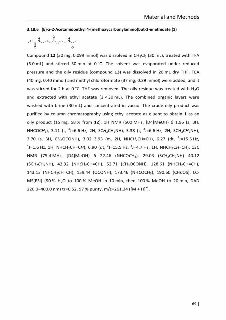
Material and Methods
69 |
3.18.6 (E)-S-2-Acetamidoethyl 4-(methoxycarbonylamino)but-2-enethioate (1)
Compound 12 (30 mg, 0.099 mmol) was dissolved in CH2Cl2 (30 mL), treated with TFA
(5.0 mL) and stirred 30 min at 0 °C. The solvent was evaporated under reduced
pressure and the oily residue (compound 13) was dissolved in 20 mL dry THF. TEA
(40 mg, 0.40 mmol) and methyl chloroformate (37 mg, 0.39 mmol) were added, and it
was stirred for 2 h at 0 °C. THF was removed. The oily residue was treated with H2O
and extracted with ethyl acetate (3 × 30 mL). The combined organic layers were
washed with brine (30 mL) and concentrated in vacuo. The crude oily product was
purified by column chromatography using ethyl acetate as eluent to obtain 1 as an
oily product (15 mg, 58 % from 12). 1H NMR (500 M , [D4]MeO ) δ 1.96 (s, 3 ,
NHCOCH3), 3.11 (t, 3J=6.6 Hz, 2H, SCH2CH2NH), 3.38 (t, 3J=6.6 Hz, 2H, SCH2CH2NH),
3.70 (s, 3H, CH3OCONH), 3.92–3.93 (m, 2H, NHCH2CH=CH), 6.27 (dt, 3J=15.5 Hz,
4J=1.6 Hz, 1H, NHCH2CH=CH), 6.90 (dt, 3J=15.5 Hz, 3J=4.7 Hz, 1H, NHCH2CH=CH); 13C
NMR (75.4 M , [D4]MeO ) δ 22.46 (N COCH3), 29.03 (SCH2CH2NH) 40.12
(SCH2CH2NH), 42.32 (NHCH2CH=CH), 52.71 (CH3OCONH), 128.61 (NHCH2CH=CH),
143.13 (NHCH2CH=CH), 159.44 (OCONH), 173.46 (NHCOCH3), 190.60 (CHCOS). LC-
MS(ESI) (90 % H2O to 100 % MeOH in 10 min, then 100 % MeOH to 20 min, DAD
220.0–400.0 nm) tr=6.52, 97 % purity, m/z=261.34 ([M + H]+).
OHN
O
S
OHN
O

70 |
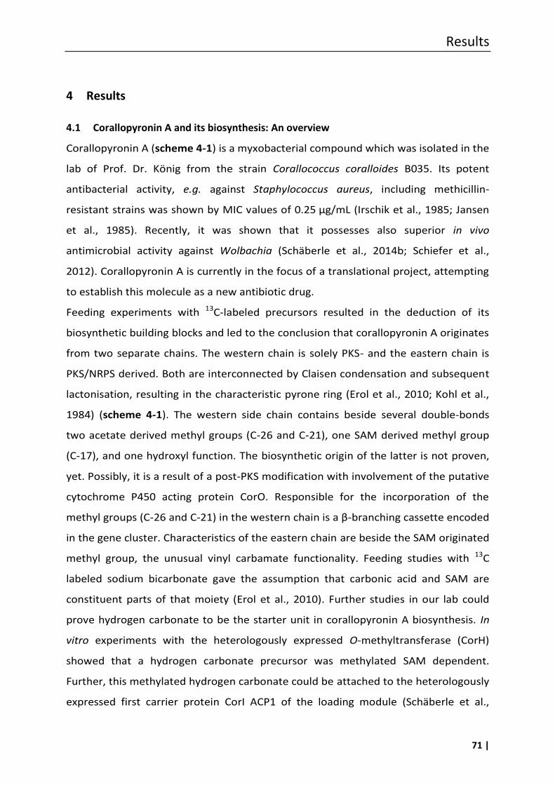
Results
71 |
4 Results
4.1 Corallopyronin A and its biosynthesis: An overview
Corallopyronin A (scheme 4-1) is a myxobacterial compound which was isolated in the
lab of Prof. Dr. König from the strain Corallococcus coralloides B035. Its potent
antibacterial activity, e.g. against Staphylococcus aureus, including methicillin-
resistant strains was shown by MIC values of 0.25 µg/mL (Irschik et al., 1985; Jansen
et al., 1985). Recently, it was shown that it possesses also superior in vivo
antimicrobial activity against Wolbachia (Schäberle et al., 2014b; Schiefer et al.,
2012). Corallopyronin A is currently in the focus of a translational project, attempting
to establish this molecule as a new antibiotic drug.
Feeding experiments with 13C-labeled precursors resulted in the deduction of its
biosynthetic building blocks and led to the conclusion that corallopyronin A originates
from two separate chains. The western chain is solely PKS- and the eastern chain is
PKS/NRPS derived. Both are interconnected by Claisen condensation and subsequent
lactonisation, resulting in the characteristic pyrone ring (Erol et al., 2010; Kohl et al.,
1984) (scheme 4-1). The western side chain contains beside several double-bonds
two acetate derived methyl groups (C-26 and C-21), one SAM derived methyl group
(C-17), and one hydroxyl function. The biosynthetic origin of the latter is not proven,
yet. Possibly, it is a result of a post-PKS modification with involvement of the putative
cytochrome P450 acting protein CorO. Responsible for the incorporation of the
methyl groups (C-26 and C-21) in the western chain is a β-branching cassette encoded
in the gene cluster. Characteristics of the eastern chain are beside the SAM originated
methyl group, the unusual vinyl carbamate functionality. Feeding studies with 13C
labeled sodium bicarbonate gave the assumption that carbonic acid and SAM are
constituent parts of that moiety (Erol et al., 2010). Further studies in our lab could
prove hydrogen carbonate to be the starter unit in corallopyronin A biosynthesis. In
vitro experiments with the heterologously expressed O-methyltransferase (CorH)
showed that a hydrogen carbonate precursor was methylated SAM dependent.
Further, this methylated hydrogen carbonate could be attached to the heterologously
expressed first carrier protein CorI ACP1 of the loading module (Schäberle et al.,
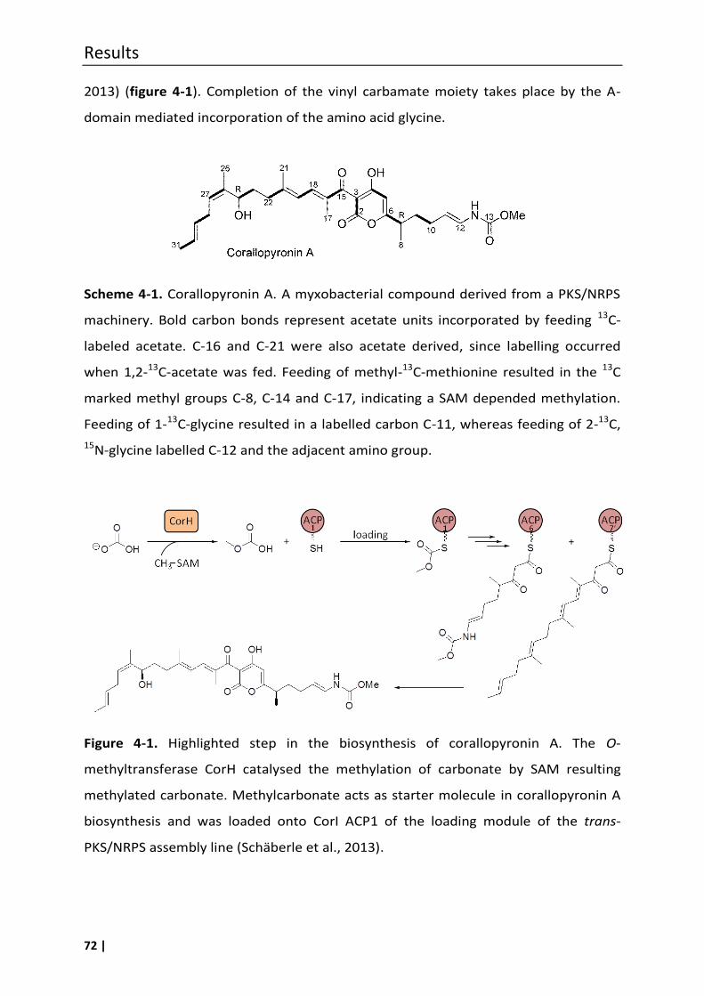
Results
72 |
2013) (figure 4-1). Completion of the vinyl carbamate moiety takes place by the A-
domain mediated incorporation of the amino acid glycine.
Scheme 4-1. Corallopyronin A. A myxobacterial compound derived from a PKS/NRPS
machinery. Bold carbon bonds represent acetate units incorporated by feeding 13C-
labeled acetate. C-16 and C-21 were also acetate derived, since labelling occurred
when 1,2-13C-acetate was fed. Feeding of methyl-13C-methionine resulted in the 13C
marked methyl groups C-8, C-14 and C-17, indicating a SAM depended methylation.
Feeding of 1-13C-glycine resulted in a labelled carbon C-11, whereas feeding of 2-13C,
15N-glycine labelled C-12 and the adjacent amino group.
Figure 4-1. Highlighted step in the biosynthesis of corallopyronin A. The O-
methyltransferase CorH catalysed the methylation of carbonate by SAM resulting
methylated carbonate. Methylcarbonate acts as starter molecule in corallopyronin A
biosynthesis and was loaded onto CorI ACP1 of the loading module of the trans-
PKS/NRPS assembly line (Schäberle et al., 2013).
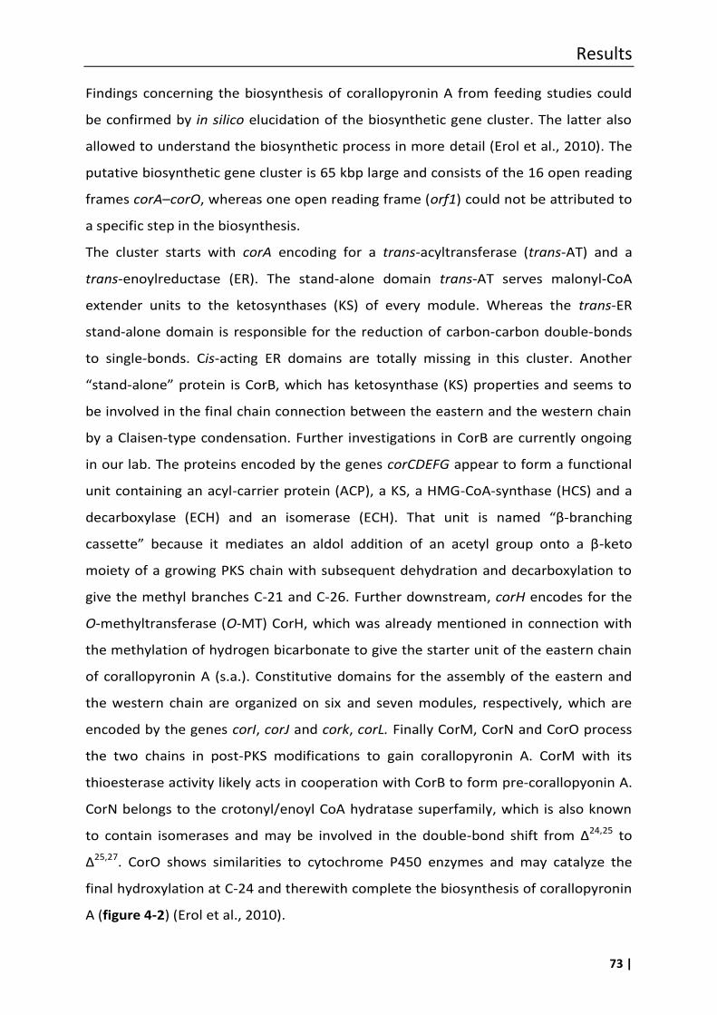
Results
73 |
Findings concerning the biosynthesis of corallopyronin A from feeding studies could
be confirmed by in silico elucidation of the biosynthetic gene cluster. The latter also
allowed to understand the biosynthetic process in more detail (Erol et al., 2010). The
putative biosynthetic gene cluster is 65 kbp large and consists of the 16 open reading
frames corA–corO, whereas one open reading frame (orf1) could not be attributed to
a specific step in the biosynthesis.
The cluster starts with corA encoding for a trans-acyltransferase (trans-AT) and a
trans-enoylreductase (ER). The stand-alone domain trans-AT serves malonyl-CoA
extender units to the ketosynthases (KS) of every module. Whereas the trans-ER
stand-alone domain is responsible for the reduction of carbon-carbon double-bonds
to single-bonds. Cis-acting ER domains are totally missing in this cluster. Another
“stand-alone” protein is CorB, which has ketosynthase ( S) properties and seems to
be involved in the final chain connection between the eastern and the western chain
by a Claisen-type condensation. Further investigations in CorB are currently ongoing
in our lab. The proteins encoded by the genes corCDEFG appear to form a functional
unit containing an acyl-carrier protein (ACP), a KS, a HMG-CoA-synthase (HCS) and a
decarboxylase (ECH) and an isomerase (ECH). That unit is named “β-branching
cassette” because it mediates an aldol addition of an acetyl group onto a β-keto
moiety of a growing PKS chain with subsequent dehydration and decarboxylation to
give the methyl branches C-21 and C-26. Further downstream, corH encodes for the
O-methyltransferase (O-MT) CorH, which was already mentioned in connection with
the methylation of hydrogen bicarbonate to give the starter unit of the eastern chain
of corallopyronin A (s.a.). Constitutive domains for the assembly of the eastern and
the western chain are organized on six and seven modules, respectively, which are
encoded by the genes corI, corJ and cork, corL. Finally CorM, CorN and CorO process
the two chains in post-PKS modifications to gain corallopyronin A. CorM with its
thioesterase activity likely acts in cooperation with CorB to form pre-corallopyonin A.
CorN belongs to the crotonyl/enoyl CoA hydratase superfamily, which is also known
to contain isomerases and may be involved in the double-bond shift from Δ24,25 to
Δ25,27. CorO shows similarities to cytochrome P450 enzymes and may catalyze the
final hydroxylation at C-24 and therewith complete the biosynthesis of corallopyronin
A (figure 4-2) (Erol et al., 2010).
Results
74 |
Figure 4-2. Complete corallopyronin A gene cluster and the hypothetical biosynthetic
pathway. A: adenylation domain; ACP: acyl-carrier protein; AT: acyltransferase; DH:
dehydratase domain; KR: ketoreductase domain; KS: ketosynthase domain; MT:
methyltransferase domain. KS0: inactive KS; AT*, ACP* and KR* are presumably
inactive due to mutations. DH*: putative “shift domain”. Methyl groups introduced by
the β-branching cassette are marked with an asterisk. Figure is modified after Erol et
al. 2010.
Results
75 |
4.2 Attemps to prove the putative biosynthetic gene cluster of coralloyronin A
In the beginning of this thesis in 2009 it was envisaged to prove the still putative
biosynthetic gene cluster of corallopyronin A. To do this, two approaches were
planned to be exploited: first, a knock-out or disruption of a gene related to the
respective biosynthesis in the producer strain Corallococcus coralloides B035 and
second, expression of parts of the gene cluster (corA–corJ) in the heterologous host
Pseudomonas putida KT2440. The basis of these molecular biological strategies was a
cosmid library of the genome of C. coralloides B035, which was established by Erol et
al. during their investigation relating to the early work on the biosynthetic cluster of
corallopyronin A. During the latter study they found cosmid FJ7 to contain 39 kbp
sequence information putatively related to the biosynthesis of corallopyronin A,
namely the genes corA–corJ (Erol et al., 2010).
The first experiments aimed to disrupt the trans-acyltransferase (AT) coding
sequences within the gene corA in the host strain C. coralloides B035. Therefore the
gene sequence of the trans-AT was replaced by a streptomycin resistance cassette
(aadA) in cosmid FJ7 using the lambda-red recombineering technique (figure 4-3 A, B)
(Gust et al., 2003). Analogously the recombinant cosmid FJ7 AT::aac(3)IV containing
an apramycin resistance cassette was prepared with the same purpose. Subsequently
the producer strain C. coralloides B035 was transformed with the generated construct
e.g. FJ7 AT::aadA with was inteneted to recombine homologously with the
corallopyronin A gene cluster in the host strain (figure 4-3 B, C). A disruption of the
trans-AT locus in corA in C. coralloides B035 would result in a mutated strain which
could not produce corallopyronin A anymore. A lack of corallopyronin A production
might have been detected via LCMS of the extracts of the bacterial cells. All efforts to
gain a knock-out mutant of the producer strain, however, failed so far. The crucial
point seemed to be the transformation of C. coralloides cells by electroporation,
which has been described in literature as a successful method for transformation of
myxobacteria (Magrini et al., 1998; Simunovic and Müller, 2007b). In the meantime,
in 2013, Sucipto et al. published the evidence of the putative biosynthetic gene
cluster of the antibiotic myxopyronin A, closely related to corallopyonin A, by gene
deletion experiments in the producer strain Myxococcus fulvus Mx f50 (Sucipto et al.,
2013).
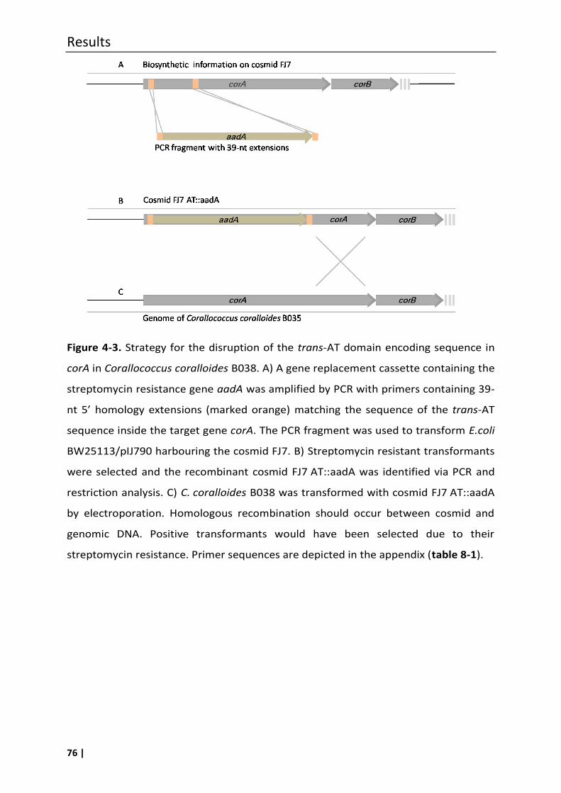
Results
76 |
Figure 4-3. Strategy for the disruption of the trans-AT domain encoding sequence in
corA in Corallococcus coralloides B038. A) A gene replacement cassette containing the
streptomycin resistance gene aadA was amplified by PCR with primers containing 39-
nt 5’ homology extensions (marked orange) matching the sequence of the trans-AT
sequence inside the target gene corA. The PCR fragment was used to transform E.coli
BW25113/pIJ790 harbouring the cosmid FJ7. B) Streptomycin resistant transformants
were selected and the recombinant cosmid FJ7 AT::aadA was identified via PCR and
restriction analysis. C) C. coralloides B038 was transformed with cosmid FJ7 AT::aadA
by electroporation. Homologous recombination should occur between cosmid and
genomic DNA. Positive transformants would have been selected due to their
streptomycin resistance. Primer sequences are depicted in the appendix (table 8-1).
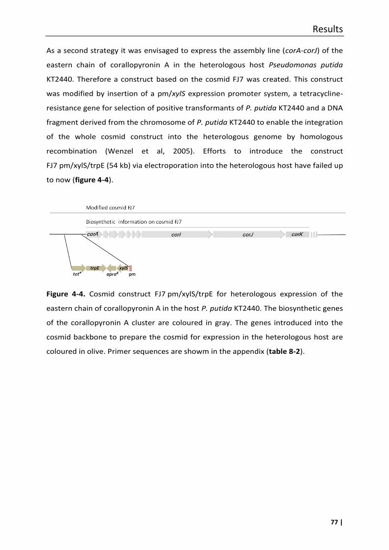
Results
77 |
As a second strategy it was envisaged to express the assembly line (corA-corJ) of the
eastern chain of corallopyronin A in the heterologous host Pseudomonas putida
KT2440. Therefore a construct based on the cosmid FJ7 was created. This construct
was modified by insertion of a pm/xylS expression promoter system, a tetracycline-
resistance gene for selection of positive transformants of P. putida KT2440 and a DNA
fragment derived from the chromosome of P. putida KT2440 to enable the integration
of the whole cosmid construct into the heterologous genome by homologous
recombination (Wenzel et al, 2005). Efforts to introduce the construct
FJ7 pm/xylS/trpE (54 kb) via electroporation into the heterologous host have failed up
to now (figure 4-4).
Figure 4-4. Cosmid construct FJ7 pm/xylS/trpE for heterologous expression of the
eastern chain of corallopyronin A in the host P. putida KT2440. The biosynthetic genes
of the corallopyronin A cluster are coloured in gray. The genes introduced into the
cosmid backbone to prepare the cosmid for expression in the heterologous host are
coloured in olive. Primer sequences are showm in the appendix (table 8-2).
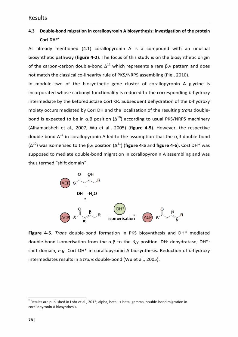
Results
78 |
4.3 Double-bond migration in corallopyronin A biosynthesis: investigation of the protein
CorJ DH*2
As already mentioned (4.1) corallopyronin A is a compound with an unusual
biosynthetic pathway (figure 4-2). The focus of this study is on the biosynthetic origin
of the carbon-carbon double-bond Δ11 which represents a rare β,γ pattern and does
not match the classical co-linearity rule of PKS/NRPS assembling (Piel, 2010).
In module two of the biosynthetic gene cluster of corallopyronin A glycine is
incorporated whose carbonyl functionality is reduced to the corresponding D-hydroxy
intermediate by the ketoreductase CorI KR. Subsequent dehydration of the D-hydroxy
moiety occurs mediated by CorI DH and the localization of the resulting trans double-
bond is expected to be in α,β position (Δ10) according to usual PKS/NRPS machinery
(Alhamadsheh et al., 2007; Wu et al., 2005) (figure 4-5). However, the respective
double-bond Δ11 in corallopyronin A led to the assumption that the α,β double-bond
(Δ10) was isomerised to the β,γ position (Δ11) (figure 4-5 and figure 4-6). CorJ DH* was
supposed to mediate double-bond migration in corallopyronin A assembling and was
thus termed “shift domain”.
Figure 4-5. Trans double-bond formation in PKS biosynthesis and DH* mediated
double-bond isomerisation from the α,β to the β,γ position. DH: dehydratase; DH*:
shift domain, e.g. CorJ DH* in corallopyronin A biosynthesis. Reduction of D-hydroxy
intermediates results in a trans double-bond (Wu et al., 2005).
2 Results are published in Lohr et al., 2013; alpha, beta –> beta, gamma, bouble-bond migration in
corallopyronin A biosynthesis.
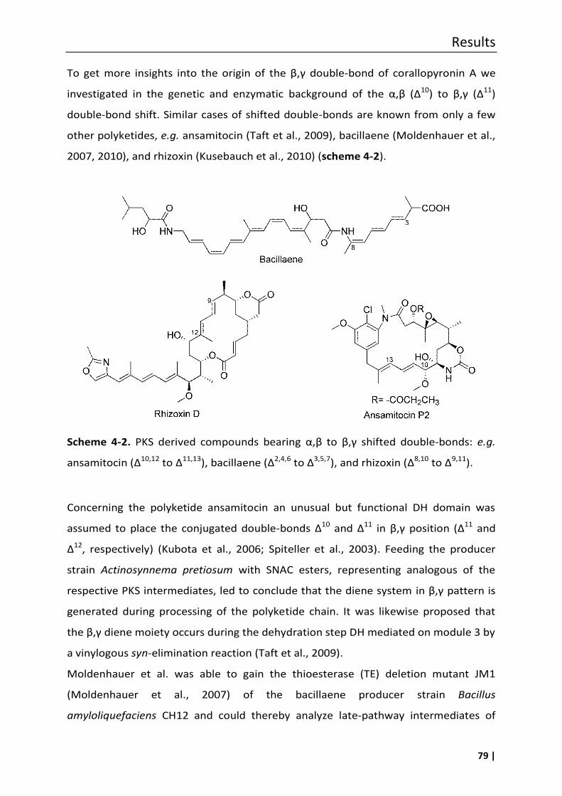
Results
79 |
To get more insights into the origin of the β,γ double-bond of corallopyronin A we
investigated in the genetic and enzymatic background of the α,β (Δ10) to β,γ (Δ11)
double-bond shift. Similar cases of shifted double-bonds are known from only a few
other polyketides, e.g. ansamitocin (Taft et al., 2009), bacillaene (Moldenhauer et al.,
2007, 2010), and rhizoxin (Kusebauch et al., 2010) (scheme 4-2).
Scheme 4-2. PKS derived compounds bearing α,β to β,γ shifted double-bonds: e.g.
ansamitocin (Δ10,12 to Δ11,13), bacillaene (Δ2,4,6 to Δ3,5,7), and rhizoxin (Δ8,10 to Δ9,11).
Concerning the polyketide ansamitocin an unusual but functional DH domain was
assumed to place the conjugated double-bonds Δ10 and Δ11 in β,γ position (Δ11 and
Δ12, respectively) (Kubota et al., 2006; Spiteller et al., 2003). Feeding the producer
strain Actinosynnema pretiosum with SNAC esters, representing analogous of the
respective PKS intermediates, led to conclude that the diene system in β,γ pattern is
generated during processing of the polyketide chain. It was likewise proposed that
the β,γ diene moiety occurs during the dehydration step DH mediated on module 3 by
a vinylogous syn-elimination reaction (Taft et al., 2009).
Moldenhauer et al. was able to gain the thioesterase (TE) deletion mutant JM1
(Moldenhauer et al., 2007) of the bacillaene producer strain Bacillus
amyloliquefaciens CH12 and could thereby analyze late-pathway intermediates of
Results
80 |
bacillaene. It could be shown that the β,γ double-bond installation of the double-
bonds Δ3 and Δ5 was performed on the same modules that performed the elongation
of the PKS chain (module 14 and 15, respectively). However, the origin of the third
double-bond Δ7of the triene system, introduced on module 16, could not be figured
out clearly. In that respect the authors consider a α,β to β,γ isomerisation by module
16, including the shift domain BaeR DH*, as alternative to β,γ desaturation as
demonstrated for module 14 and 15 (Moldenhauer et al., 2010).
In the biosynthesis of the PKS/NRPS derived compound rhizoxin both mechanisms
could be evidenced to give the β,γ diene moieties Δ11 and Δ9. The first unsaturation
Δ11 is introduced simultaneously to the double-bond shift in module 7 of the
biosynthetic gene cluster which corresponds to a formal β,γ dehydration. Regarding
double-bond Δ9 it was shown that first elongation of the nascent polyketide chain
takes place and second a distinct “shift module” (module 9) downstream mediates
the double-bond migration resulting in a β,γ double-bond (Δ9). The corresponding
shift domain appeared to be the protein RhiE DH* (Kusebauch et al., 2010).
For the β,γ double-bond Δ11 in corallopyronin A we proposed a similar process as
shown for double-bond Δ9 in the rhizoxin pathway. We proposed a distinct domain in
the assembly line performing the double-bond shift. Therefore, this study mainly
focuses on module 3 of the biosynthetic gene cluster of corallopyronin A which
consists of a ketosynthase (CorI KS), an unusual dehydratase (CorJ DH*) and an acyl-
carrier protein (CorJ ACP) (figure 4-6). A peculiarity of module 3 is that it is encoded
on two genes, subdividing the domains of one module from each other, and is
therefore named “split module” (Silakowski et al., 2001). So that the KS domain is
localized on the protein CorI and the domains DH* and ACP are part of the protein
CorJ (figure 4-6). The ketosynthase CorI KS is assumed to be inactive, despite of the
presence of a catalytic triad (Erol et al., 2010), that means no elongation occurs of the
nascent PKS chain in module 3. The further investigations ignore the role of CorI KS
and focus on the functional role of CorJ DH* and the corresponding carrier protein
CorJ ACP.

Results
81 |
Figure 4-6. Part of the corallopyronin A biosynthetic gene cluster with the focus on
the double-bond shift performed by the “shift module” (marked in orange). Module 3
consist of a ketosynthase (KS), a putative shift domain (DH*) and an acyl-carrier
protein (ACP) encoded by the genes corI and corJ, respectively.
Comparison of CorJ DH* to other shift domains (DH*) and to a dehydratase domain
(DH). To classify the putative corallopyronin A shift domain (CorJ DH*) its protein
sequence was compared with a classical dehydratase domain (EryAII DH) and with
two known shift domains (RhiE DH*, BaeR-DH*) and with one suspected shift domain
(DifK DH*). For bioinformatic analysis multiple sequence alignment using ClustalW
was performed with the amino acid sequences of CorJ DH* (Erol et al., 2010), RhiE
DH* (Kusebauch et al., 2010), BaeR-DH* (Moldenhauer et al., 2010) and DifK DH*
(Chen et al., 2006). The isomerisation activity of RhiE DH* and BaeR DH* had been
proven indirectly by knock-out experiments of the respective gene locus and
subsequent analysis of the PKS intermediates of rhizoxin D and bacillaene,
respectively (as described above) (Kusebauch et al., 2010; Moldenhauer et al., 2007).
DifK DH* is suspected to be a similar shift domain but detailed insight referring its
function as double-bond mediating enzyme have not been published, yet (Piel, 2010).
To depict the differences between shift domains and a classical dehydratase domain
(EryAII DH), the primary sequence of EryAII DH, derived from the erythromycin
biosynthesis gene cluster, was included into the alignment (figure 4-7). The amino
acid sequences compared in the alignment were extracted from the complete
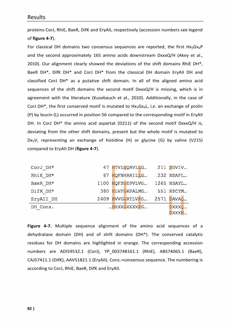
Results
82 |
proteins CorJ, RhiE, BaeR, DifK and EryAII, respectively (accession numbers see legend
of figure 4-7).
For classical DH domains two consensus sequences are reported, the first Hx3Gx4P
and the second approximately 165 amino acids downstream DxxxQ/H (Akey et al.,
2010). Our alignment clearly showed the deviations of the shift domains RhiE DH*,
BaeR DH*, DifK DH* and CorJ DH* from the classical DH domain EryAII DH and
classified CorJ DH* as a putative shift domain. In all of the aligned amino acid
sequences of the shift domains the second motif DxxxQ/H is missing, which is in
agreement with the literature (Kusebauch et al., 2010). Additionally, in the case of
CorJ DH*, the first conserved motif is mutated to Hx3Gx4L, i.e. an exchange of prolin
(P) by leucin (L) occurred in position 56 compared to the corresponding motif in EryAII
DH. In CorJ DH* the amino acid aspartat (D211) of the second motif DxxxQ/H is,
deviating from the other shift domains, present but the whole motif is mutated to
Dx3V, representing an exchange of histidine (H) or glycine (G) by valine (V215)
compared to EryAII DH (figure 4-7).
Figure 4-7. Multiple sequence alignment of the amino acid sequences of a
dehydratase domain (DH) and of shift domains (DH*). The conserved catalytic
residues for DH domains are highlighted in orange. The corresponding accession
numbers are ADI59532.1 (CorJ), YP_003748161.1 (RhiE), ABS74065.1 (BaeR),
CAJ57411.1 (DifK), AAV51821.1 (EryAII). Cons.=consensus sequence. The numbering is
according to CorJ, RhiE, BaeR, DifK and EryAII.
Results
83 |
4.4 In vitro assays envisaged to investigate the functional role of CorJ DH*
To prove the functionality of CorJ DH* as a double-bond migration catalyzing protein, two
enzyme assays were planned to be established by which the isomerisation of the respective
carbon-carbon double-bond could be traced. The first assay (4.7.1) was to base on a mass
spectrometric tracing of the CorJ DH* mediated double-bond shift of an ACP-bound
substrate (2). The second assay (4.7.2) was to rely on a comparison between the NMR
spectra of (i) the educt (substrate of CorJ DH*, 1), (ii) the reference compound 7 containing a
double-bond in β,γ pattern and (iii) of the reaction product of the assay with CorJ DH* and 1.
Both assays require the heterologously expressed proteins involved in the Δ10,11Δ11,12
double-bond shift in corallopyronin A biosynthesis: the suspected shift domain CorJ DH* and
its corresponding carrier protein CorJ ACP. CorJ DH* and CorJ ACP were planned to be
expressed as individual domains, as well as a didomain CorJ DH*-ACP which closely
resembled the natural arrangement. The didomain CorJ DH*-ACP was envisaged to be
expressed as well in its holo form (CorJ DH* holo-ACP) using E. coli Bap-1 cells as in its apo
form (CorJ DH* apo-ACP) feasible due to expression in the standard expression host E. coli
BL21. CorJ DH*holo-ACP should be applied in the first in vitro assay with subsequent mass
spectroscopic analysis, whereas CorJ DH*apo-ACP thought to be used in the second in vitro
assay followed by an NMR based analysis.
Furthermore, there was the need of a suitable substrate for CorJ DH*. On the one
hand it should be an analogue of the respective intermediate of corallopyronin A
biosynthesis, and on the other hand it should be able to be transferrable to the
phosphopantetheine residue of the CorJ holo-ACP domain. In order to fulfil the latter
requirement a N-acetylcysteamine (SNAC) thioester analogue of the respective
biosynthetic intermediate of corallopyronin A was synthesised (1; figure 4-14 and
scheme 4-5).
Results
84 |
4.5 Heterologous expression of CorJ DH*-ACP, CorJ DH* and CorJ ACP
The DNA of cosmid FJ7 was used for amplification of the respective primary sequence
of the proteins. The generation of the cosmid library of C. coralloides B035 is
described in Erol et al., 2010. In order to obtain the expression construct for the CorJ
DH*-ACP didomain a segment harbouring the start of corJ was amplified with the
primer pair (8.1, table 8-3) CorJ_start/ corJ_end_ACP yielding a fragment of 1227 bp.
The corresponding coding sequence of the CorJ ACP and of the single shift domain
CorJ DH* were amplified as well. Here the primer pairs ACP_1_upstream/
corJ_end_ACP and corJ_start/ DH_shift_neu were used to obtain a fragment of
309 bp and 736 bp, respectively. Each fragment was ligated into the cloning vector
pGEM-T (Promega) and checked for identity by sequencing with standard primers
(SP6 and T7). From this plasmid the desired fragment was cut out using the restriction
sites EcoRI/HindIII, which were introduced beforehand by the primers used. The
fragment was ligated in the likewise restricted expression vector pET28a which links
the protein to an N-terminal 6x His-tag upon expression. After ligation E. coli XL1 Blue
cells were transformed with the ligation mixture and plated on LB supplemented with
kanamycin for selection. After identifying positive clones by plasmid isolation and test
restriction the corresponding plasmid was isolated and transferred into the respective
expression host. Cloning and transformation procedures were applied according to
described methods (3.11.1) (figure 4-8). Respective protein sequences are depicted in
the appendix 8.2.
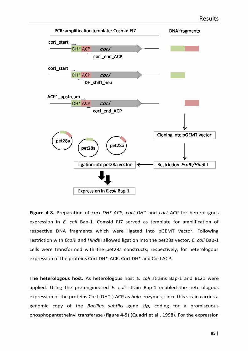
Results
85 |
Figure 4-8. Preparation of corJ DH*-ACP, corJ DH* and corJ ACP for heterologous
expression in E. coli Bap-1. Comsid FJ7 served as template for amplification of
respective DNA fragments which were ligated into pGEMT vector. Following
restriction with EcoRI and HindIII allowed ligation into the pet28a vector. E. coli Bap-1
cells were transformed with the pet28a constructs, respectively, for heterologous
expression of the proteins CorJ DH*-ACP, CorJ DH* and CorJ ACP.
The heterologous host. As heterologous host E. coli strains Bap-1 and BL21 were
applied. Using the pre-engineered E. coli strain Bap-1 enabled the heterologous
expression of the proteins CorJ (DH*-) ACP as holo-enzymes, since this strain carries a
genomic copy of the Bacillus subtilis gene sfp, coding for a promiscuous
phosphopantetheinyl transferase (figure 4-9) (Quadri et al., 1998). For the expression
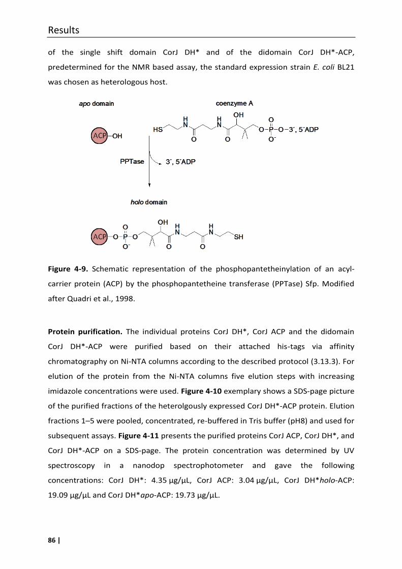
Results
86 |
of the single shift domain CorJ DH* and of the didomain CorJ DH*-ACP,
predetermined for the NMR based assay, the standard expression strain E. coli BL21
was chosen as heterologous host.
Figure 4-9. Schematic representation of the phosphopantetheinylation of an acyl-
carrier protein (ACP) by the phosphopantetheine transferase (PPTase) Sfp. Modified
after Quadri et al., 1998.
Protein purification. The individual proteins CorJ DH*, CorJ ACP and the didomain
CorJ DH*-ACP were purified based on their attached his-tags via affinity
chromatography on Ni-NTA columns according to the described protocol (3.13.3). For
elution of the protein from the Ni-NTA columns five elution steps with increasing
imidazole concentrations were used. Figure 4-10 exemplary shows a SDS-page picture
of the purified fractions of the heterolgously expressed CorJ DH*-ACP protein. Elution
fractions 1–5 were pooled, concentrated, re-buffered in Tris buffer (pH8) and used for
subsequent assays. Figure 4-11 presents the purified proteins CorJ ACP, CorJ DH*, and
CorJ DH*-ACP on a SDS-page. The protein concentration was determined by UV
spectroscopy in a nanodop spectrophotometer and gave the following
concentrations: CorJ DH*: 4.35 µg/µL, CorJ ACP: 3.04 µg/µL, CorJ DH*holo-ACP:
19.09 µg/µL and CorJ DH*apo-ACP: 19.73 µg/µL.
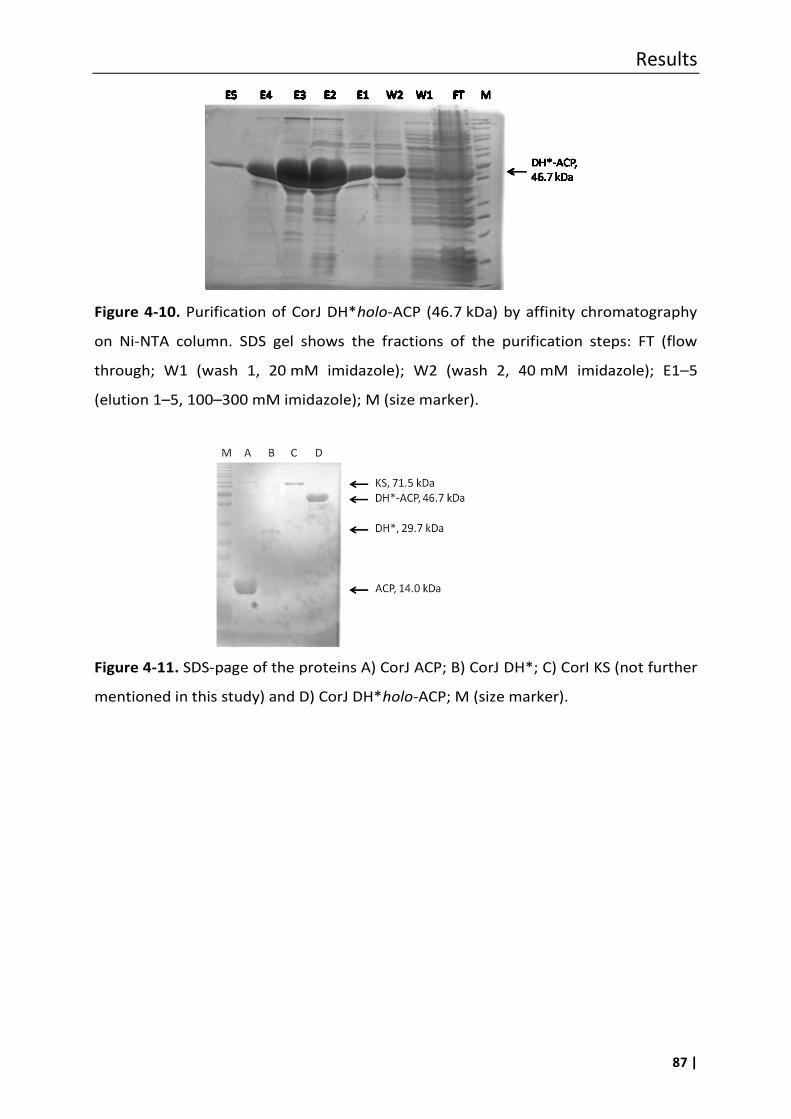
Results
87 |
Figure 4-10. Purification of CorJ DH*holo-ACP (46.7 kDa) by affinity chromatography
on Ni-NTA column. SDS gel shows the fractions of the purification steps: FT (flow
through; W1 (wash 1, 20 mM imidazole); W2 (wash 2, 40 mM imidazole); E1–5
(elution 1–5, 100–300 mM imidazole); M (size marker).
Figure 4-11. SDS-page of the proteins A) CorJ ACP; B) CorJ DH*; C) CorI KS (not further
mentioned in this study) and D) CorJ DH*holo-ACP; M (size marker).
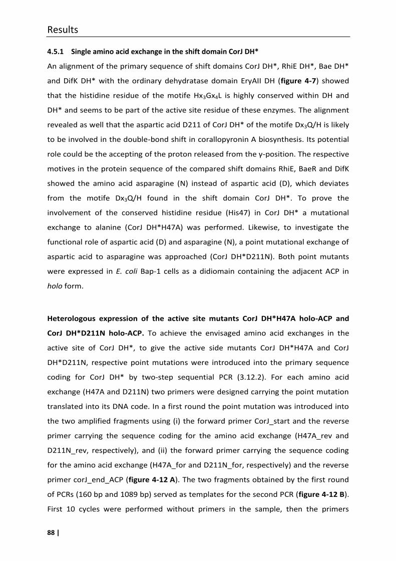
Results
88 |
4.5.1 Single amino acid exchange in the shift domain CorJ DH*
An alignment of the primary sequence of shift domains CorJ DH*, RhiE DH*, Bae DH*
and DifK DH* with the ordinary dehydratase domain EryAII DH (figure 4-7) showed
that the histidine residue of the motife Hx3Gx4L is highly conserved within DH and
DH* and seems to be part of the active site residue of these enzymes. The alignment
revealed as well that the aspartic acid D211 of CorJ DH* of the motife Dx3Q/H is likely
to be involved in the double-bond shift in corallopyronin A biosynthesis. Its potential
role could be the accepting of the proton released from the γ-position. The respective
motives in the protein sequence of the compared shift domains RhiE, BaeR and DifK
showed the amino acid asparagine (N) instead of aspartic acid (D), which deviates
from the motife Dx3Q/H found in the shift domain CorJ DH*. To prove the
involvement of the conserved histidine residue (His47) in CorJ DH* a mutational
exchange to alanine (CorJ DH*H47A) was performed. Likewise, to investigate the
functional role of aspartic acid (D) and asparagine (N), a point mutational exchange of
aspartic acid to asparagine was approached (CorJ DH*D211N). Both point mutants
were expressed in E. coli Bap-1 cells as a didiomain containing the adjacent ACP in
holo form.
Heterologous expression of the active site mutants CorJ DH*H47A holo-ACP and
CorJ DH*D211N holo-ACP. To achieve the envisaged amino acid exchanges in the
active site of CorJ DH*, to give the active side mutants CorJ DH*H47A and CorJ
DH*D211N, respective point mutations were introduced into the primary sequence
coding for CorJ DH* by two-step sequential PCR (3.12.2). For each amino acid
exchange (H47A and D211N) two primers were designed carrying the point mutation
translated into its DNA code. In a first round the point mutation was introduced into
the two amplified fragments using (i) the forward primer CorJ_start and the reverse
primer carrying the sequence coding for the amino acid exchange (H47A_rev and
D211N_rev, respectively), and (ii) the forward primer carrying the sequence coding
for the amino acid exchange (H47A_for and D211N_for, respectively) and the reverse
primer corJ_end_ACP (figure 4-12 A). The two fragments obtained by the first round
of PCRs (160 bp and 1089 bp) served as templates for the second PCR (figure 4-12 B).
First 10 cycles were performed without primers in the sample, then the primers
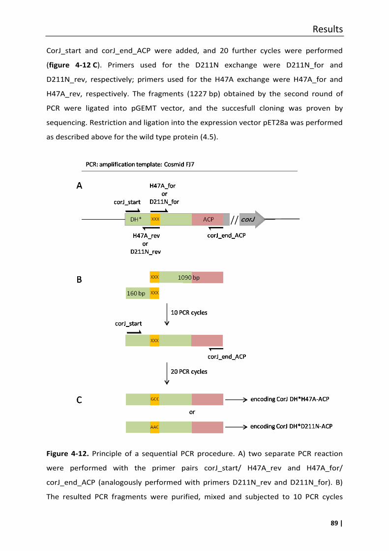
Results
89 |
CorJ_start and corJ_end_ACP were added, and 20 further cycles were performed
(figure 4-12 C). Primers used for the D211N exchange were D211N_for and
D211N_rev, respectively; primers used for the H47A exchange were H47A_for and
H47A_rev, respectively. The fragments (1227 bp) obtained by the second round of
PCR were ligated into pGEMT vector, and the succesfull cloning was proven by
sequencing. Restriction and ligation into the expression vector pET28a was performed
as described above for the wild type protein (4.5).
Figure 4-12. Principle of a sequential PCR procedure. A) two separate PCR reaction
were performed with the primer pairs corJ_start/ H47A_rev and H47A_for/
corJ_end_ACP (analogously performed with primers D211N_rev and D211N_for). B)
The resulted PCR fragments were purified, mixed and subjected to 10 PCR cycles
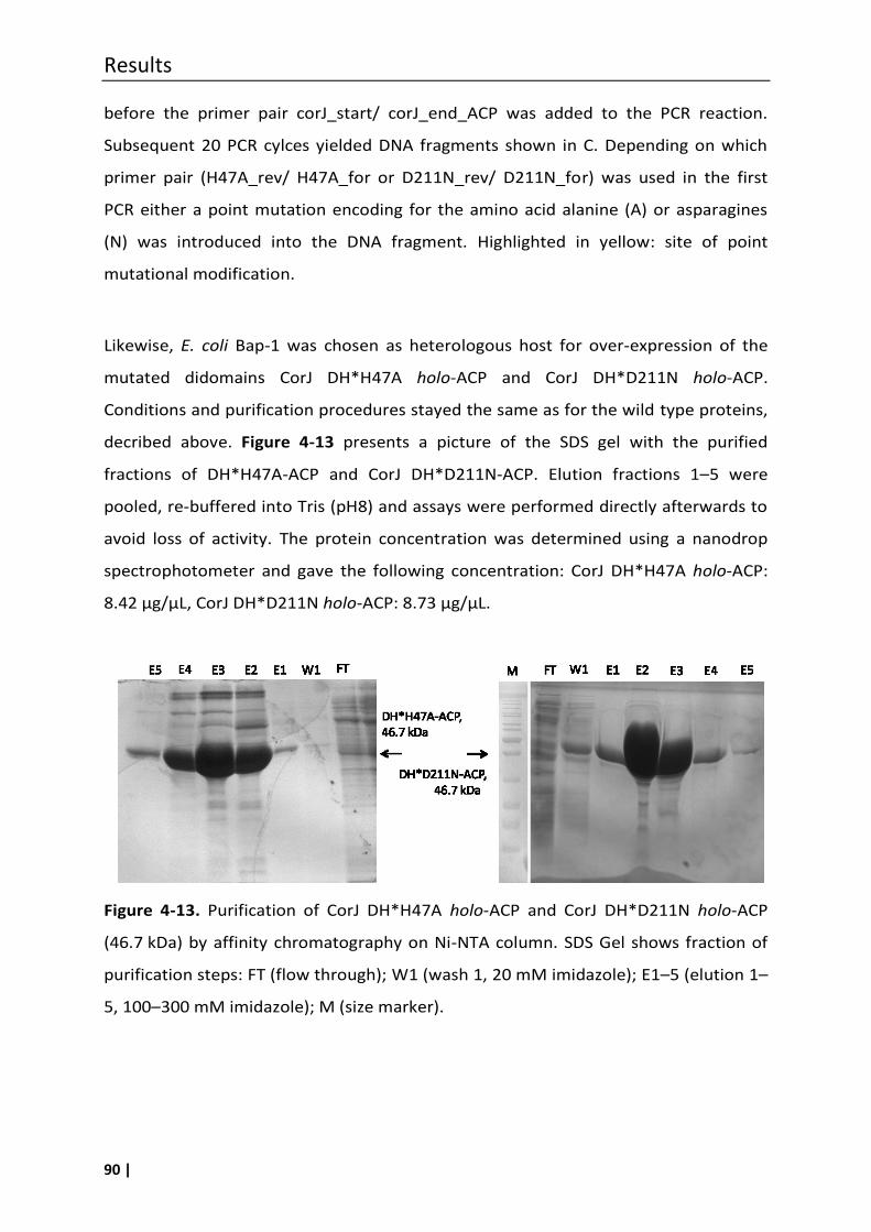
Results
90 |
before the primer pair corJ_start/ corJ_end_ACP was added to the PCR reaction.
Subsequent 20 PCR cylces yielded DNA fragments shown in C. Depending on which
primer pair (H47A_rev/ H47A_for or D211N_rev/ D211N_for) was used in the first
PCR either a point mutation encoding for the amino acid alanine (A) or asparagines
(N) was introduced into the DNA fragment. Highlighted in yellow: site of point
mutational modification.
Likewise, E. coli Bap-1 was chosen as heterologous host for over-expression of the
mutated didomains CorJ DH*H47A holo-ACP and CorJ DH*D211N holo-ACP.
Conditions and purification procedures stayed the same as for the wild type proteins,
decribed above. Figure 4-13 presents a picture of the SDS gel with the purified
fractions of DH*H47A-ACP and CorJ DH*D211N-ACP. Elution fractions 1–5 were
pooled, re-buffered into Tris (pH8) and assays were performed directly afterwards to
avoid loss of activity. The protein concentration was determined using a nanodrop
spectrophotometer and gave the following concentration: CorJ DH*H47A holo-ACP:
8.42 µg/µL, CorJ DH*D211N holo-ACP: 8.73 µg/µL.
Figure 4-13. Purification of CorJ DH*H47A holo-ACP and CorJ DH*D211N holo-ACP
(46.7 kDa) by affinity chromatography on Ni-NTA column. SDS Gel shows fraction of
purification steps: FT (flow through); W1 (wash 1, 20 mM imidazole); E1–5 (elution 1–
5, 100–300 mM imidazole); M (size marker).
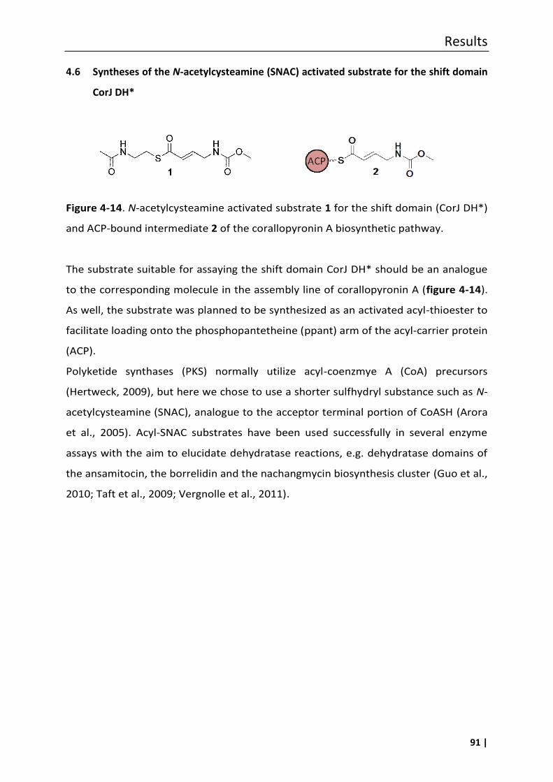
Results
91 |
4.6 Syntheses of the N-acetylcysteamine (SNAC) activated substrate for the shift domain
CorJ DH*
Figure 4-14. N-acetylcysteamine activated substrate 1 for the shift domain (CorJ DH*)
and ACP-bound intermediate 2 of the corallopyronin A biosynthetic pathway.
The substrate suitable for assaying the shift domain CorJ DH* should be an analogue
to the corresponding molecule in the assembly line of corallopyronin A (figure 4-14).
As well, the substrate was planned to be synthesized as an activated acyl-thioester to
facilitate loading onto the phosphopantetheine (ppant) arm of the acyl-carrier protein
(ACP).
Polyketide synthases (PKS) normally utilize acyl-coenzmye A (CoA) precursors
(Hertweck, 2009), but here we chose to use a shorter sulfhydryl substance such as N-
acetylcysteamine (SNAC), analogue to the acceptor terminal portion of CoASH (Arora
et al., 2005). Acyl-SNAC substrates have been used successfully in several enzyme
assays with the aim to elucidate dehydratase reactions, e.g. dehydratase domains of
the ansamitocin, the borrelidin and the nachangmycin biosynthesis cluster (Guo et al.,
2010; Taft et al., 2009; Vergnolle et al., 2011).
Results
92 |
4.6.1 Synthesis of compound 7
The first approach to obtain 1 yielded in compound 7, which represents the isomer of
1 with the double-bond in β,γ position (scheme 4-3).
In the first reaction step crotonic acid 3 was converted into the corresponding 4-
bromocrotonic acid 4 via a radical mechanism using the radical forming agent
azobisisobutyronitrile (AIBN) and N-bromosuccinimide (NBS). After recrystallisation
from a mixture of petroleum ether and ethylacetate compound 4 was obtained
(43 %). In the following step the bromine residue was replaced by an amino moiety
using aqueous ammonia to yield 10 % of 4-aminocrotonic acid (5) after purification
using a cation exchange resin. The conversion of the amino functionality to a
carbamate moiety was performed under basic conditions using methyl chloroformate
to give 4-methoxycarbonylaminobut 2-enoic acid (6) in yields of about 74 %. The
analytical data of the compounds 4 and 5 were in accordance with data from
literature (Höfling et al., 2008). It has to be mentioned that compounds 3–6 are
commercial available, however due to the need of greater amounts it was preferred
to synthesise them by ourselves. Finally, a coupling of 6 with N-acetylcysteamine
(SNAC) via 1-Ethyl-3-(3-dimethylaminopropyl)carbodiimide (EDC)/4-
Dimethylaminopyridine (DMAP) standard procedure (Neises and Steglich, 1978)
should have resulted in the desired substrate 1. However, a mixture of different
products was obtained from which compound 7 was isolated.
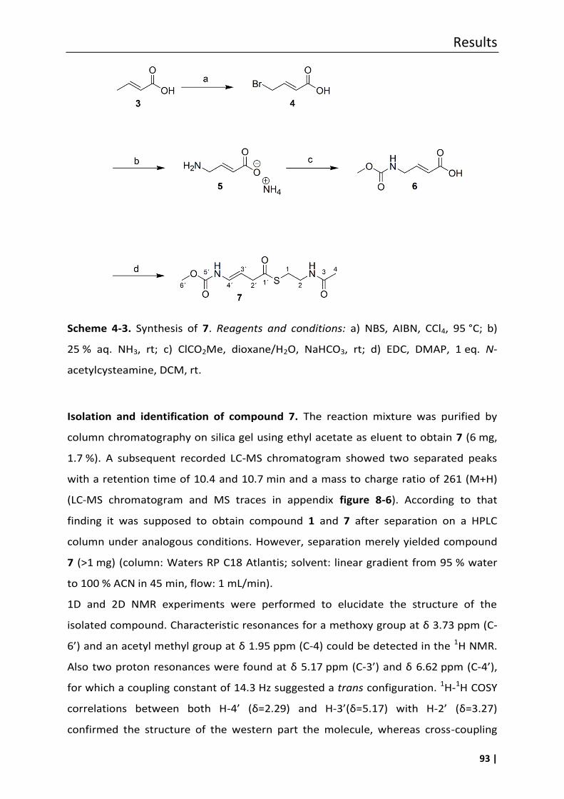
Results
93 |
Scheme 4-3. Synthesis of 7. Reagents and conditions: a) NBS, AIBN, CCl4, 95 °C; b)
25 % aq. NH3, rt; c) ClCO2Me, dioxane/H2O, NaHCO3, rt; d) EDC, DMAP, 1 eq. N-
acetylcysteamine, DCM, rt.
Isolation and identification of compound 7. The reaction mixture was purified by
column chromatography on silica gel using ethyl acetate as eluent to obtain 7 (6 mg,
1.7 %). A subsequent recorded LC-MS chromatogram showed two separated peaks
with a retention time of 10.4 and 10.7 min and a mass to charge ratio of 261 (M+H)
(LC-MS chromatogram and MS traces in appendix figure 8-6). According to that
finding it was supposed to obtain compound 1 and 7 after separation on a HPLC
column under analogous conditions. However, separation merely yielded compound
7 (>1 mg) (column: Waters RP C18 Atlantis; solvent: linear gradient from 95 % water
to 100 % ACN in 45 min, flow: 1 mL/min).
1D and 2D NMR experiments were performed to elucidate the structure of the
isolated compound. Characteristic resonances for a methoxy group at δ 3.73 ppm (C-
6’) and an acetyl methyl group at δ 1.95 ppm (C-4) could be detected in the 1H NMR.
Also two proton resonances were found at δ 5.17 ppm (C-3’) and δ 6.62 ppm (C-4’),
for which a coupling constant of 14.3 Hz suggested a trans configuration. 1H-1H COSY
correlations between both H-4’ (δ=2.29) and H-3’(δ=5.17) with H-2’ (δ=3.27)
confirmed the structure of the western part the molecule, whereas cross-coupling
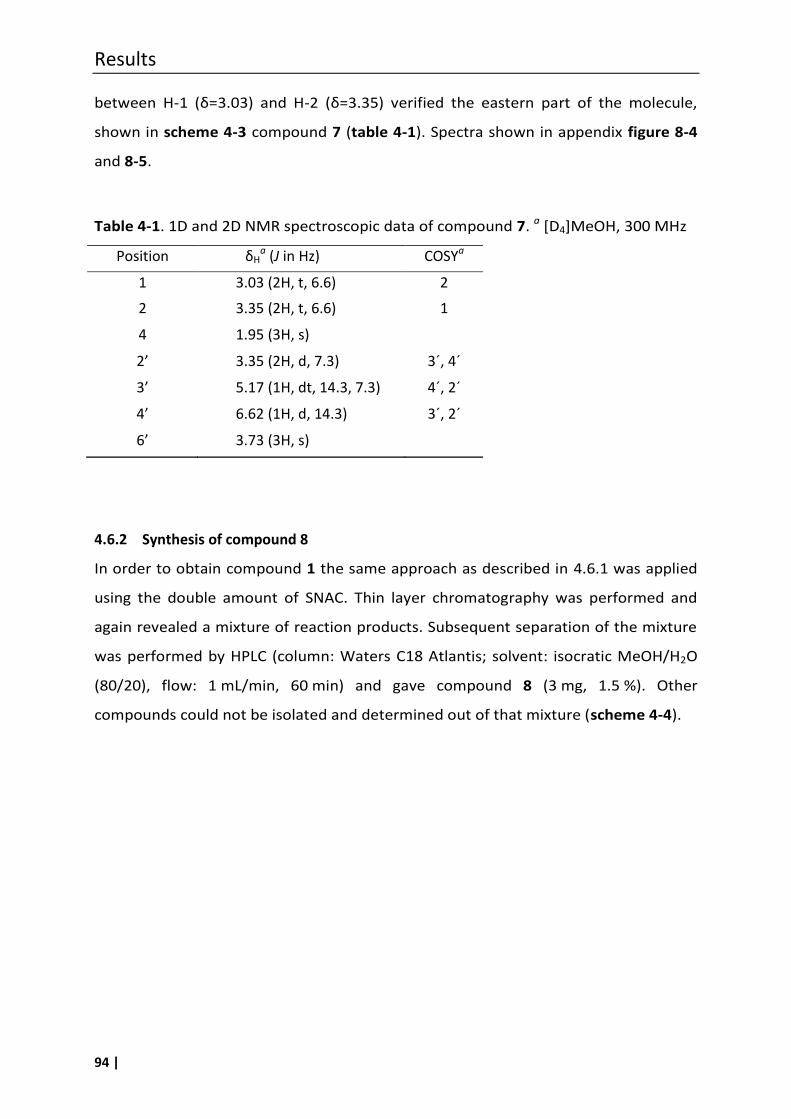
Results
94 |
between H-1 (δ=3.03) and H-2 (δ=3.35) verified the eastern part of the molecule,
shown in scheme 4-3 compound 7 (table 4-1). Spectra shown in appendix figure 8-4
and 8-5.
Table 4-1. 1D and 2D NMR spectroscopic data of compound 7. a [D4]MeOH, 300 MHz
Position δHa (J in Hz) COSYa
1 3.03 (2H, t, 6.6) 2
2 3.35 (2H, t, 6.6) 1
4 1.95 (3H, s)
2’ 3.35 (2H, d, 7.3) 3´, 4´
3’ 5.17 (1H, dt, 14.3, 7.3) 4´, 2´
4’ 6.62 (1H, d, 14.3) 3´, 2´
6’ 3.73 (3H, s)
4.6.2 Synthesis of compound 8
In order to obtain compound 1 the same approach as described in 4.6.1 was applied
using the double amount of SNAC. Thin layer chromatography was performed and
again revealed a mixture of reaction products. Subsequent separation of the mixture
was performed by HPLC (column: Waters C18 Atlantis; solvent: isocratic MeOH/H2O
(80/20), flow: 1 mL/min, 60 min) and gave compound 8 (3 mg, 1.5 %). Other
compounds could not be isolated and determined out of that mixture (scheme 4-4).
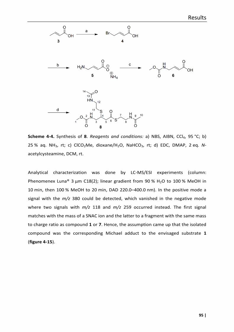
Results
95 |
Scheme 4-4. Synthesis of 8. Reagents and conditions: a) NBS, AIBN, CCl4, 95 °C; b)
25 % aq. NH3, rt; c) ClCO2Me, dioxane/H2O, NaHCO3, rt; d) EDC, DMAP, 2 eq. N-
acetylcysteamine, DCM, rt.
Analytical characterization was done by LC-MS/ESI experiments (column:
Phenomenex Luna® 3 µm C18(2); linear gradient from 90 % H2O to 100 % MeOH in
10 min, then 100 % MeOH to 20 min, DAD 220.0–400.0 nm). In the positive mode a
signal with the m/z 380 could be detected, which vanished in the negative mode
where two signals with m/z 118 and m/z 259 occurred instead. The first signal
matches with the mass of a SNAC ion and the latter to a fragment with the same mass
to charge ratio as compound 1 or 7. Hence, the assumption came up that the isolated
compound was the corresponding Michael adduct to the envisaged substrate 1
(figure 4-15).
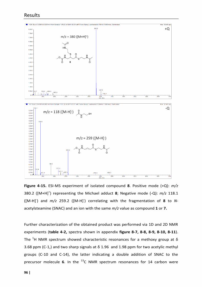
Results
96 |
Figure 4-15. ESI-MS experiment of isolated compound 8. Positive mode (+Q): m/z
380.2 ([M+H]+) representing the Michael adduct 8; Negative mode (-Q): m/z 118.1
([M-H]-) and m/z 259.2 ([M-H]-) correlating with the fragmentation of 8 to N-
acetylsteamine (SNAC) and an ion with the same m/z value as compound 1 or 7.
Further characterization of the obtained product was performed via 1D and 2D NMR
experiments (table 4-2, spectra shown in appendix figure 8-7, 8-8, 8-9, 8-10, 8-11).
The 1H NMR spectrum showed characteristic resonances for a methoxy group at δ
3.68 ppm (C-1,) and two sharp signals at δ 1.96 and 1.98 ppm for two acetylic methyl
groups (C-10 and C-14), the latter indicating a double addition of SNAC to the
precursor molecule 6. In the 13C NMR spectrum resonances for 14 carbon were
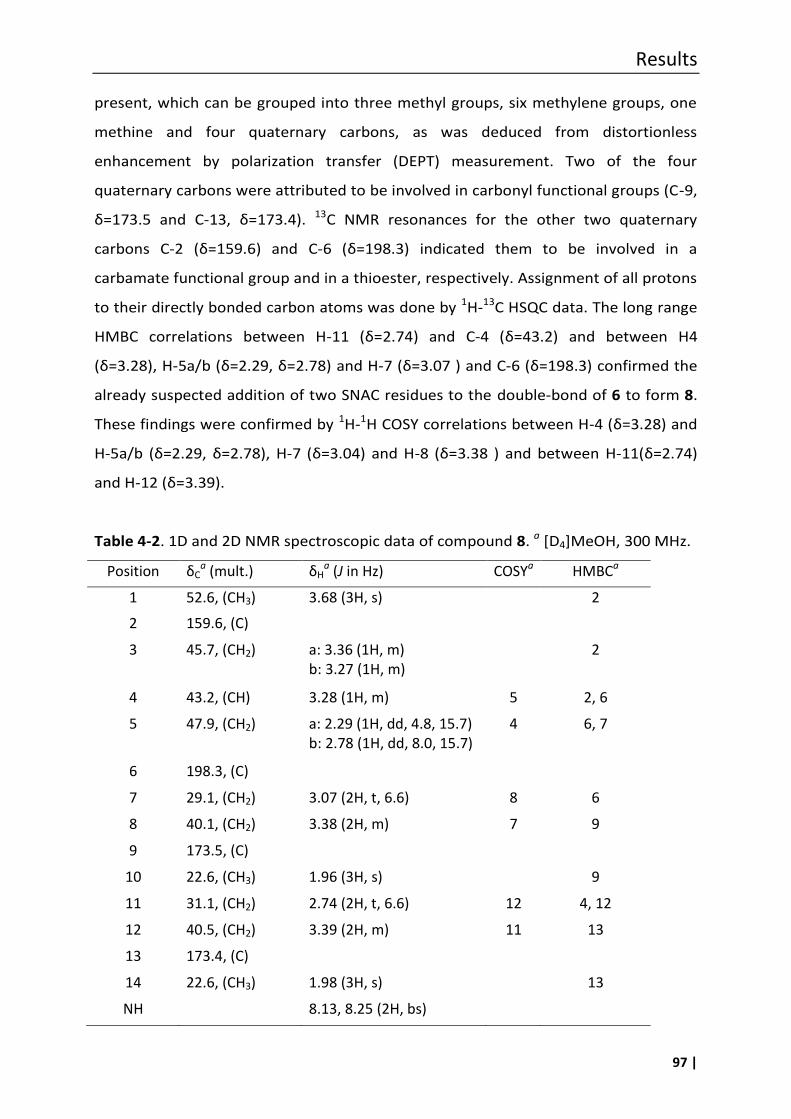
Results
97 |
present, which can be grouped into three methyl groups, six methylene groups, one
methine and four quaternary carbons, as was deduced from distortionless
enhancement by polarization transfer (DEPT) measurement. Two of the four
quaternary carbons were attributed to be involved in carbonyl functional groups (C-9,
δ=173.5 and C-13, δ=173.4). 13C NMR resonances for the other two quaternary
carbons C-2 (δ=159.6) and C-6 (δ=198.3) indicated them to be involved in a
carbamate functional group and in a thioester, respectively. Assignment of all protons
to their directly bonded carbon atoms was done by 1H-13C HSQC data. The long range
HMBC correlations between H-11 (δ=2.74) and C-4 (δ=43.2) and between H4
(δ=3.28), H-5a/b (δ=2.29, δ=2.78) and H-7 (δ=3.07 ) and C-6 (δ=198.3) confirmed the
already suspected addition of two SNAC residues to the double-bond of 6 to form 8.
These findings were confirmed by 1H-1H COSY correlations between H-4 (δ=3.28) and
H-5a/b (δ=2.29, δ=2.78), H-7 (δ=3.04) and -8 (δ=3.38 ) and between H-11(δ=2.74)
and H-12 (δ=3.39).
Table 4-2. 1D and 2D NMR spectroscopic data of compound 8. a [D4]MeOH, 300 MHz.
Position δCa (mult.) δH
a (J in Hz) COSYa HMBCa
1 52.6, (CH3) 3.68 (3H, s) 2
2 159.6, (C)
3 45.7, (CH2) a: 3.36 (1H, m) b: 3.27 (1H, m)
2
4 43.2, (CH) 3.28 (1H, m) 5 2, 6
5 47.9, (CH2) a: 2.29 (1H, dd, 4.8, 15.7) b: 2.78 (1H, dd, 8.0, 15.7)
15
jjjjjh888880) 15.7)
4 6, 7
6 198.3, (C)
7
8
9
10
11
12
13
29.1, (CH2)
40.5, (CH2)
173.5, (C)
22.6, (CH3)
29.4, (CH2)
40.1, (CH2)
173.4, (C)
3.07 (2H, t, 6.6)
3.38 (2H, t)
1.96 (3H, s)b
3.07 (2H, t)
3.38 (2H, t)
8
7
12
11
6
9
6
8 40.1, (CH2)
3.38 (2H, m)
7
9
9 173.5, (C)
10 22.6, (CH3)
1.96 (3H, s)
9
11 31.1, (CH2)
2.74 (2H, t, 6.6)
12
4, 12
12 40.5, (CH2)
3.39 (2H, m) 11 13
13 173.4, (C)
14 22.6, (CH3) 1.98 (3H, s) 13
NH 8.13, 8.25 (2H, bs)
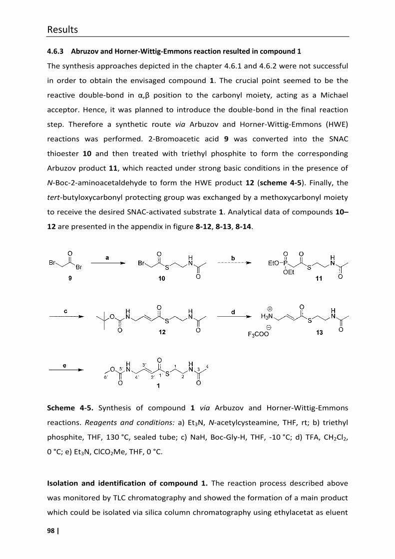
Results
98 |
4.6.3 Abruzov and Horner-Wittig-Emmons reaction resulted in compound 1
The synthesis approaches depicted in the chapter 4.6.1 and 4.6.2 were not successful
in order to obtain the envisaged compound 1. The crucial point seemed to be the
reactive double-bond in α,β position to the carbonyl moiety, acting as a Michael
acceptor. Hence, it was planned to introduce the double-bond in the final reaction
step. Therefore a synthetic route via Arbuzov and Horner-Wittig-Emmons (HWE)
reactions was performed. 2-Bromoacetic acid 9 was converted into the SNAC
thioester 10 and then treated with triethyl phosphite to form the corresponding
Arbuzov product 11, which reacted under strong basic conditions in the presence of
N-Boc-2-aminoacetaldehyde to form the HWE product 12 (scheme 4-5). Finally, the
tert-butyloxycarbonyl protecting group was exchanged by a methoxycarbonyl moiety
to receive the desired SNAC-activated substrate 1. Analytical data of compounds 10–
12 are presented in the appendix in figure 8-12, 8-13, 8-14.
Scheme 4-5. Synthesis of compound 1 via Arbuzov and Horner-Wittig-Emmons
reactions. Reagents and conditions: a) Et3N, N-acetylcysteamine, THF, rt; b) triethyl
phosphite, THF, 130 °C, sealed tube; c) NaH, Boc-Gly-H, THF, -10 °C; d) TFA, CH2Cl2,
0 °C; e) Et3N, ClCO2Me, THF, 0 °C.
Isolation and identification of compound 1. The reaction process described above
was monitored by TLC chromatography and showed the formation of a main product
which could be isolated via silica column chromatography using ethylacetat as eluent
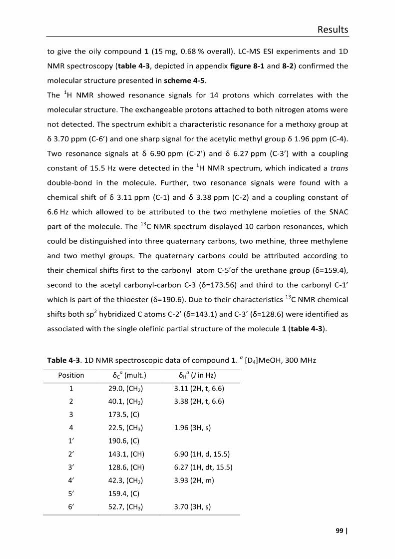
Results
99 |
to give the oily compound 1 (15 mg, 0.68 % overall). LC-MS ESI experiments and 1D
NMR spectroscopy (table 4-3, depicted in appendix figure 8-1 and 8-2) confirmed the
molecular structure presented in scheme 4-5.
The 1H NMR showed resonance signals for 14 protons which correlates with the
molecular structure. The exchangeable protons attached to both nitrogen atoms were
not detected. The spectrum exhibit a characteristic resonance for a methoxy group at
δ 3.70 ppm (C-6’) and one sharp signal for the acetylic methyl group δ 1.96 ppm (C-4).
Two resonance signals at δ 6.90 ppm (C-2’) and δ 6.27 ppm (C-3’) with a coupling
constant of 15.5 Hz were detected in the 1H NMR spectrum, which indicated a trans
double-bond in the molecule. Further, two resonance signals were found with a
chemical shift of δ 3.11 ppm (C-1) and δ 3.38 ppm (C-2) and a coupling constant of
6.6 Hz which allowed to be attributed to the two methylene moieties of the SNAC
part of the molecule. The 13C NMR spectrum displayed 10 carbon resonances, which
could be distinguished into three quaternary carbons, two methine, three methylene
and two methyl groups. The quaternary carbons could be attributed according to
their chemical shifts first to the carbonyl atom C-5’of the urethane group (δ=159.4),
second to the acetyl carbonyl-carbon C-3 (δ=173.56) and third to the carbonyl C-1’
which is part of the thioester (δ=190.6). Due to their characteristics 13C NMR chemical
shifts both sp2 hybridized C atoms C-2’ (δ=143.1) and C-3’ (δ=128.6) were identified as
associated with the single olefinic partial structure of the molecule 1 (table 4-3).
Table 4-3. 1D NMR spectroscopic data of compound 1. a [D4]MeOH, 300 MHz
Position δCa (mult.) δH
a (J in Hz)
1 29.0, (CH2) 3.11 (2H, t, 6.6)
2 40.1, (CH2) 3.38 (2H, t, 6.6)
3 173.5, (C)
4 22.5, (CH3) 1.96 (3H, s)
1’ 190.6, (C)
2’ 143.1, (CH) 6.90 (1H, d, 15.5)
3’ 128.6, (CH) 6.27 (1H, dt, 15.5)
7.3) 4’ 42.3, (CH2) 3.93 (2H, m)
5’ 159.4, (C)
6’ 52.7, (CH3) 3.70 (3H, s)
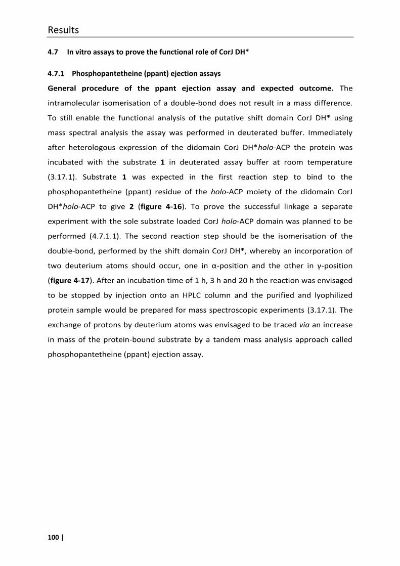
Results
100 |
4.7 In vitro assays to prove the functional role of CorJ DH*
4.7.1 Phosphopantetheine (ppant) ejection assays
General procedure of the ppant ejection assay and expected outcome. The
intramolecular isomerisation of a double-bond does not result in a mass difference.
To still enable the functional analysis of the putative shift domain CorJ DH* using
mass spectral analysis the assay was performed in deuterated buffer. Immediately
after heterologous expression of the didomain CorJ DH*holo-ACP the protein was
incubated with the substrate 1 in deuterated assay buffer at room temperature
(3.17.1). Substrate 1 was expected in the first reaction step to bind to the
phosphopantetheine (ppant) residue of the holo-ACP moiety of the didomain CorJ
DH*holo-ACP to give 2 (figure 4-16). To prove the successful linkage a separate
experiment with the sole substrate loaded CorJ holo-ACP domain was planned to be
performed (4.7.1.1). The second reaction step should be the isomerisation of the
double-bond, performed by the shift domain CorJ DH*, whereby an incorporation of
two deuterium atoms should occur, one in α-position and the other in γ-position
(figure 4-17). After an incubation time of 1 h, 3 h and 20 h the reaction was envisaged
to be stopped by injection onto an HPLC column and the purified and lyophilized
protein sample would be prepared for mass spectroscopic experiments (3.17.1). The
exchange of protons by deuterium atoms was envisaged to be traced via an increase
in mass of the protein-bound substrate by a tandem mass analysis approach called
phosphopantetheine (ppant) ejection assay.
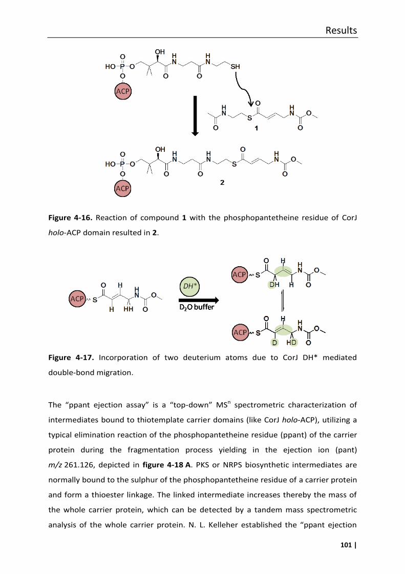
Results
101 |
Figure 4-16. Reaction of compound 1 with the phosphopantetheine residue of CorJ
holo-ACP domain resulted in 2.
Figure 4-17. Incorporation of two deuterium atoms due to CorJ DH* mediated
double-bond migration.
The “ppant ejection assay” is a “top-down” MSn spectrometric characterization of
intermediates bound to thiotemplate carrier domains (like CorJ holo-ACP), utilizing a
typical elimination reaction of the phosphopantetheine residue (ppant) of the carrier
protein during the fragmentation process yielding in the ejection ion (pant)
m/z 261.126, depicted in figure 4-18 A. PKS or NRPS biosynthetic intermediates are
normally bound to the sulphur of the phosphopantetheine residue of a carrier protein
and form a thioester linkage. The linked intermediate increases thereby the mass of
the whole carrier protein, which can be detected by a tandem mass spectrometric
analysis of the whole carrier protein. N. L. elleher established the “ppant ejection
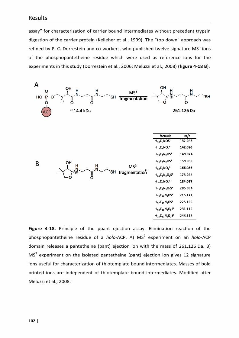
Results
102 |
assay” for characteri ation of carrier bound intermediates without precedent trypsin
digestion of the carrier protein (Kelleher et al., 1999). The “top down” approach was
refined by P. C. Dorrestein and co-workers, who published twelve signature MS3 ions
of the phosphopantetheine residue which were used as reference ions for the
experiments in this study (Dorrestein et al., 2006; Meluzzi et al., 2008) (figure 4-18 B).
Figure 4-18. Principle of the ppant ejection assay. Elimination reaction of the
phosphopantetheine residue of a holo-ACP. A) MS2 experiment on an holo-ACP
domain releases a pantetheine (pant) ejection ion with the mass of 261.126 Da. B)
MS3 experiment on the isolated pantetheine (pant) ejection ion gives 12 signature
ions useful for characterization of thiotemplate bound intermediates. Masses of bold
printed ions are independent of thiotemplate bound intermediates. Modified after
Meluzzi et al., 2008.
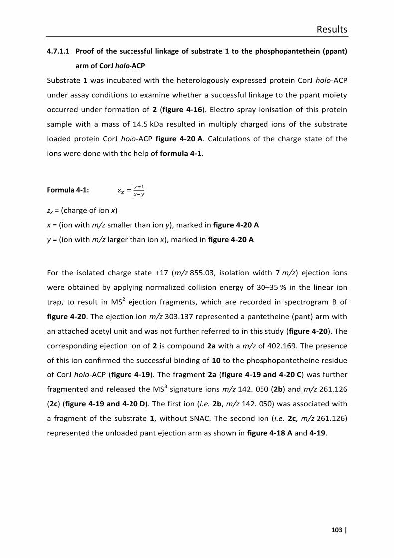
Results
103 |
4.7.1.1 Proof of the successful linkage of substrate 1 to the phosphopantethein (ppant)
arm of CorJ holo-ACP
Substrate 1 was incubated with the heterologously expressed protein CorJ holo-ACP
under assay conditions to examine whether a successful linkage to the ppant moiety
occurred under formation of 2 (figure 4-16). Electro spray ionisation of this protein
sample with a mass of 14.5 kDa resulted in multiply charged ions of the substrate
loaded protein CorJ holo-ACP figure 4-20 A. Calculations of the charge state of the
ions were done with the help of formula 4-1.
Formula 4-1:
zx = (charge of ion x)
x = (ion with m/z smaller than ion y), marked in figure 4-20 A
y = (ion with m/z larger than ion x), marked in figure 4-20 A
For the isolated charge state +17 (m/z 855.03, isolation width 7 m/z) ejection ions
were obtained by applying normalized collision energy of 30–35 % in the linear ion
trap, to result in MS2 ejection fragments, which are recorded in spectrogram B of
figure 4-20. The ejection ion m/z 303.137 represented a pantetheine (pant) arm with
an attached acetyl unit and was not further referred to in this study (figure 4-20). The
corresponding ejection ion of 2 is compound 2a with a m/z of 402.169. The presence
of this ion confirmed the successful binding of 10 to the phosphopantetheine residue
of CorJ holo-ACP (figure 4-19). The fragment 2a (figure 4-19 and 4-20 C) was further
fragmented and released the MS3 signature ions m/z 142. 050 (2b) and m/z 261.126
(2c) (figure 4-19 and 4-20 D). The first ion (i.e. 2b, m/z 142. 050) was associated with
a fragment of the substrate 1, without SNAC. The second ion (i.e. 2c, m/z 261.126)
represented the unloaded pant ejection arm as shown in figure 4-18 A and 4-19.
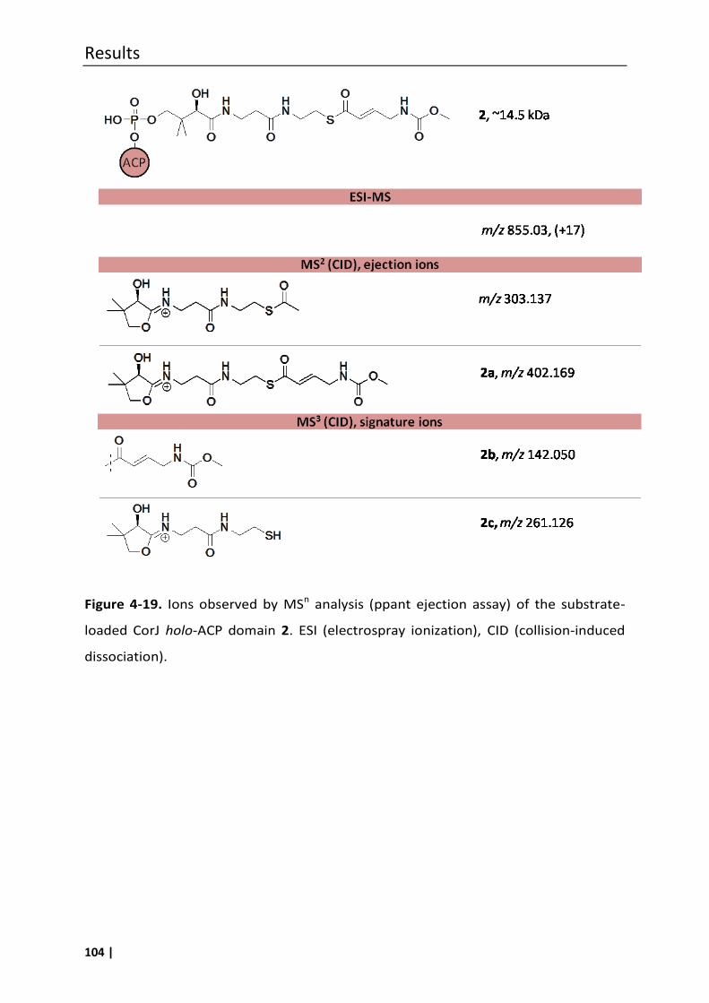
Results
104 |
Figure 4-19. Ions observed by MSn analysis (ppant ejection assay) of the substrate-
loaded CorJ holo-ACP domain 2. ESI (electrospray ionization), CID (collision-induced
dissociation).
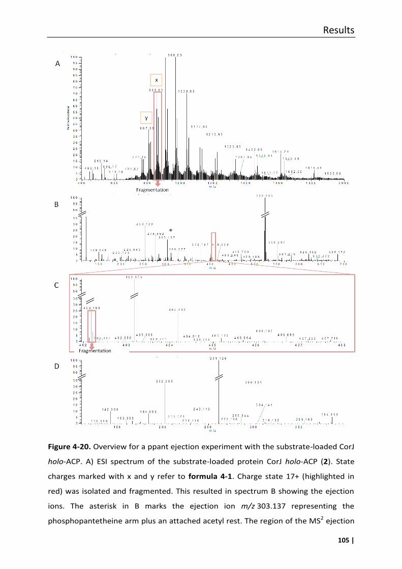
Results
105 |
Figure 4-20. Overview for a ppant ejection experiment with the substrate-loaded CorJ
holo-ACP. A) ESI spectrum of the substrate-loaded protein CorJ holo-ACP (2). State
charges marked with x and y refer to formula 4-1. Charge state 17+ (highlighted in
red) was isolated and fragmented. This resulted in spectrum B showing the ejection
ions. The asterisk in B marks the ejection ion m/z 303.137 representing the
phosphopantetheine arm plus an attached acetyl rest. The region of the MS2 ejection
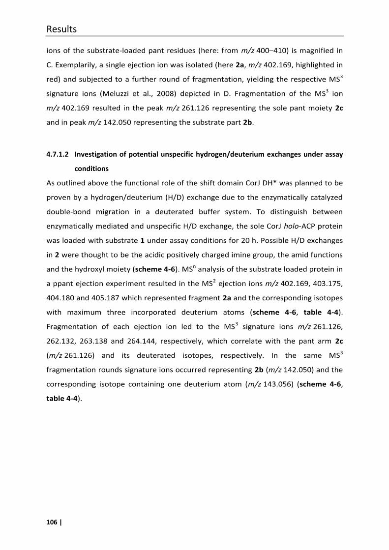
Results
106 |
ions of the substrate-loaded pant residues (here: from m/z 400–410) is magnified in
C. Exemplarily, a single ejection ion was isolated (here 2a, m/z 402.169, highlighted in
red) and subjected to a further round of fragmentation, yielding the respective MS3
signature ions (Meluzzi et al., 2008) depicted in D. Fragmentation of the MS3 ion
m/z 402.169 resulted in the peak m/z 261.126 representing the sole pant moiety 2c
and in peak m/z 142.050 representing the substrate part 2b.
4.7.1.2 Investigation of potential unspecific hydrogen/deuterium exchanges under assay
conditions
As outlined above the functional role of the shift domain CorJ DH* was planned to be
proven by a hydrogen/deuterium (H/D) exchange due to the enzymatically catalyzed
double-bond migration in a deuterated buffer system. To distinguish between
enzymatically mediated and unspecific H/D exchange, the sole CorJ holo-ACP protein
was loaded with substrate 1 under assay conditions for 20 h. Possible H/D exchanges
in 2 were thought to be the acidic positively charged imine group, the amid functions
and the hydroxyl moiety (scheme 4-6). MSn analysis of the substrate loaded protein in
a ppant ejection experiment resulted in the MS2 ejection ions m/z 402.169, 403.175,
404.180 and 405.187 which represented fragment 2a and the corresponding isotopes
with maximum three incorporated deuterium atoms (scheme 4-6, table 4-4).
Fragmentation of each ejection ion led to the MS3 signature ions m/z 261.126,
262.132, 263.138 and 264.144, respectively, which correlate with the pant arm 2c
(m/z 261.126) and its deuterated isotopes, respectively. In the same MS3
fragmentation rounds signature ions occurred representing 2b (m/z 142.050) and the
corresponding isotope containing one deuterium atom (m/z 143.056) (scheme 4-6,
table 4-4).
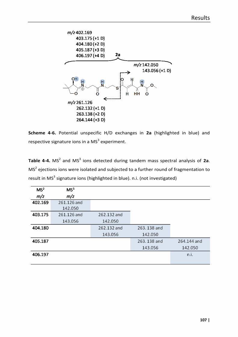
Results
107 |
Scheme 4-6. Potential unspecific H/D exchanges in 2a (highlighted in blue) and
respective signature ions in a MS3 experiment.
Table 4-4. MS2 and MS3 ions detected during tandem mass spectral analysis of 2a.
MS2 ejections ions were isolated and subjected to a further round of fragmentation to
result in MS3 signature ions (highlighted in blue). n.i. (not investigated)
Results
108 |
4.7.1.3 Proof of the β,γ double-bond migration using the ppant ejection assay
To prove the double-bond shift activity of CorJ DH*, the didomain CorJ DH*holo-ACP
was loaded with substrate 1 and incubated in deuterated buffer at room temperature
for 1 h, 3 h and 20 h. Due to a CorJ DH* mediated double-bond isomerisation an
specific exchange of two hydrogen atoms by two deuterium atoms was expected.
Electro spray ionization of the 47.2 kDa large substrate-loaded protein CorJ DH*holo-
ACP did not result in single charge states of the protein. Thus, MS2 ejection ions were
obtained by in-source fragmentation, applying 50–65 V fragmentation energy. The
mass of the non-deuterated form of ejection ion 2a (m/z 402.169) increased to
403.175, 404.182, and surprisingly to 405.188, indicating the number of incorporated
deuterium atoms (figure 4-21, table 4-5). MS3 fragmentation of each of these ejection
ions yielded clear pant (i.e. 2c, m/z 261.126), pant+substrate (i.e. m/z 300.102,
301.10, 302.114, 303.114) and substrate signature peaks (i.e. m/z 142.050, 143.056,
144.062, 145.068), presenting a mass increase up to +3, respectively (figure 4-21).
The substrate derived signature ions (i.e m/z 142.050, 143.056, 144.062, 145.068)
were each subjected to a further round of fragmentation yielding in the
corresponding MS4 fragments m/z 110.024, 111.030, 112.037, 113.043, respectively,
as depicted in figure 4-21. The importance of the MS4 fragments is that all of their
protons or deuterium atoms are bound to carbon atoms. Hence, observed mass
increase due to deuterium incorporation can be correlated directly to the double-
bond shifting activity of CorJ DH* and a mass increase associated with an unspecific
H/D exchange at the amide function, as depicted in scheme 4-6 can be excluded.
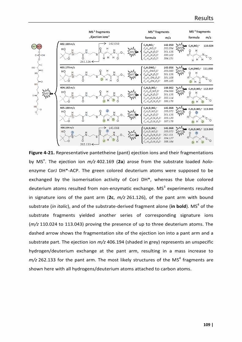
Results
109 |
Figure 4-21. Representative pantetheine (pant) ejection ions and their fragmentations
by MSn. The ejection ion m/z 402.169 (2a) arose from the substrate loaded holo-
enzyme CorJ DH*-ACP. The green colored deuterium atoms were supposed to be
exchanged by the isomerisation activity of CorJ DH*, whereas the blue colored
deuterium atoms resulted from non-enzymatic exchange. MS3 experiments resulted
in signature ions of the pant arm (2c, m/z 261.126), of the pant arm with bound
substrate (in italic), and of the substrate-derived fragment alone (in bold). MS4 of the
substrate fragments yielded another series of corresponding signature ions
(m/z 110.024 to 113.043) proving the presence of up to three deuterium atoms. The
dashed arrow shows the fragmentation site of the ejection ion into a pant arm and a
substrate part. The ejection ion m/z 406.194 (shaded in grey) represents an unspecific
hydrogen/deuterium exchange at the pant arm, resulting in a mass increase to
m/z 262.133 for the pant arm. The most likely structures of the MS4 fragments are
shown here with all hydrogens/deuterium atoms attached to carbon atoms.
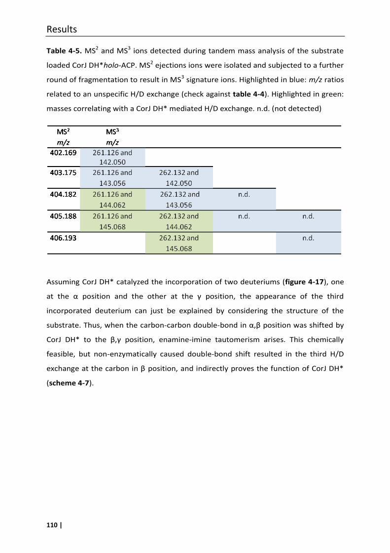
Results
110 |
Table 4-5. MS2 and MS3 ions detected during tandem mass analysis of the substrate
loaded CorJ DH*holo-ACP. MS2 ejections ions were isolated and subjected to a further
round of fragmentation to result in MS3 signature ions. Highlighted in blue: m/z ratios
related to an unspecific H/D exchange (check against table 4-4). Highlighted in green:
masses correlating with a CorJ DH* mediated H/D exchange. n.d. (not detected)
Assuming CorJ DH* catalyzed the incorporation of two deuteriums (figure 4-17), one
at the α position and the other at the γ position, the appearance of the third
incorporated deuterium can just be explained by considering the structure of the
substrate. Thus, when the carbon-carbon double-bond in α,β position was shifted by
CorJ D * to the β,γ position, enamine-imine tautomerism arises. This chemically
feasible, but non-enzymatically caused double-bond shift resulted in the third H/D
exchange at the carbon in β position, and indirectly proves the function of CorJ DH*
(scheme 4-7).
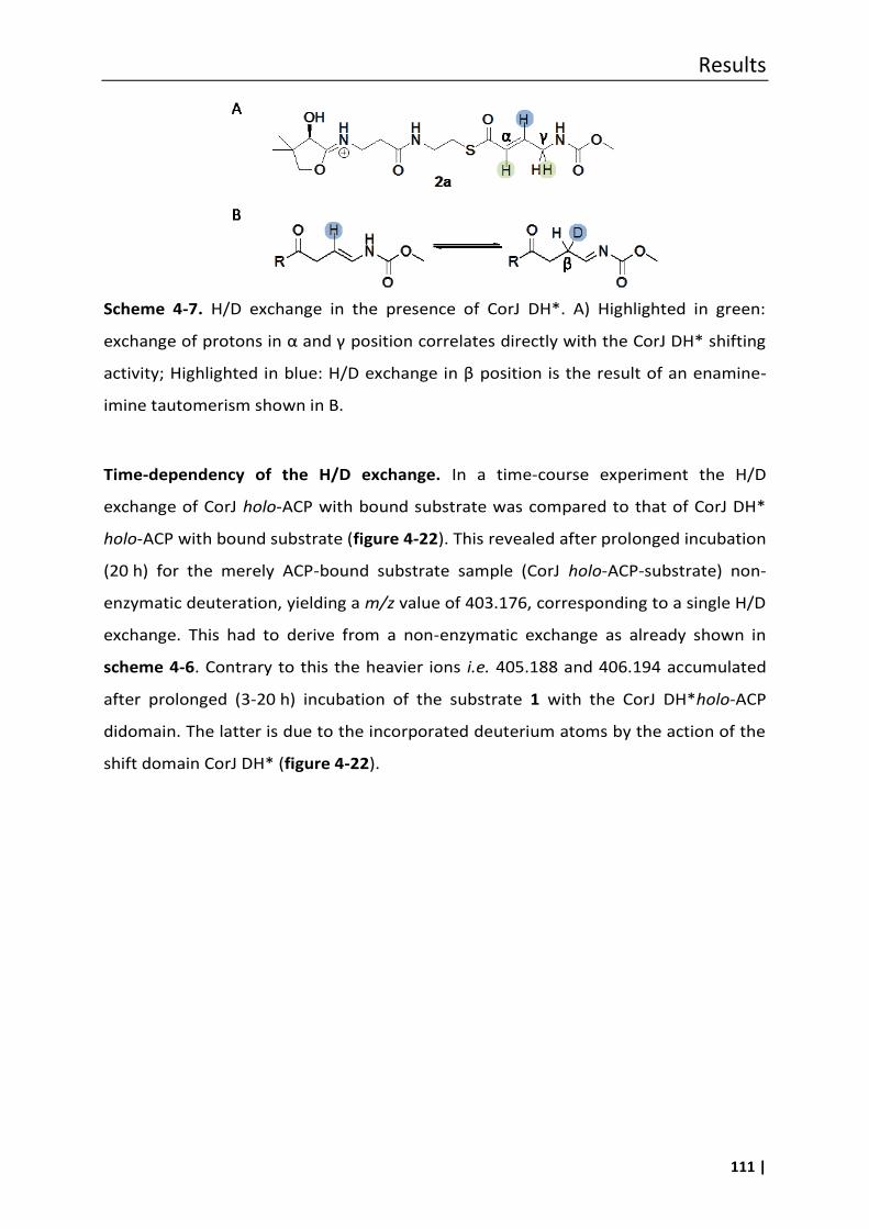
Results
111 |
Scheme 4-7. H/D exchange in the presence of CorJ DH*. A) Highlighted in green:
exchange of protons in α and γ position correlates directly with the CorJ DH* shifting
activity; Highlighted in blue: /D exchange in β position is the result of an enamine-
imine tautomerism shown in B.
Time-dependency of the H/D exchange. In a time-course experiment the H/D
exchange of CorJ holo-ACP with bound substrate was compared to that of CorJ DH*
holo-ACP with bound substrate (figure 4-22). This revealed after prolonged incubation
(20 h) for the merely ACP-bound substrate sample (CorJ holo-ACP-substrate) non-
enzymatic deuteration, yielding a m/z value of 403.176, corresponding to a single H/D
exchange. This had to derive from a non-enzymatic exchange as already shown in
scheme 4-6. Contrary to this the heavier ions i.e. 405.188 and 406.194 accumulated
after prolonged (3-20 h) incubation of the substrate 1 with the CorJ DH*holo-ACP
didomain. The latter is due to the incorporated deuterium atoms by the action of the
shift domain CorJ DH* (figure 4-22).
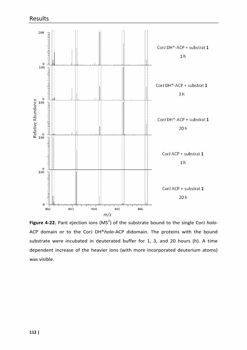
Results
112 |
Figure 4-22. Pant ejection ions (MS2) of the substrate bound to the single CorJ holo-
ACP domain or to the CorJ DH*holo-ACP didomain. The proteins with the bound
substrate were incubated in deuterated buffer for 1, 3, and 20 hours (h). A time
dependent increase of the heavier ions (with more incorporated deuterium atoms)
was visible.
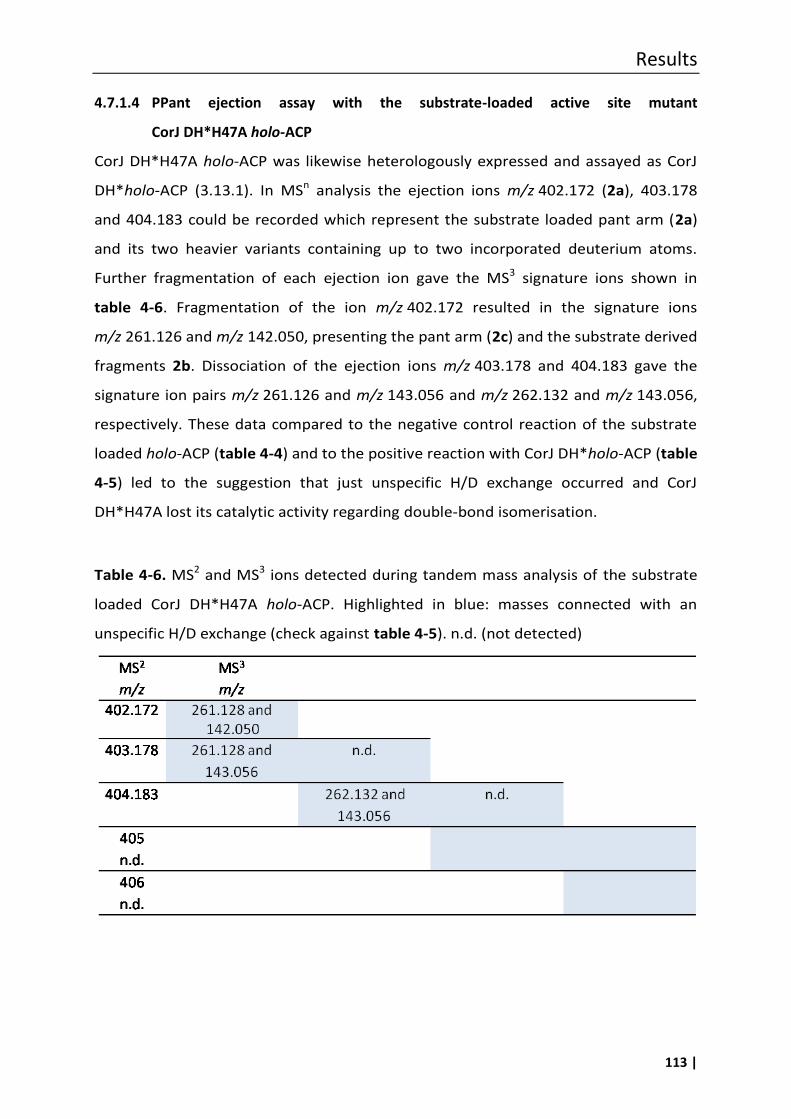
Results
113 |
4.7.1.4 PPant ejection assay with the substrate-loaded active site mutant
CorJ DH*H47A holo-ACP
CorJ DH*H47A holo-ACP was likewise heterologously expressed and assayed as CorJ
DH*holo-ACP (3.13.1). In MSn analysis the ejection ions m/z 402.172 (2a), 403.178
and 404.183 could be recorded which represent the substrate loaded pant arm (2a)
and its two heavier variants containing up to two incorporated deuterium atoms.
Further fragmentation of each ejection ion gave the MS3 signature ions shown in
table 4-6. Fragmentation of the ion m/z 402.172 resulted in the signature ions
m/z 261.126 and m/z 142.050, presenting the pant arm (2c) and the substrate derived
fragments 2b. Dissociation of the ejection ions m/z 403.178 and 404.183 gave the
signature ion pairs m/z 261.126 and m/z 143.056 and m/z 262.132 and m/z 143.056,
respectively. These data compared to the negative control reaction of the substrate
loaded holo-ACP (table 4-4) and to the positive reaction with CorJ DH*holo-ACP (table
4-5) led to the suggestion that just unspecific H/D exchange occurred and CorJ
DH*H47A lost its catalytic activity regarding double-bond isomerisation.
Table 4-6. MS2 and MS3 ions detected during tandem mass analysis of the substrate
loaded CorJ DH*H47A holo-ACP. Highlighted in blue: masses connected with an
unspecific H/D exchange (check against table 4-5). n.d. (not detected)
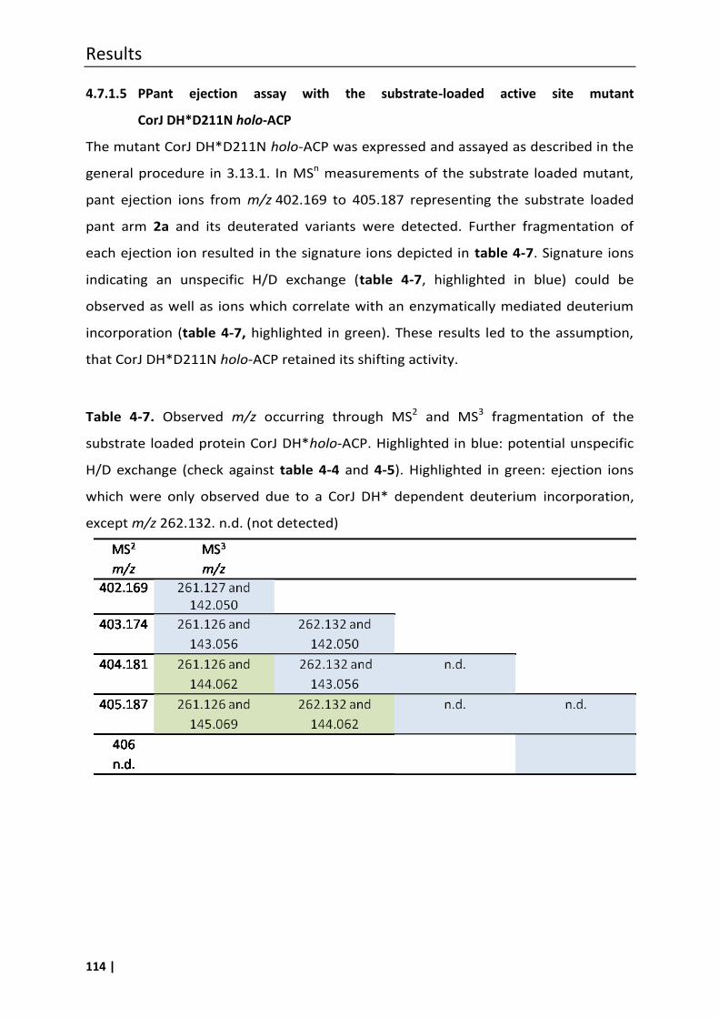
Results
114 |
4.7.1.5 PPant ejection assay with the substrate-loaded active site mutant
CorJ DH*D211N holo-ACP
The mutant CorJ DH*D211N holo-ACP was expressed and assayed as described in the
general procedure in 3.13.1. In MSn measurements of the substrate loaded mutant,
pant ejection ions from m/z 402.169 to 405.187 representing the substrate loaded
pant arm 2a and its deuterated variants were detected. Further fragmentation of
each ejection ion resulted in the signature ions depicted in table 4-7. Signature ions
indicating an unspecific H/D exchange (table 4-7, highlighted in blue) could be
observed as well as ions which correlate with an enzymatically mediated deuterium
incorporation (table 4-7, highlighted in green). These results led to the assumption,
that CorJ DH*D211N holo-ACP retained its shifting activity.
Table 4-7. Observed m/z occurring through MS2 and MS3 fragmentation of the
substrate loaded protein CorJ DH*holo-ACP. Highlighted in blue: potential unspecific
H/D exchange (check against table 4-4 and 4-5). Highlighted in green: ejection ions
which were only observed due to a CorJ DH* dependent deuterium incorporation,
except m/z 262.132. n.d. (not detected)
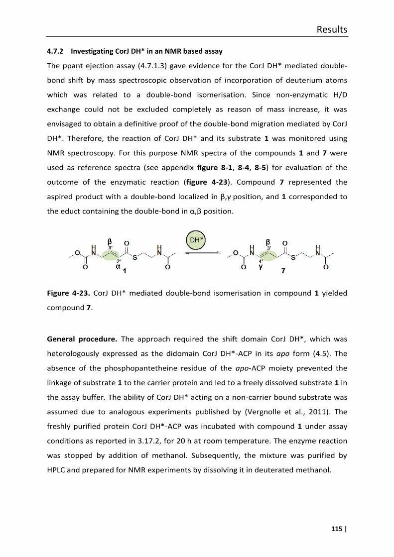
Results
115 |
4.7.2 Investigating CorJ DH* in an NMR based assay
The ppant ejection assay (4.7.1.3) gave evidence for the CorJ DH* mediated double-
bond shift by mass spectroscopic observation of incorporation of deuterium atoms
which was related to a double-bond isomerisation. Since non-enzymatic H/D
exchange could not be excluded completely as reason of mass increase, it was
envisaged to obtain a definitive proof of the double-bond migration mediated by CorJ
DH*. Therefore, the reaction of CorJ DH* and its substrate 1 was monitored using
NMR spectroscopy. For this purpose NMR spectra of the compounds 1 and 7 were
used as reference spectra (see appendix figure 8-1, 8-4, 8-5) for evaluation of the
outcome of the enzymatic reaction (figure 4-23). Compound 7 represented the
aspired product with a double-bond locali ed in β,γ position, and 1 corresponded to
the educt containing the double-bond in α,β position.
Figure 4-23. CorJ DH* mediated double-bond isomerisation in compound 1 yielded
compound 7.
General procedure. The approach required the shift domain CorJ DH*, which was
heterologously expressed as the didomain CorJ DH*-ACP in its apo form (4.5). The
absence of the phosphopantetheine residue of the apo-ACP moiety prevented the
linkage of substrate 1 to the carrier protein and led to a freely dissolved substrate 1 in
the assay buffer. The ability of CorJ DH* acting on a non-carrier bound substrate was
assumed due to analogous experiments published by (Vergnolle et al., 2011). The
freshly purified protein CorJ DH*-ACP was incubated with compound 1 under assay
conditions as reported in 3.17.2, for 20 h at room temperature. The enzyme reaction
was stopped by addition of methanol. Subsequently, the mixture was purified by
HPLC and prepared for NMR experiments by dissolving it in deuterated methanol.
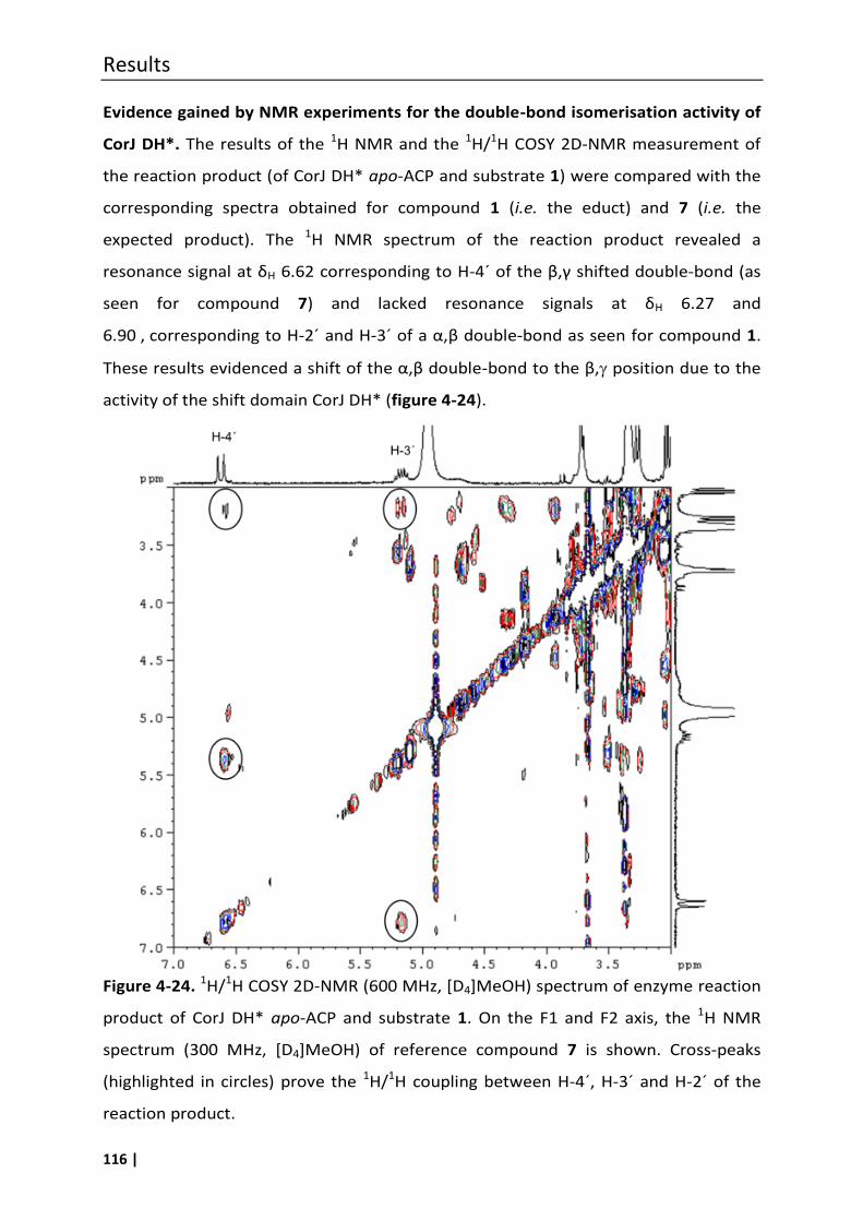
Results
116 |
Evidence gained by NMR experiments for the double-bond isomerisation activity of
CorJ DH*. The results of the 1H NMR and the 1H/1H COSY 2D-NMR measurement of
the reaction product (of CorJ DH* apo-ACP and substrate 1) were compared with the
corresponding spectra obtained for compound 1 (i.e. the educt) and 7 (i.e. the
expected product). The 1H NMR spectrum of the reaction product revealed a
resonance signal at δH 6.62 corresponding to H-4´ of the β,γ shifted double-bond (as
seen for compound 7) and lacked resonance signals at δH 6.27 and
6.90 , corresponding to H-2´ and H-3´ of a α,β double-bond as seen for compound 1.
These results evidenced a shift of the α,β double-bond to the β, position due to the
activity of the shift domain CorJ DH* (figure 4-24).
Figure 4-24. 1H/1H COSY 2D-NMR (600 MHz, [D4]MeOH) spectrum of enzyme reaction
product of CorJ DH* apo-ACP and substrate 1. On the F1 and F2 axis, the 1H NMR
spectrum (300 MHz, [D4]MeOH) of reference compound 7 is shown. Cross-peaks
(highlighted in circles) prove the 1H/1H coupling between H-4´, H-3´ and H-2´ of the
reaction product.
Conclusion
117 |
5 Conclusion
5.1 Drug discovery from natural products
Many of our most valuable drugs today derive from natural products, e.g.
cyclosporine, lovastatine. Above all, antibiotic drug discovery is dependent on natural
product research. This is even more true since the increasing emergence of bacteria
resistant to antibiotics is a serious threat to modern medicine (Schäberle and Hack,
2014). Already in the 1990s it became obvious that resistances of bacteria towards
established antibiotic therapies were developing much faster than new antibiotics
could be discovered. Thus, pharmaceutical industry altered its discovery approaches
and used “high-tech” methods like genomics, combinatorial chemistry and high
throughput screening (HTS) to identify new antibacterial compounds and targets
unfortunately with little success, yet (Lewis, 2013). By now 14 antibiotic classes are
known (Lewis, 2013) but the number of new antibiotically active drugs introduced
into therapy is dramatically dwindling (Schäberle et al., 2014b). In the light of urgently
needed new antibacterial lead structures a revival of natural product drug discovery is
currently taking place, and the whole cell (i.e. agar diffusion assay) screening
approach introduced by S. Waksman in the 1940s does appear modern again (Lewis,
2013). There is, however, the need for innovative assays, e.g. using transgenic test
organisms, and the discovery of novel natural product producer strains (Donadio et
al., 2007).
Traditional antibiotic producer strains are often members of the actinobacteria or
bacilli. In the last two decades δ-proteobacteria, especially myxobacteria got into the
focus as alternative providers of antimicrobial natural products. Myxobacteria have
been shown to produce a wide range of secondary metabolites with unique structural
features and rare or novel modes of action, which make them attractive for
pharmaceutical research (Weissman and Müller, 2009). A broad overview of
antibacterially active myxobacterial compounds is given in a recent review (Schäberle
et al., 2014c) (see also introduction of this thesis).
The observation of antimicrobially active compounds produced by myxobacteria
started already in 1947 (Oxford, 1947). The compound, 1-hydroxy-6-
methoxyphenazine-N5,N10-dioxide (myxin) was reported as one of the first
Conclusion
118 |
antibiotically active compounds from a Sorangium species (Peterson et al., 1966), but
proved to be toxic (Chowdhury et al., 2012). In 1962 Norén and Raper published that
several myxobacteria secreted natural products able to inhibit the growth of Gram-
positive bacteria whereas none of the Gram-negative test organisms were found to
be sensitive (Norén and Raper, 1962). This is still true today, even though very few
myxobacterial metabolites being able to inhibit Gram-negatives were described.
Corallopyronin A, the focus of this study, is a myxobacterial secondary metabolite
whose rare mode of action as RNA-polymerase inhibitor, good antibacterial activity
towards, e.g. MSRA and Wolbachia and low toxicity, qualify it as a promising
candidate for pre-clinical evaluation (Erol et al., 2010; Schäberle et al., 2014b).
5.2 Biosynthesis of myxobacterial natural products focussing on corallopyronin A
Understanding the biosynthetic process of bioactive natural products is of importance
for their further development, e.g. sustaining their supply, bioengineering of
derivatives. Myxobacterial secondary metabolites such as corallopyronin A are
characterized by a high structural complexity including multiple chiral centres, ring
formations and functional groups. The basic structure of most of these diverse
compounds is synthesized by large multi enzymes, i.e. polyketide synthases (PKS),
non-ribosomal peptide synthases (NRPS) and mixed-type PKS/NRPS systems (Wenzel
and Müller, 2009). Post-NRPS or post-PKS enzymes like glycosyl transferases,
methyltransferases, acylases, cyclases and oxidative enzymes further modify these
basic structures (Staunton and Wilkinson, 1998).
Corallopyronin A is the product of a type 1 PKS/NRPS, which produces pre-
corallopyronin A, while further enzymes perform post-PKS/NRPS reactions. The latter
include, e.g. the isomerisation of the double-bond Δ26 to Δ25 putatively mediated by
CorN and the incorporation of a hydroxyl moiety at C-25 probably catalyzed by the
cytochrome P450 oxidase CorO (figure 4-1).
In modular PKS and linear NRPS systems such as the one responsible for
corallopyronin A biosynthesis a one-to-one correspondence between the architecture
of the PKS or NRPS assembly line and the backbone of the assembled intermediate
exists. This correlation is named “co-linearity rule” and in the case of corallopyronin A
it helped in the identification and characterization of the biosynthetic gene cluster.
Conclusion
119 |
Corallopyronin A biosynthesis, however, also shows some non-canonical features, (i)
the core structure is formed by head to head connection of two separate chains
derived from a PKS and a NRPS/PKS system, respectively, (ii) two of the methyl groups
are incorporated by a β-branching cassette, (iii) the ER functionality is encoded in
trans-position as a bifunctional enzyme together with the trans-AT. Additionally, the
starter unit was surprisingly shown to be hydrogen carbonate. Therefore, not all
processes of this biosynthesis are easily predictable since the classical co-linearity rule
does not apply (Piel, 2010).
Such an exception to the co-linearity rule in corallopyronin A biosynthesis is the
position of the C-11/C-12 carbon-carbon double-bond, representing a rare β,γ pattern
(figure 4-6). The usual PKS-NRPS machinery places such double-bonds in α,β position
by elimination of water from the respective β-hydroxy intermediate. Similar cases are
exemplified in very few other polyketides, e.g. rhizoxin (Kusebauch et al., 2010),
ansamitocin (Taft et al., 2009) and bacillaene (Moldenhauer et al., 2010). Concerning
ansamitocin an unusual but functional DH domain was assumed to place the double-
bond in β,γ position (Taft et al., 2009). A yet different mechanism was recently found
to occur in the biosynthesis of bacillaene and rhizoxin. By the construction of
thioesterase knockout mutants, late-pathway intermediates had been identified in
which double-bonds were present in α,β and in the shifted β,γ position (Kusebauch et
al., 2010; Moldenhauer et al., 2010). These experiments showed that isomerisation is
taking place during polyketide assembly, and due to the structure of the
intermediates the timing of the isomerisation reaction was assigned to specific
modules of the assembly line. Thus, for rhizoxin it was shown that the double-bond Δ8
was shifted to Δ9 after elongation of the nascent polyketide chain by a downstream to
the respective P S module encoded “shift module (including D *)” (Kusebauch et al.,
2010). We proposed a similar process for corallopyronin A biosynthesis and provide
here evidence that a distinct domain of the respective assembly line is responsible for
the double-bond shift.
The current study describes two different in vitro assays to prove the functional role
of the CorJ DH* domain within the respective shift module of the corallopyronin A
biosynthesis cluster. For this purpose the respective protein domains (CorJ DH* and
CorJ ACP, figure 4-11) involved in corallopyronin A biosynthesis were heterologously
Conclusion
120 |
expressed, and a suitable substrate 1 for the isomerization reaction was synthesized
as an N-acetylcysteamine (SNAC) thioester derivative (figure 4-14).
For the initial detection of a double-bond migration within the substrate the
phosphopantetheine (PPant) ejection assay was used, which enables the analysis of
carrier protein bound intermediates (Dorrestein et al., 2006; Meluzzi et al., 2008)
(figure 4-18). To allow a mass spectroscopic monitoring of the CorJ DH* mediated
double-bond shift the enzyme assay was performed in deuterated buffer to allow an
H/D exchange correlating with the double-bond migration (figure 4-17). The
corresponding increase in mass was observed by applying high resolution tandem MS
technique. As expected a mass increase up to three could be observed in the
obtained ions of the ACP-bound substrate and indicated thereby the catalytic activity
of CorJ DH* (figure 4-21). However, non-enzymatical H/D exchange surely also
occurred (scheme 4-6). Even though MS4 experiments clearly pointed out the
enzymatic reaction, we sought additional evidence using NMR spectroscopy.
For the NMR experiment the enzyme, i.e. CorJ DH* was incubated with substrate 1.
1D and 2D NMR spectra, recorded of the educt of the enzyme-substrate reaction and
of the product, clearly revealed that the double-bond had shifted to the β,γ position
(figure 4-24). This provided a second proof of the double-bond isomerisation activity
of CorJ DH*.
In order to get insight into the mechanisms of this double-bond migration, the protein
CorJ DH* was mutated. It was assumed that one of the active site residues of the
enzyme is a histidine, which is highly conserved within DH and DH* domains (figure 4-
7). Both domain variants, i.e. DH and DH* have the same basic structure and the
conserved residues in the same positions as was indicated by aligning and threading
the sequences to the solved crystal structure of the DEBS DH (Keatinge-Clay, 2008;
Kusebauch et al., 2010). To prove the involvement of the conserved histidine (H)
residue a mutational exchange to alanine (A) was performed in CorJ DH* to give CorJ
DH*H47A. This enzyme was likewise expressed and assayed as CorJ DH* using the
masspectrometric assay. Indeed, the only H/D exchange which could be observed was
unspecific and not at the site of the carbon double-bond (table 4-6). These results
provided evidence that the histidine residue at position 47 is of major importance for
the enzymatic reaction and possibly acts as a proton donor and acceptor in the course
Conclusion
121 |
of the back and forward isomerisation reaction. A further amino acid within the
enzyme should function as a proton acceptor, i.e. it should be accepting the proton
from the γ-position. We expected the conserved aspartic acid D211 in CorJ DH* to
represent this proton acceptor, but the mutated enzyme containing an asparagine (N)
instead of aspartic acid (D) CorJ DH*D211N retained the shifting activity (table 4-7).
Our experiments clearly evidenced that under assay conditions the carbon-carbon
double-bond migration is reversible and resembles the described mechanism for DH
domains (Keatinge-Clay, 2008). In the case of the CorJ DH* domain H47 serves as
proton donor and a still unknown residue as acceptor. This is in agreement with the
mechanism postulated earlier by Hertweck and co-workers (Kusebauch et al., 2010).
It is assumed that in PKS-systems KSs act as gatekeepers preventing incorporation of
erroneous building blocks (Taft et al., 2009). This arrangement assures that only the
correct substrate, in this case the rearranged β,γ double-bond, can be passed on for
further processing. In line with this all corallopyronin-type molecules known to date
possess the β,γ pattern. Only under in vitro conditions the isomerase-like CorJ DH*
domain acts reversibly, since the equilibrium is not affected by further processing of
the intermediates.
5.3 Outlook
In vitro analysis of the enzymatic activity of wild type and mutated variants of CorJ
DH* encoded within the corallopyronin A cluster provides evidence that this domain
is responsible for the unusual carbon-carbon double bond migration during the
biosynthesis of this antibiotic. Our experiments thus clarified the genetic and
biochemical basis underlying double-bond isomerisations in polyketide biosynthesis.
This will allow a better prediction of polyketide structures from DNA sequence
information, which is of major importance considering the increasing number of
sequenced microbial genomes and biosynthetic gene clusters, and will enlarge the
tool box for the rational design of metabolites in genetic engineering.
Future studies will focus on the crystallization of the respective proteins, which will
aid to reveal the molecular basis for the double bond migration reaction, in particular
the still unknown proton acceptor within the enzyme.

122 |

Summary
123 |
6 Summary
Corallopyronin A is a myxobacterial compound, which was isolated in our lab from the
strain Corallococcus coralloides B035. It is a potent in vivo active antibiotic, which is
currently undergoing preclinical studies. Regarding its biosynthesis, corallopyronin A
was found to originate from two chains, one being solely PKS- and the other
NRPS/PKS dervived.
Scheme 6-1. Corallopyronin A
In polyketide biosynthesis the reduction of β-carbonyl groups to an alkene moiety
usually results in a α,β positioned double-bond. However, in a few polyketides the
rare case of such a carbon-carbon double-bond in β,γ position is depicted, e.g. in the
biosynthesis of ansamitocin (Taft et al., 2009), bacillaene (Moldenhauer et al., 2010)
and rhizoxin (Kusebauch et al., 2010). For rhizoxin it was shown that the respective
double-bond (Δ8) was shifted to the β,γ position after elongation of the nascent
polyketide chain by a distinct “shift module” including an unusual dehydratase-like
domain (DH*) downstream in the PKS assembly line (Kusebauch et al., 2010). We
proposed a similar process for the antibiotic corallopyronin A and provided here
evidence that a distinct domain (CorJ DH*) catalyses the carbon-carbon double-bond
shift from α,β to β,γ position during corallopyronin A biosynthesis.
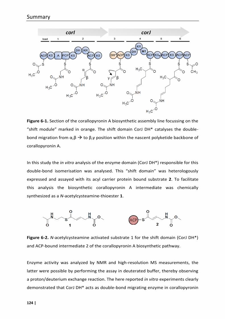
Summary
124 |
Figure 6-1. Section of the corallopyronin A biosynthetic assembly line focussing on the
“shift module” marked in orange. The shift domain CorJ D * catalyses the double-
bond migration from α,β to β,γ position within the nascent polyketide backbone of
corallopyronin A.
In this study the in vitro analysis of the enzyme domain (CorJ DH*) responsible for this
double-bond isomerisation was analysed. This “shift domain” was heterologously
expressed and assayed with its acyl carrier protein bound substrate 2. To facilitate
this analysis the biosynthetic corallopyronin A intermediate was chemically
synthesized as a N-acetylcysteamine-thioester 1.
Figure 6-2. N-acetylcysteamine activated substrate 1 for the shift domain (CorJ DH*)
and ACP-bound intermediate 2 of the corallopyronin A biosynthetic pathway.
Enzyme activity was analyzed by NMR and high-resolution MS measurements, the
latter were possible by performing the assay in deuterated buffer, thereby observing
a proton/deuterium exchange reaction. The here reported in vitro experiments clearly
demonstrated that CorJ DH* acts as double-bond migrating enzyme in corallopyronin
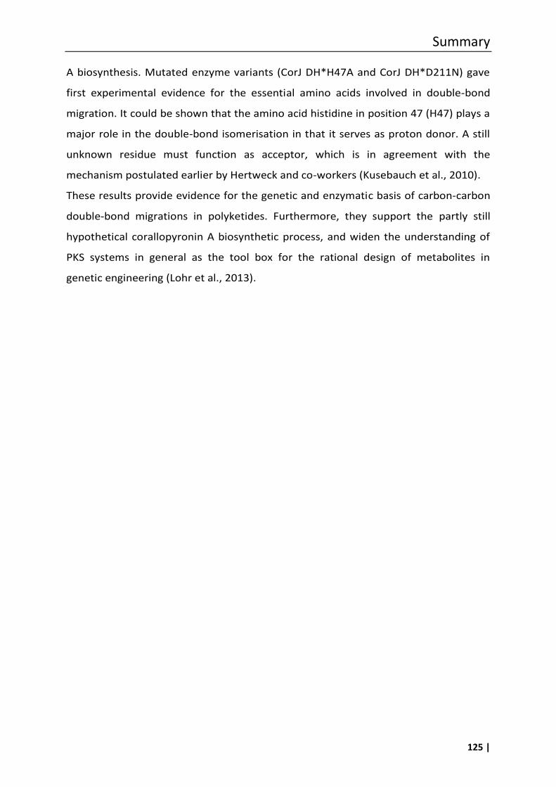
Summary
125 |
A biosynthesis. Mutated enzyme variants (CorJ DH*H47A and CorJ DH*D211N) gave
first experimental evidence for the essential amino acids involved in double-bond
migration. It could be shown that the amino acid histidine in position 47 (H47) plays a
major role in the double-bond isomerisation in that it serves as proton donor. A still
unknown residue must function as acceptor, which is in agreement with the
mechanism postulated earlier by Hertweck and co-workers (Kusebauch et al., 2010).
These results provide evidence for the genetic and enzymatic basis of carbon-carbon
double-bond migrations in polyketides. Furthermore, they support the partly still
hypothetical corallopyronin A biosynthetic process, and widen the understanding of
PKS systems in general as the tool box for the rational design of metabolites in
genetic engineering (Lohr et al., 2013).

126 |
References
127 |
7 References
Ahn, J.-W., Jang, K.H., Chung, S.-C., Oh, K.-B., and Shin, J. (2008). Sorangiadenosine, a new sesquiterpene adenoside from the myxobacterium Sorangium cellulosum. Org. Lett. 10, 1167–1169.
Akey, D.L., Razelun, J.R., Tehranisa, J., Sherman, D.H., Gerwick, W.H., and Smith, J.L. (2010). Crystal structures of dehydratase domains from the curacin polyketide biosynthetic pathway. Structure 18, 94–105.
Alhamadsheh, M.M., Palaniappan, N., DasChouduri, S., and Reynolds, K.A. (2007). Modular polyketide synthases and cis double-bond formation: establishment of activated cis -3-cyclohexylpropenoic acid as the diketide intermediate in phoslactomycin biosynthesis. J. Am. Chem. Soc. 129, 1910–1911.
Altendorfer, M., Irschik, H., and Menche, D. (2012). Design, synthesis and biological evaluation of simplified side chains of the macrolide antibiotic etnangien. Bioorg. Med. Chem. Lett. 22, 5731–5734.
Altendorfer, M., Raja, A., Sasse, F., Irschik, H., and Menche, D. (2013). Modular synthesis of polyene side chain analogues of the potent macrolide antibiotic etnangien by a flexible coupling strategy based on hetero-bis-metallated alkenes. Org. Biomol. Chem. 11, 2116–2139.
Apel, C., Barg, A., Rheinberg, A., Conrads, G., and Wagner-Döbler, I. (2013). Dental composite materials containing carolacton inhibit biofilm growth of Streptococcus mutans. Dent. Mater. 29, 1188–1199.
Arora, P., Vats, A., Saxena, P., Mohanty, D., and Gokhale, R.S. (2005). Promiscuous fatty acyl CoA ligases produce acyl-CoA and acyl-SNAC precursors for polyketide biosynthesis. J. Am. Chem. Soc. 127, 9388–9389.
Artsimovitch, I., Seddon, J., and Sears, P. (2012). Fidaxomicin Is an Inhibitor of the initiation of bacterial RNA synthesis. Clin. Infect. Dis. 55, S127–S131.
Augustiniak, H., Höfle, G., Irschik, H., and Reichenbach, H. (1996). Antibiotics from gliding bacteria, LXXVIII. Ripostatin A, B, and C: isolation and structure and structure elucidation of novel metabolites from Sorangium cellulosum. Liebigs Ann. 1996, 1657–1663.
Ayele, W.Y., Neill, S.D., Zinsstag, J., Weiss, M.G., and Pavlik, I. (2004). Bovine tuberculosis: an old disease but a new threat to Africa. Int. J. Tuberc. Lung Dis. 8, 924–937.
Banu, L.D., Conrads, G., Rehrauer, H., Hussain, H., Allan, E., and van der Ploeg, J.R. (2010). The Streptococcus mutans serine/threonine kinase, PknB, regulates competence development, bacteriocin production, and cell wall metabolism. Infect. Immun. 78, 2209–2220.
References
128 |
Belogurov, G.A., Vassylyeva, M.N., Sevostyanova, A., Appleman, J.R., Xiang, A.X., Lira, R., Webber, S.E., Klyuyev, S., Nudler, E., Artsimovitch, I., et al. (2008). Transcription inactivation through local refolding of the RNA polymerase structure. Nature 457, 332–335.
Bergeron, R.J., and Phanstiel, O. (1992). The total synthesis of nannochelin: a novel cinnamoyl hydroxamate-containing siderophore. J. Org. Chem. 57, 7140–7143.
Bock, M., Buntin, K., Müller, R., and Kirschning, A. (2008). Stereochemical determination of thuggacins A–C, highly active antibiotics from the myxobacterium Sorangium cellulosum. Angew. Chem. Int. Ed. 47, 2308–2311.
Buedenbender, S., Rachid, S., Müller, R., and Schulz, G.E. (2009). Structure and action of the myxobacterial chondrochloren halogenase CndH: a new variant of FAD-dependent halogenases. J. Mol. Biol. 385, 520–530.
Buntin, K., Irschik, H., Weissman, K.J., Luxenburger, E., Blöcker, H., and Müller, R. (2010). Biosynthesis of thuggacins in myxobacteria: comparative cluster analysis reveals basis for natural product structural diversity. Chem. Biol. 17, 342–356.
Buurman, E.T., Foulk, M.A., Gao, N., Laganas, V.A., McKinney, D.C., Moustakas, D.T., Rose, J.A., Shapiro, A.B., and Fleming, P.R. (2012). Novel rapidly diversifiable antimicrobial RNA polymerase switch region inhibitors withconfirmed mode of action in Haemophilus influenzae. J. Bacteriol. 194, 5504–5512.
Campbell, E.A., Pavlova, O., Zenkin, N., Leon, F., Irschik, H., Jansen, R., Severinov, K., and Darst, S.A. (2005). Structural, functional, and genetic analysis of sorangicin inhibition of bacterial RNA polymerase. EMBO J. 24, 674–682.
Chen, X.-H., Vater, J., Piel, J., Franke, P., Scholz, R., Schneider, K., Koumoutsi, A., Hitzeroth, G., Grammel, N., Strittmatter, A.W., et al. (2006). Structural and functional characterization of three polyketide synthase geneclusters in Bacillus amyloliquefaciens FZB 42. J. Bacteriol. 188, 4024–4036.
Chopra, I. (2007). Bacterial RNA polymerase: A promising target for the discovery of new antimicrobial agents. Curr. Opin. Investig. Drugs 600–607.
Chowdhury, G., Sarkar, U., Pullen, S., Wilson, W.R., Rajapakse, A., Fuchs-Knotts, T., and Gates, K.S. (2012). DNA strand cleavage by the phenazine di- N -oxide natural product myxin under both aerobic and anaerobic conditions. Chem. Res. Toxicol. 25, 197–206.
Content, S., Dutton, C.J., and Roberts, L. (2003). Myxovirescin analogues via macrocyclic ring-closing metathesis. Bioorg. Med. Chem. Lett. 13, 321–325.
Cortina, N.S., Revermann, O., Krug, D., and Müller, R. (2011). Identification and characterization of the althiomycin biosynthetic gene cluster in Myxococcus xanthus DK897. ChemBioChem 12, 1411–1416.
Crimmins, M.T., aley, M.W., and O’Bryan, E.A. (2011). Formal synthesis of (+)-sorangicin A. Org. Lett. 13, 4712–4715.
References
129 |
Datsenko, K.A., and Wanner, B.L. (2000). One-step inactivation of chromosomal genes in Escherichia coli K-12 using PCR products. Proc. Natl. Acad. Sci. 97, 6640–6645.
Donadio, S., Brandi, L., Monciardini, P., Sosio, M., and Gualerzi, C.O. (2007). Novel assays and novel strains ? Promising routes to new antibiotics? Expert Opin. Drug Discov. 2, 789–798.
Dorrestein, P.C., Bumpus, S.B., Calderone, C.T., Garneau-Tsodikova, S., Aron, Z.D., Straight, P.D., Kolter, R., Walsh, C.T., and Kelleher, N.L. (2006). Facile detection of acyl and peptidyl intermediates on thiotemplate carrier domains via phosphopantetheinyl elimination reactions during tandem mass spectrometry. Biochemistry (Mosc.) 45, 12756–12766.
Doundoulakis, T., Xiang, A.X., Lira, R., Agrios, K.A., Webber, S.E., Sisson, W., Aust, R.M., Shah, A.M., Showalter, R.E., Appleman, J.R., et al. (2004). Myxopyronin B analogs as inhibitors of RNA polymerase, synthesis and biological evaluation. Bioorg. Med. Chem. Lett. 14, 5667–5672.
Erol, Ö., Schäberle, T.F., Schmitz, A., Rachid, S., Gurgui, C., El Omari, M., Lohr, F., Kehraus, S., Piel, J., Müller, R., et al. (2010). Biosynthesis of the myxobacterial antibiotic corallopyronin A. ChemBioChem 11, 1253–1265.
Felder, S., Kehraus, S., Neu, E., Bierbaum, G., Schäberle, T.F., and König, G.M. (2013a). Salimyxins and enhygrolides: antibiotic, sponge-related metabolites from the obligate marine myxobacterium Enhygromyxa salina. ChemBioChem 14, 1363–1371.
Felder, S., Dreisigacker, S., Kehraus, S., Neu, E., Bierbaum, G., Wright, P.R., Menche, D., Schäberle, T.F., and König, G.M. (2013b). Salimabromide: unexpected chemistry from the obligate marine myxobacterium Enhygromxya salina. Chem. - Eur. J. 19, 9319–9324.
Fujimoto, H., Kinoshita, T., Suzuki, H., and Umezawa, H. (1970). Studiess in the mode of action of althiomycin. J. Antibiot. (Tokyo) 23, 271–275.
Fürstner, A., Bonnekessel, M., Blank, J.T., Radkowski, K., Seidel, G., Lacombe, F., Gabor, B., and Mynott, R. (2007). Total Ssynthesis of myxovirescin A1. Chem. - Eur. J. 13, 8762–8783.
Gerc, A.J., Song, L., Challis, G.L., Stanley-Wall, N.R., and Coulthurst, S.J. (2012). The insect pathogen Serratia marcescens Db10 uses a hybrid Non-Ribosomal Peptide Synthetase-Polyketide Synthase to produce the antibiotic althiomycin. PLoS ONE 7, e44673.
Gerth, K., Irschik, H., Reichenbach, H., and Trowitzsch, W. (1982). The myxovirescins, a family of antibiotics from Myxococcus virescens (Myxobacterales). J. Antibiot. (Tokyo) 35, 1454–1459.
Giddens, A.C., Nielsen, L., Boshoff, H.I., Tasdemir, D., Perozzo, R., Kaiser, M., Wang, F., Sacchettini, J.C., and Copp, B.R. (2008). Natural product inhibitors of fatty acid biosynthesis: synthesis of the marine microbial metabolites pseudopyronines A and B and evaluation of their anti-infective activities. Tetrahedron 64, 1242–1249.
Gieseler, M.T., and Kalesse, M. (2013). Synthesis of angiolam A. Org. Lett. 16, 548–551.
Glaus, F., and Altmann, K.-H. (2012). Total synthesis of the bacterial RNA polymerase inhibitor ripostatin B. Angew. Chem. Int. Ed. 51, 3405–3409.
References
130 |
Guo, H., Naser, S.A., Ghobrial, G., and Phanstiel (2002). Synthesis and biological evaluation of new citrate-based siderophores as potential probes for the mechanism of Iron uptake in mycobacteria. J. Med. Chem. 45, 2056–2063.
Guo, X., Liu, T., Valenzano, C.R., Deng, Z., and Cane, D.E. (2010). Mechanism and stereospecificity of a fully saturating polyketide synthase module: nanchangmycin synthase module 2 and its dehydratase domain. J. Am. Chem. Soc. 132, 14694–14696.
Gust, B., Challis, G.L., Fowler, K., Kieser, T., and Chater, K.F. (2003). PCR-targeted streptomyces gene replacement identifies a protein domain needed for biosynthesis of the sesquiterpene soil odor geosmin. Proc. Natl. Acad. Sci. 100, 1541–1546.
Haebich, D., and von Nussbaum, F. (2009). Lost in transcription-inhibition of RNA polymerase. Angew. Chem. Int. Ed. 48, 3397–3400.
Herrmann, M., Böhlendorf, B., Irschik, H., Reichenbach, H., and Höfle, G. (1998). Maracin and maracen: new types of ethynyl vinyl ether andα-chloro divinyl ether antibiotics from Sorangium cellulosum with specific activity against mycobacteria. Angew. Chem. Int. Ed. 37, 1253–1255.
Hertweck, C. (2009). The biosynthetic logic of polyketide diversity. Angew. Chem. Int. Ed. 48, 4688–4716.
Ho, M.X., Hudson, B.P., Das, K., Arnold, E., and Ebright, R.H. (2009). Structures of RNA polymerase–antibiotic complexes. Curr. Opin. Struct. Biol. 19, 715–723.
Höfle, G. (1998). Sci. Annu. Rep. of the GBF.
e, G., and Kunze, B. (2008). Biosynthesis of aurachins A−L in Stigmatella aurantiaca : a feeding study. J. Nat. Prod. 71, 1843–1849.
e, ., B hlendorf, B., Fecker, T., Sasse, F., and Kunze, B. (2008). Semisynthesis and antiplasmodial activity of the quinoline alkaloid aurachin E. J. Nat. Prod. 71, 1967–1969.
Höfling, S.B., Hultsch, C., Wester, H.-J., and Heinrich, M.R. (2008). Radiochemical 18F-fluoroarylation of unsaturated α-, β- and γ-amino acids, application to a radiolabelled analogue of baclofen. Tetrahedron 64, 11846–11851.
Hu, T., Schaus, J.V., Lam, K., Palfreyman, M.G., Wuonola, M., Gustafson, G., and Panek, J.S. (1998). Total Synthesis and Preliminary Antibacterial Evaluation of the RNA Polymerase Inhibitors (±)-Myxopyronin A and B. J. Org. Chem. 63, 2401–2406.
Hutchings, M.I., Palmer, T., Harrington, D.J., and Sutcliffe, I.C. (2009). Lipoprotein biogenesis in Gram-positive bacteria: knowing when to hold ‘em, knowing when to fold ‘em. Trends Microbiol. 17, 13–21.
Inami, K., and Shiba, T. (1986). Syntheses of althiomycin analogs in relation to antibacterial activities. Bull. Chem. Soc. Jpn. 59, 2185–2189.
Irschik, H., and Reichenbach, H. (1985). The mechanism of action of myxovalargin A, a peptide antibiotic from Myxococcus fulvus. J. Antibiot. (Tokyo) 38, 1237–1245.
References
131 |
Irschik, H., Gerth, K., Höfle, G., Kohl, W., and Reichenbach, H. (1983a). The myxopyronins, new inhibitors of bacterial RNA synthesis from Myxococcus fulvus (Myxobacterales). J. Antibiot. (Tokyo) 36, 1651–1658.
Irschik, H., Gerth, K., Kemmer, T., Steinmetz, H., and Reichenbach, H. (1983b). The myxovalargins, new peptide antibiotics from Myxococcus fulvus (myxobacterales). I. cultivation, isolation, and some chemical and biological properties. J. Antibiot. (Tokyo) 36, 6–12.
Irschik, H., Jansen, R., Höfle, G., Gerth, K., and Reichenbach, H. (1985). The corallopyronins, new inhibitors of bacterial RNA synthesis from Myxobacteria. J. Antibiot. (Tokyo) 38, 145–152.
Irschik, H., Jansen, R., Gerth, K., Höfle, G., and Reichenbach, H. (1987). The sorangicins, novel and powerful inhibitors of eubacterial RNA polymerase isolated from myxobacteria. J. Antibiot. (Tokyo) 40, 7–13.
Irschik, H., Augustiniak, H., Gerth, K., HöFle, G., and Reichenbach, H. (1995). Antibiotics from gliding bacteria. No. 68. The Ripostatins, Novel Inhibitors of Eubacterial RNA Polymerase Isolated from myxobacteria. J. Antibiot. (Tokyo) 48, 787–792.
Irschik, H., Schummer, D., Höfle, G., Reichenbach, H., Steinmetz, H., and Jansen, R. (2007a). Etnangien, a macrolide-polyene antibiotic from Sorangium cellulosum that inhibits nucleic acid polymerases. J. Nat. Prod. 70, 1060–1063.
Irschik, H., Reichenbach, H., Höfle, G., and Jansen, R. (2007b). The thuggacins, novel antibacterial macrolides from Sorangium cellulosum acting against selected Gram-positive bacteria. J. Antibiot. (Tokyo) 60, 733–738.
Irschik, H., Kopp, M., Weissman, K.J., Buntin, K., Piel, J., and Müller, R. (2010). Analysis of the sorangicin gene cluster reinforces the utility of a combined phylogenetic/retrobiosynthetic analysis for deciphering natural product assembly by trans-AT PKS. ChemBioChem 11, 1840–1849.
Ishihara, M., Iizuka, T., Fudou, R., Yamanaka, S., Ojika, M., Suzuki, Y., and Sakagami, Y. (2002). Macrolide fungicides manufacture with Sorangium. Patent WO 2002099113 A1.
Jansen, R., Höfle, G., Irschik, H., and Reichenbach, H. (1985). Antibiotika aus gleitenden Bakterien, XXIV. Corallopyronin A, B und C – drei neue Antibiotika ausCorallococcus coralloides Cc c127 (Myxobacterales). Liebigs Ann. Chem. 1985, 822–836.
Jansen, R., Höfle, G., Irschik, H., Reichenbach, H., and Wray, V. (1989a). Antibiotika aus gleitenden Bakterien, XXXVIII. Natürliche Strukturvarianten von sorangicin A aus Sorangium cellulosum, So ce12. Liebigs Ann. Chem. 1989, 213–222.
Jansen, R., Höfle, G., Irschik, H., Reichenbach, H., and Wray, V. (1989b). Antibiotika aus gleitenden Bakterien, XXXIX. Biosynthese von Sorangicin A insorangium cellulosum, So ce12. Liebigs Ann. Chem. 1989, 309–313.
References
132 |
Jansen, R., Kunze, B., Reichenbach, H., and Höfle, G. (2003). Chondrochloren A and B, new β-amino styrenes from chondromyces crocatus (Myxobacteria). Eur. J. Org. Chem. 2003, 2684–2689.
Jansen, R., Irschik, H., Huch, V., Schummer, D., Steinmetz, H., Bock, M., Schmidt, T., Kirschning, A., and Müller, R. (2010). Carolacton - a macrolide ketocarbonic acid that reduces biofilm formation by the caries- and endocarditis-associated bacterium Streptococcus mutans. Eur. J. Org. Chem. 2010, 1284–1289.
Jansen, R., Kunze, B., and Irschik, H. (2012). Thuggacin-type macrolide antibiotics and their use as antimycobacterial agents. Patent EP2089025 B1.
Jiang, Z.-D., and Gerwick, W.H. (1997). Novel oxylipins from the temperate red alga Polyneura latissima: Evidence for an arachidonate 9(S)-lipoxygenase. Lipids 32, 231–235.
Jünemann, S., Wrigglesworth, J.M., and Rich, P.R. (1997). Effects of decyl-aurachin d and reversed electron transfer in cytochrome bd. Biochemistry (Mosc.) 36, 9323–9331.
Katsuyama, Y., Harmrolfs, K., Pistorius, D., Li, Y., and Müller, R. (2012). A semipinacol rearrangement directed by an enzymatic system featuring dual-function fad-dependent monooxygenase. Angew. Chem. Int. Ed. 51, 9437–9440.
Keatinge-Clay, A. (2008). Crystal structure of the erythromycin polyketide synthase dehydratase. J. Mol. Biol. 384, 941–953.
Kelleher, N.L., Lin, H.Y., Valaskovic, G.A., Aaserud, D.J., Fridriksson, E.K., and McLafferty, F.W. (1999). Top down versus bottom up protein characterization by tandem high-resolution mass spectrometry. J. Am. Chem. Soc. 121, 806–812.
Kirst, H.A., Szymanski, E.F., Dorman, D.E., Occolowitz, J.L., Jones, N.D., Chaney, M.O., Hamill, R.L., and Hoehn, M.M. (1975). Structure of althiomycin. J. Antibiot. (Tokyo) 28, 286–291.
Kitagawa, W., Ozaki, T., Nishioka, T., Yasutake, Y., Hata, M., Nishiyama, M., Kuzuyama, T., and Tamura, T. (2013). Cloning and heterologous expression of the aurachin RE biosynthesis gene cluster afford a new cytochrome p450 for quinoline N-hydroxylation. ChemBioChem 14, 1085–1093.
Kitsche, A., and Kalesse, M. (2013). Configurational assignment of secondary hydroxyl groups and methyl branches in polyketide natural products through bioinformatic analysis of the ketoreductase domain. ChemBioChem 14, 851–861.
Kohl, W., Irschik, H., Reichenbach, H., and Höfle, G. (1983). Antibiotika aus gleitenden Bakterien, XVII. Myxopyronin A und B – zwei neue Antibiotika ausMyxococcus fulvus Stamm Mx f50. Liebigs Ann. Chem. 1983, 1656–1667.
Kohl, W., Irschik, H., Reichenbach, H., and Höfle, G. (1984). Antibiotika aus gleitenden Bakterien, XXII. Die Biosynthese des Antibiotikums Myxopyronin A ausMyxococcus fulvus Stamm Mx f50. Liebigs Ann. Chem. 1984, 1088–1093.
References
133 |
Kohl, W., Witte, B., Höfle, G., Kunze, B., Reichenbach, H., Wray, V., and Schomburg, D. (1985). Antibiotika aus gleitenden Bakterien, XXVII. Angiolam A – ein neues Antibiotikum aus Angiococcus disciformis (Myxobacterales). Liebigs Ann. Chem. 1985, 2088–2097.
Kopp, M., Rupprath, C., Irschik, H., Bechthold, A., Elling, L., and Müller, R. (2007). SorF: a glycosyltransferase with promiscuous donor substrate specificity in vitro. ChemBioChem 8, 813–819.
Kovacs-Simon, A., Titball, R.W., and Michell, S.L. (2011). Lipoproteins of bacterial pathogens. Infect. Immun. 79, 548–561.
Krug, D., and Müller, R. (2009). Discovery of additional members of the tyrosine aminomutase enzyme family and the mutational analysis of CmdF. ChemBioChem 10, 741–750.
Kubota, T., Brünjes, M., Frenzel, T., Xu, J., Kirschning, A., and Floss, H.G. (2006). Determination of the cryptic stereochemistry of the first PKS chain-extension step in ansamitocin biosynthesis by Actinosynnema pretiosum. ChemBioChem 7, 1221–1225.
Kunze, B., Reichenbach, H., Augustiniak, H., and HöFle, G. (1982). Isolation and identification of althiomycin from Cystobacter fuscus (Myxobacterales). J. Antibiot. (Tokyo) 35, 635–636.
Kunze, B., Kohl, W., HOFLE, G., and REICHENBACH, H. (1985). Antibiotics from gliding bacteria - production, isolation, physicochemical and biological properties of angiolam-a, a new antibiotic from Angiococcus-disciformis (myxobacterales). J. Antibiot. (Tokyo) 38, 1649–1654.
Kunze, B., HöFle, G., and Reichenbach, H. (1987). The aurachins, new quinoline antibiotics from myxobacteria : production, physico-chemical and biological properties. J. Antibiot. (Tokyo) 40, 258–265.
Kunze, B., Trowitzsch-Kienast, W., HöFle, G., and Reichenbach, H. (1992). Nannochelins A, B and C, new iron-chelating compounds from Nannocystis exedens (myxobacteria). production, isolation,physico-chemical and biological properties. J. Antibiot. (Tokyo) 45, 147–150.
Kunze, B., Reck, M., Dötsch, A., Lemme, A., Schummer, D., Irschik, H., Steinmetz, H., and Wagner-Döbler, I. (2010). Damage of Streptococcus mutans biofilms by carolacton, a secondary metabolite from the myxobacterium Sorangium cellulosum. BMC Microbiol. 10, 199.
Kusebauch, B., Busch, B., Scherlach, K., Roth, M., and Hertweck, C. (2010). Functionally distinct modules operate two consecutive α,β→β,γ double-bond shifts in the rhizoxin polyketide assembly line. Angew. Chem. Int. Ed. 49, 1460–1464.
Lee, K., Kim, H., and Hong, J. (2012). Stereoselective synthesis of tetrahydropyrans through tandem and organocatalytic oxa-michael reactions: synthesis of the tetrahydropyran cores of ent-(+)-sorangicin A. Eur. J. Org. Chem. 2012, 1025–1032.
Lewis, K. (2013). Platforms for antibiotic discovery. Nat. Rev. Drug Discov. 12, 371–387.
References
134 |
Li, X.-W., Herrmann, J., Zang, Y., Grellier, P., Prado, S., Müller, R., and Nay, B. (2013). Synthesis and biological activities of the respiratory chain inhibitor aurachin D and new ring versus chain analogues. Beilstein J. Org. Chem. 9, 1551–1558.
Lira, R., Xiang, A.X., Doundoulakis, T., Biller, W.T., Agrios, K.A., Simonsen, K.B., Webber, S.E., Sisson, W., Aust, R.M., Shah, A.M., et al. (2007). Syntheses of novel myxopyronin B analogs as potential inhibitors of bacterial RNA polymerase. Bioorg. Med. Chem. Lett. 17, 6797–6800.
Lohr, F., Jenniches, I., Frizler, M., Meehan, M.J., Sylvester, M., Schmitz, A., Gütschow, M., Dorrestein, P.C., König, G.M., and Schäberle, T.F. (2013). α,β → β,γ double bond migration in corallopyronin A biosynthesis. Chem. Sci. 4, 4175.
Magrini, V., Creighton, C., White, D., Hartzell, P.L., and Youderian, P. (1998). The aadA gene of plasmid R100 confers resistance to spectinomycin and streptomycin in Myxococcus xanthus. J. Bacteriol. 180, 6757–6760.
Manor, A., Eli, I., Varon, M., Judes, H., and Rosenberg, E. (1989). Effect of adhesive antibiotic TA on plaque and gingivitis in man. J. Clin. Periodontol. 16, 621–624.
Mariani, R., and Maffioli, S.I. (2009). Bacterial RNA polymerase inhibitors: an organized overview of their structure, derivatives, biological activity and current development status. Curr. Med. Chem. 430–454.
Mariner, ., McPhillie, M., Trowbridge, R., Smith, C., O’Neill, A.J., Fishwick, C.W. ., and Chopra, I. (2011). Activity of and development of resistance to corallopyronin a, an inhibitor of RNA polymerase. Antimicrob. Agents Chemother. 55, 2413–2416.
Marsh, E.N.G., Patwardhan, A., and Huhta, M.S. (2004). S-Adenosylmethionine radical enzymes. Bioorganic Chem. 32, 326–340.
Martinez, J.P., Hinkelmann, B., Fleta-Soriano, E., Steinmetz, H., Jansen, R., Diez, J., Frank, R., Sasse, F., and Meyerhans, A. (2013). Identification of myxobacteria-derived HIV inhibitors by a high-throughput two-step infectivity assay. Microb. Cell Factories 12, 85.
McPhillie, M.J., Trowbridge, R., Mariner, .R., O’Neill, A.J., Johnson, A.P., Chopra, I., and Fishwick, C.W.G. (2011). Structure-based ligand design of novel bacterial RNA polymerase inhibitors. ACS Med. Chem. Lett. 2, 729–734.
Meluzzi, D., Zheng, W.H., Hensler, M., Nizet, V., and Dorrestein, P.C. (2008). Top-down mass spectrometry on low-resolution instruments: Characterization of phosphopantetheinylated carrier domains in polyketide and non-ribosomal biosynthetic pathways. Bioorg. Med. Chem. Lett. 18, 3107–3111.
Menche, D., Arikan, F., Perlova, O., orstmann, N., Ahlbrecht, W., Wen el, S.C., Jansen, R., Irschik, ., and M ller, R. (2008). Stereochemical determination and complex biosynthetic assembly of etnangien, a highly potent RNA polymerase inhibitor from the myxobacterium Sorangium cellulosum. J. Am. Chem. Soc. 130, 14234–14243.
References
135 |
Menche, D., Li, P., and Irschik, H. (2010). Design, synthesis and biological evaluation of simplified analogues of the RNA polymerase inhibitor etnangien. Bioorg. Med. Chem. Lett. 20, 939–941.
Meunier, B., Madgwick, S.A., Reil, E., Oettmeier, W., and Rich, P.R. (1995). New inhibitors of the quinol oxidation sites of bacterial cytochromes bo and bd. Biochemistry (Mosc.) 34, 1076–1083.
Milhous, W.K., Weatherly, N.F., Bowdre, J.H., and Desjardins, R.E. (1985). In vitro activities of and mechanisms of resistance to antifol antimalarial drugs. Antimicrob. Agents Chemother. 27, 525–530.
Mogi, T., Akimoto, S., Endou, S., Watanabe-Nakayama, T., Mizuochi-Asai, E., and Miyoshi, H. (2006). Probing the ubiquinol-binding site in cytochrome bd by site-directed mutagenesis. Biochemistry (Mosc.) 45, 7924–7930.
Mohapatra, D.K., Das, P.P., Pattanayak, M.R., and Yadav, J.S. (2010). Iodine-catalyzed highly diastereoselective synthesis of trans-2,6-disubstituted-3,4-dihydropyrans: application to concise construction of c28-c37 bicyclic core of (+)-sorangicin A. Chem. - Eur. J. 16, 2072–2078.
Moldenhauer, J., Chen, X.-H., Borriss, R., and Piel, J. (2007). Biosynthesis of the antibiotic bacillaene, the product of a giant polyketide synthase complex of the trans-AT family. Angew. Chem. Int. Ed. 46, 8195–8197.
Moldenhauer, J., Götz, D.C.G., Albert, C.R., Bischof, S.K., Schneider, K., Süssmuth, R.D., Engeser, M., Gross, H., Bringmann, G., and Piel, J. (2010). The final steps of bacillaene biosynthesis in Bacillus amyloliquefaciens FZB42: direct evidence for β,γ dehydration by a trans-acyltransferase polyketide synthase. Angew. Chem. 122, 1507–1509.
Moy, T.I., Daniel, A., Hardy, C., Jackson, A., Rehrauer, O., Hwang, Y.S., Zou, D., Nguyen, K., Silverman, J.A., Li, Q., et al. (2011). Evaluating the activity of the RNA polymerase inhibitor myxopyronin B against Staphylococcus aureus: characterization of myxopyronin B in Staphylococcus aureus. FEMS Microbiol. Lett. 319, 176–179.
Mukhopadhyay, J., Das, K., Ismail, S., Koppstein, D., Jang, M., Hudson, B., Sarafianos, S., Tuske, S., Patel, J., Jansen, R., et al. (2008). The RNA polymerase “switch region” is a target for inhibitors. Cell 135, 295–307.
Nachtigall, J., Schneider, K., Nicholson, G., Goodfellow, M., Zinecker, H., Imhoff, J.F., Süssmuth, R.D., and Fiedler, H.-P. (2010). Two new aurachins from Rhodococcus sp. Acta 2259*. J. Antibiot. (Tokyo) 63, 567–569.
Neises, B., and Steglich, W. (1978). Simple method for the esterification of carboxylic acids. Angew. Chem. Int. Ed. Engl. 17, 522–524.
Norén, B., and Raper, K.B. (1962). Antibiotic activity of myxobacteria in relation to their bacteriolytic capacity. J. Bacteriol. 84, 157–162.
References
136 |
O’Neill, A., Oliva, B., Storey, C., oyle, A., Fishwick, C., and Chopra, I. (2000). RNA polymerase inhibitors with activity against rifampin-resistant mutants of Staphylococcus aureus. Antimicrob. Agents Chemother. 44, 3163–3166.
Onishi, N., Izaki, K., and Takahashi, H. (1984). A macrocyclic antibiotic M-230B produced by Myxococcus xanthus. isolation and characterization. J. Antibiot. (Tokyo) 37, 13–19.
Oxford, A.E. (1947). Observations concerning the growth and metabolic activities of myxococci in a simple protein-free liquid medium. J. Bacteriol. 53, 129–138.
Peach, K.C., Cheng, A.T., Oliver, A.G., Yildiz, F.H., and Linington, R.G. (2013). Discovery and biological characterization of the auromomycin chromophore as an inhibitor of biofilm formation in Vibrio cholerae. ChemBioChem 14, 2209–2215.
Peterson, E.A., Gillespie, D.C., and Cook, F.D. (1966). A wide-spectrum antibiotic produced by a species of Sorangium. Can. J. Microbiol. 12, 221–230.
Pfeifer, B.A., and Khosla, C. (2001). Biosynthesis of polyketides in heterologous hosts. Microbiol. Mol. Biol. Rev. 65, 106–118.
Piel, J. (2010). Biosynthesis of polyketides by trans-AT polyketide synthases. Nat. Prod. Rep. 27, 996.
Pistorius, D., Li, Y., Sandmann, A., and Müller, R. (2011). Completing the puzzle of aurachin biosynthesis in Stigmatella aurantiaca Sg a15. Mol. Biosyst. 7, 3308–3315.
Quade, N., Huo, L., Rachid, S., Heinz, D.W., and Müller, R. (2011). Unusual carbon fixation gives rise to diverse polyketide extender units. Nat. Chem. Biol. 8, 117–124.
Quadri, L.E.N., Weinreb, P.H., Lei, M., Nakano, M.M., Zuber, P., and Walsh, C.T. (1998). Characterization of Sfp, a Bacillus subtilis phosphopantetheinyl transferase for peptidyl carrier protein domains in peptide synthetases. Biochemistry (Mosc.) 37, 1585–1595.
Rachid, S., Krug, D., Kunze, B., Kochems, I., Scharfe, M., Zabriskie, T.M., Blöcker, H., and Müller, R. (2006). Molecular and biochemical studies of chondramide formation—highly cytotoxic natural products from Chondromyces crocatus Cm c5. Chem. Biol. 13, 667–681.
Rachid, S., Scharfe, M., Blöcker, H., Weissman, K.J., and Müller, R. (2009). Unusual chemistry in the biosynthesis of the antibiotic chondrochlorens. Chem. Biol. 16, 70–81.
Rachid, S., Revermann, O., Dauth, C., Kazmaier, U., and Muller, R. (2010). Characterization of a novel type of oxidative decarboxylase involved in the biosynthesis of the styryl moiety of chondrochloren from an acylated tyrosine. J. Biol. Chem. 285, 12482–12489.
Reck, M., Rutz, K., Kunze, B., Tomasch, J., Surapaneni, S.K., Schulz, S., and Wagner-Dobler, I. (2011). The biofilm inhibitor carolacton disturbs membrane integrity and cell division of Streptococcus mutans through the serine/threonine protein kinase PknB. J. Bacteriol. 193, 5692–5706.
Rentsch, A., and Kalesse, M. (2012). The total synthesis of corallopyronin A and myxopyronin B. Angew. Chem. Int. Ed. 51, 11381–11384.
References
137 |
Roblot, G., Wylde, R., Martin, A., and Parello, J. (1993). Regioselective synthesis of inhibitors of histone acetyl transferase covalently linking spermidine to the s-terminus of coenzyme a and fragments. Tetrahedron 49, 6381–6398.
Rosenberg, E., and Dworkin, M. (1996). Autocides and a paracide, antibiotic TA, produced by Myxococcus xanthus. J. Ind. Microbiol. Biotechnol. 17, 424–431.
Rosenberg, E., Porter, J.M., Nathan, P.N., Manor, A., and Varon, M. (1984). Antibiotic TA: an adherent antibiotic. Bio/Technology 2, 796–799.
Sahner, J.H., Groh, M., Negri, M., Haupenthal, J., and Hartmann, R.W. (2013). Novel small molecule inhibitors targeting the “switch region” of bacterial RNAP: structure-based optimization of a virtual screening hit. Eur. J. Med. Chem. 65, 223–231.
Sakakibara, H., Naganawa, H., Ohno, M., Maeda, K., and Umezawa, H. (1974). The structure of althiomycin. J. Antibiot. (Tokyo) 27, 897–899.
Sakamoto, T., Li, H., and Kikugawa, Y. (1996). A total synthesis of nannochelin a. a short route to optically active Nω-hydroxy-α-amino acid derivatives. J. Org. Chem. 61, 8496–8499.
Sandmann, A., Dickschat, J., Jenke-Kodama, H., Kunze, B., Dittmann, E., and Müller, R. (2007). A type II polyketide synthase from the gram-negative bacteriumstigmatella aurantiaca is involved in aurachin alkaloid biosynthesis. Angew. Chem. Int. Ed. 46, 2712–2716.
Sanger, F., Nicklen, S., and Coulson, A.R. (1977). DNA sequencing with chain-terminating inhibitors. Proc. Natl. Acad. Sci. 74, 5463–5467.
Schäberle, T.F., and Hack, I.M. (2014). Overcoming the current deadlock in antibiotic research. Trends Microbiol. 22, 165–167.
Schäberle, T.F., Mir Mohseni, M., Lohr, F., Schmitz, A., and Konig, G.M. (2014a). Function of the loading module in cori and of the o-methyltransferase corh in vinyl carbamate biosynthesis of the antibiotic corallopyronin A. Antimicrob. Agents Chemother. 58, 950–956.
Schäberle, T.F., Schiefer, A., Schmitz, A., König, G.M., Hoerauf, A., and Pfarr, K. (2014b). Corallopyronin A – a promising antibiotic for treatment of filariasis. Int. J. Med. Microbiol. 304, 72–78.
Schäberle, T.F., Lohr, F., Schmitz, A., König, G.M. (2014c). Antibiotics from myxobacteria.
DOI:10.1039/C4NP00011K
Schiefer, A., Schmitz, A., Schaberle, T.F., Specht, S., Lammer, C., Johnston, K.L., Vassylyev, D.G., Konig, G.M., Hoerauf, A., and Pfarr, K. (2012). Corallopyronin A specifically targets and depletes essential obligate wolbachia endobacteria from filarial nematodes in vivo. J. Infect. Dis. 206, 249–257.
Schleicher, K.D., and Jamison, T.F. (2013). A reductive coupling strategy towards ripostatin A. Beilstein J. Org. Chem. 9, 1533–1550.
References
138 |
Schmidt, T., and Kirschning, A. (2012). Total synthesis of carolacton, a highly potent biofilm inhibitor. Angew. Chem. Int. Ed. 51, 1063–1066.
Schmitz, A. (2013). The corallopyronin A and the corallorazines from the myxobacterium Corallococcus coalloides 035. PhD thesis.
Schmitz, A., Felder, S., Höver, T., Kehraus, S., Neu, E., Lohr, F., König, G.M., and Schäberle, T.F. (2013). Antibiotics from gliding bacteria. Phytochem. Rev. 12, 507–516.
Silakowski, B., Nordsiek, G., Kunze, B., Blöcker, H., and Müller, R. (2001). Novel features in a combined polyketide synthase/non-ribosomal peptide synthetase: the myxalamid biosynthetic gene cluster of the myxobacterium Stigmatella aurantiaca Sga1511. Chem. Biol. 8, 59–69.
Simunovic, V., and Müller, R. (2007a). 3-Hydroxy-3-methylglutaryl-coA-like synthases direct the formation of methyl and ethyl side groups in the biosynthesis of the antibiotic myxovirescin A. ChemBioChem 8, 497–500.
Simunovic, V., and Müller, R. (2007b). Mutational analysis of the myxovirescin biosynthetic gene cluster reveals novel insights into the functional elaboration of polyketide backbones. ChemBioChem 8, 1273–1280.
Simunovic, V., Zapp, J., Rachid, S., Krug, D., Meiser, P., and Müller, R. (2006). Myxovirescin A biosynthesis is directed by hybrid polyketide synthases/nonribosomal peptide synthetase, 3-hydroxy-3-methylglutaryl-coA synthases, and trans-acting acyltransferases. ChemBioChem 7, 1206–1220.
Smith, A.B., Dong, S., Brenneman, J.B., and Fox, R.J. (2009). Total synthesis of (+)-sorangicin A. J. Am. Chem. Soc. 131, 12109–12111.
Spiteller, P., Bai, L., Shang, G., Carroll, B.J., Yu, T.-W., and Floss, H.G. (2003). The post-polyketide synthase modification steps in the biosynthesis of the antitumor agent ansamitocin by Actinosynnema p retiosum. J. Am. Chem. Soc. 125, 14236–14237.
Srivastava, A., Talaue, M., Liu, S., Degen, D., Ebright, R.Y., Sineva, E., Chakraborty, A., Druzhinin, S.Y., Chatterjee, S., Mukhopadhyay, J., et al. (2011). New target for inhibition of bacterial RNA polymerase: “switch region.”Curr. Opin. Microbiol. 14, 532–543.
Staunton, J., and Wilkinson, B. (1998). The biosynthesis of aliphatic polyketides.
Stec, E., Pistorius, D., Müller, R., and Li, S.-M. (2011). AuaA, a membrane-bound farnesyltransferase from stigmatella aurantiaca, catalyzes the prenylation of 2-methyl-4-hydroxyquinoline in the biosynthesis of aurachins. ChemBioChem 12, 1724–1730.
Steinmetz, H., Irschik, H., Kunze, B., Reichenbach, H., Höfle, G., and Jansen, R. (2007). Thuggacins, macrolide antibiotics active against mycobacterium tuberculosis: isolation from myxobacteria, structure elucidation, conformation analysis and biosynthesis. Chem. - Eur. J. 13, 5822–5832.
References
139 |
Steinmetz, H., Mohr, K.I., Zander, W., Jansen, R., Gerth, K., and Müller, R. (2012). Indiacens A and B: prenyl indoles from the myxobacterium Sandaracinus amylolyticus. J. Nat. Prod. 75, 1803–1805.
Stephan, S., Heinzle, E., Wenzel, S.C., Krug, D., Müller, R., and Wittmann, C. (2006). Metabolic physiology of Pseudomonas putida for heterologous production of myxochromide. Process Biochem. 41, 2146–2152.
Sucipto, H., Wenzel, S.C., and Müller, R. (2013). Exploring chemical diversity of α-pyrone antibiotics: molecular basis of myxopyronin biosynthesis. ChemBioChem 14, 1581–1589.
Taft, F., Knobloch, T., Floss, H.G., and Kirschning, A. (2009). Timing of the Δ10,12-Δ11,13 double bond migration during ansamitocin biosynthesis in Actinosynnema pretiosum. J. Am. Chem. Soc. 131, 3812–3813.
Takayama, S., Yamanaka, S., Miyashiro, S., Yokokawa, Y., and Shibai, H. (1988). Novel macrocyclic antibiotics: megovalicins A, B, C, D, G and H. II. isolation and chemical structures of megovalicins. J. Antibiot. (Tokyo) 41, 439–445.
Tang, W., and Prusov, E.V. (2012a). Total synthesis of RNA-polymerase inhibitor ripostatin B and 15-deoxyripostatin A. Angew. Chem. Int. Ed. 51, 3401–3404.
Tang, W., and Prusov, E.V. (2012b). Total synthesis of ripostatin A. Org. Lett. 14, 4690–4693.
Taylor, M.J., Hoerauf, A., and Bockarie, M. (2010). Lymphatic filariasis and onchocerciasis. The Lancet 376, 1175–1185.
Thompson, C.A. (2007). FDA approves new breast cancer treatment. Am. J. Health. Syst. Pharm. 64, 2406–2406.
Tjalsma, H. (1999). The role of lipoprotein processing by signal peptidase II in the Gram-positive eubacterium bacillus subtilis. signal peptidase II is required for the efficient secretion of alpha -amylase, a non-lipoprotein. J. Biol. Chem. 274, 1698–1707.
Trowitzsch-Kienast, W., Schober, K., Wray, V., Gerth, K., Reichenbach, H., and Höfle, G. (1989). Antibiotika aus gleitenden Bakterien, XLI. Zur Konstitution der myxovirescine B - T und Biogenese des myxovirescins A. Liebigs Ann. Chem. 1989, 345–355.
Vergnolle, O., Hahn, F., Baerga-Ortiz, A., Leadlay, P.F., and Andexer, J.N. (2011). Stereoselectivity of isolated dehydratase domains of the borrelidin polyketide synthase: implications for cis double-bond formation. ChemBioChem 12, 1011–1014.
Villain-Guillot, P., Bastide, L., Gualtieri, M., and Leonetti, J.-P. (2007). Progress in targeting bacterial transcription. Drug Discov. Today 12, 200–208.
Walsh, C.T., Chen, H., Keating, T.A., Hubbard, B.K., Losey, H.C., Luo, L., Marshall, C.G., Miller, D.A., and Patel, H.M. (2001). Tailoring enzymes that modify nonribosomal peptides during and after chain elongation on NRPS assembly lines. Curr. Opin. Chem. Biol. 5, 525–534.
Wehrli, W., and Staehelo, M. (1971). Actions of rifamycins. Bacteriol. Rev. 35, 290–309.
References
140 |
Weissman, K.J., and Müller, R. (2009). A brief tour of myxobacterial secondary metabolism. Bioorg. Med. Chem. 17, 2121–2136.
Wenzel, S.C., Zhang, Y., Fu, J., Stewart, A.F., Müller, R. (2005). Heterologous Expression of a Myxobacterial Natural Products Assembly Line in Pseudomonads via Red/ET Recombineering Chem. Biol. 12, 349–356 Wenzel, S.C., and Müller, R. (2009). The impact of genomics on the exploitation of the myxobacterial secondary metabolome. Nat. Prod. Rep. 26, 1385–1407.
Winter, P., Hiller, W., and Christmann, M. (2012). Access to skipped polyene macrolides through ring-closing metathesis: total synthesis of the RNA polymerase inhibitor ripostatin B. Angew. Chem. Int. Ed. 51, 3396–3400.
Wu, J., Zaleski, T.J., Valenzano, C., Khosla, C., and Cane, D.E. (2005). Polyketide double-bond biosynthesis. mechanistic analysis of the dehydratase-containing module 2 of the picromycin/methymycin polyketide synthase. J. Am. Chem. Soc. 127, 17393–17404.
Xiao, Y., Gerth, K., Muller, R., and Wall, D. (2012). Myxobacterium-produced antibiotic TA (myxovirescin) inhibits type II signal peptidase. Antimicrob. Agents Chemother. 56, 2014–2021.
Xu, M., Zhou, Y.N., Goldstein, B.P., and Jin, D.J. (2005). Cross-resistance of Escherichia coli RNA polymerases conferring rifampin resistance to different antibiotics. J. Bacteriol. 187, 2783–2792.
Yakushiji, F., Miyamoto, Y., Kunoh, Y., Okamoto, R., Nakaminami, H., Yamazaki, Y., Noguchi, N., and Hayashi, Y. (2013). Novel hybrid-type antimicrobial agents targeting the switch region of bacterial RNA polymerase. ACS Med. Chem. Lett. 4, 220–224.
Yamaguchi, H., Nakayama, Y., Takeda, K., Tawara, K., Maeda, K., Takeuchi, T., and Umezawa, H. (1957). A new antibiotic, althiomycin. J. Antibiot. (Tokyo) 10, 195–200.
Zander, W., Gerth, K., Mohr, K.I., Kessler, W., Jansen, R., and Müller, R. (2011). Roimatacene: an antibiotic against Gram-negative bacteria isolated from Cystobacter ferrugineus Cb G35 (myxobacteria). Chem. - Eur. J. 17, 7875–7881.
Zander, W., Irschik, H., Augustiniak, H., Herrmann, M., Jansen, R., Steinmetz, H., Gerth, K., Kessler, W., Kalesse, M., Höfle, G., et al. (2012). Sulfangolids, macrolide sulfate esters from Sorangium cellulosum. Chem. - Eur. J. 18, 6264–6271.
Zarantonello, P., Leslie, C.P., Ferritto, R., and Kazmierski, W.M. (2002). Total synthesis and semi-synthetic approaches to analogues of antibacterial natural product althiomycin. Bioorg. Med. Chem. Lett. 12, 561–565.
Zhou, H., Qiao, K., Gao, Z., Meehan, M.J., Li, J.W.-H., Zhao, X., Dorrestein, P.C., Vederas, J.C., and Tang, Y. (2010). Enzymatic synthesis of resorcylic acid lactones by cooperation of fungal iterative polyketide synthases involved in hypothemycin biosynthesis. J. Am. Chem. Soc. 132, 4530–4531.
References
141 |
Annual report 2012 of the German Centre for Infection Research.

142 |
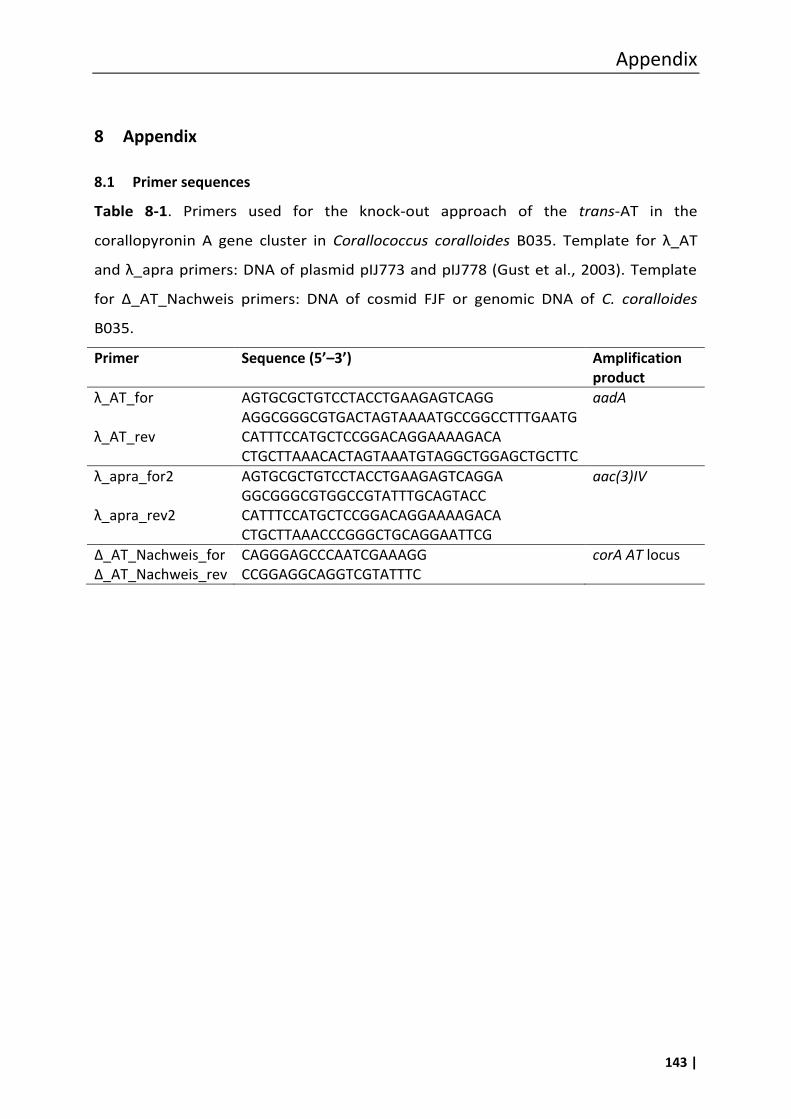
Appendix
143 |
8 Appendix
8.1 Primer sequences
Table 8-1. Primers used for the knock-out approach of the trans-AT in the
corallopyronin A gene cluster in Corallococcus coralloides B035. Template for λ_AT
and λ_apra primers: DNA of plasmid pIJ773 and pIJ778 (Gust et al., 2003). Template
for Δ_AT_Nachweis primers: DNA of cosmid FJF or genomic DNA of C. coralloides
B035.
Primer Sequence (5’–3’) Amplification product
λ_AT_for λ_AT_rev
AGTGCGCTGTCCTACCTGAAGAGTCAGG AGGCGGGCGTGACTAGTAAAATGCCGGCCTTTGAATG CATTTCCATGCTCCGGACAGGAAAAGACA CTGCTTAAACACTAGTAAATGTAGGCTGGAGCTGCTTC
aadA
λ_apra_for2 λ_apra_rev2
AGTGCGCTGTCCTACCTGAAGAGTCAGGA GGCGGGCGTGGCCGTATTTGCAGTACC CATTTCCATGCTCCGGACAGGAAAAGACA CTGCTTAAACCCGGGCTGCAGGAATTCG
aac(3)IV
Δ_AT_Nachweis_for Δ_AT_Nachweis_rev
CAGGGAGCCCAATCGAAAGG CCGGAGGCAGGTCGTATTTC
corA AT locus

Appendix
144 |
Table 8-2. Primers used for the heterologous expression approach of the eastern
chain of corallopyronin A in Pseudomonas putida KT2440. Template for λ_EcoRV_3
and λ_SpeI_3 primers: DNA of plasmid pIJ778 (Gust et al., 2003). Amplification
product aadA_EcoRV/SpeI was used to be integrated into cosmid FJ7 by Lamda Red
recombineering. Template for Apra_SpeI and Pm_EcoRV primers: DNA of the
construct pIB861_apra. Amplification product pm/xylS/aac(3)IV_EcoRV/SpeI was used
to be integrated into the restricted cosmid FJ7_aadA_EcoRV/SpeI by ligation.
Template for λ_for_tet_3 and λ_for_trpE_3 primers: DNA of the cloning construct
pGEMT_trpE_tetA. Amplification product tetA/trpE was used to be integrated into the
recombinant construct FJ7_pm/xylS by Lamda Red recombineering to give the
finished construct FJ7_pm/xylS/trpE (54 kb).
Primer Sequence (5’–3’) Amplification product
λ_EcoRV_3 λ_SpeI_3
TGACTCTTCAGGTAGGACAGCGCACTGAC GACGTACAGCGATATCTGGCGAGCGGCATCTTATTTG CGCGATTGACATGTTCACTGGCGGTCAGT ATGGATTGCGACTAGTGACGCCGTTGGATACACCAAGG
aadA
Apra_SpeI_for Pm_EcoRV_rev
ACTAGTTACGGCCCACAGAATG GATATCGCCGCAATTCACATGTTC
pm/xylS/ aac(3)IV
λ_for_tet_3 λ_rev_trpE_3
ATGATTGAACAAGATGGATTGCACGCAG GTTCTCCGGCCGCTTGGCGCCAAGCTATTTAGGTG TCAGAAGAACTCGTCAAGAAGGCGATAG AAGGCGATGCGCTGCGAATCACTATAGGGCGAATTGGG
tetA/trpE
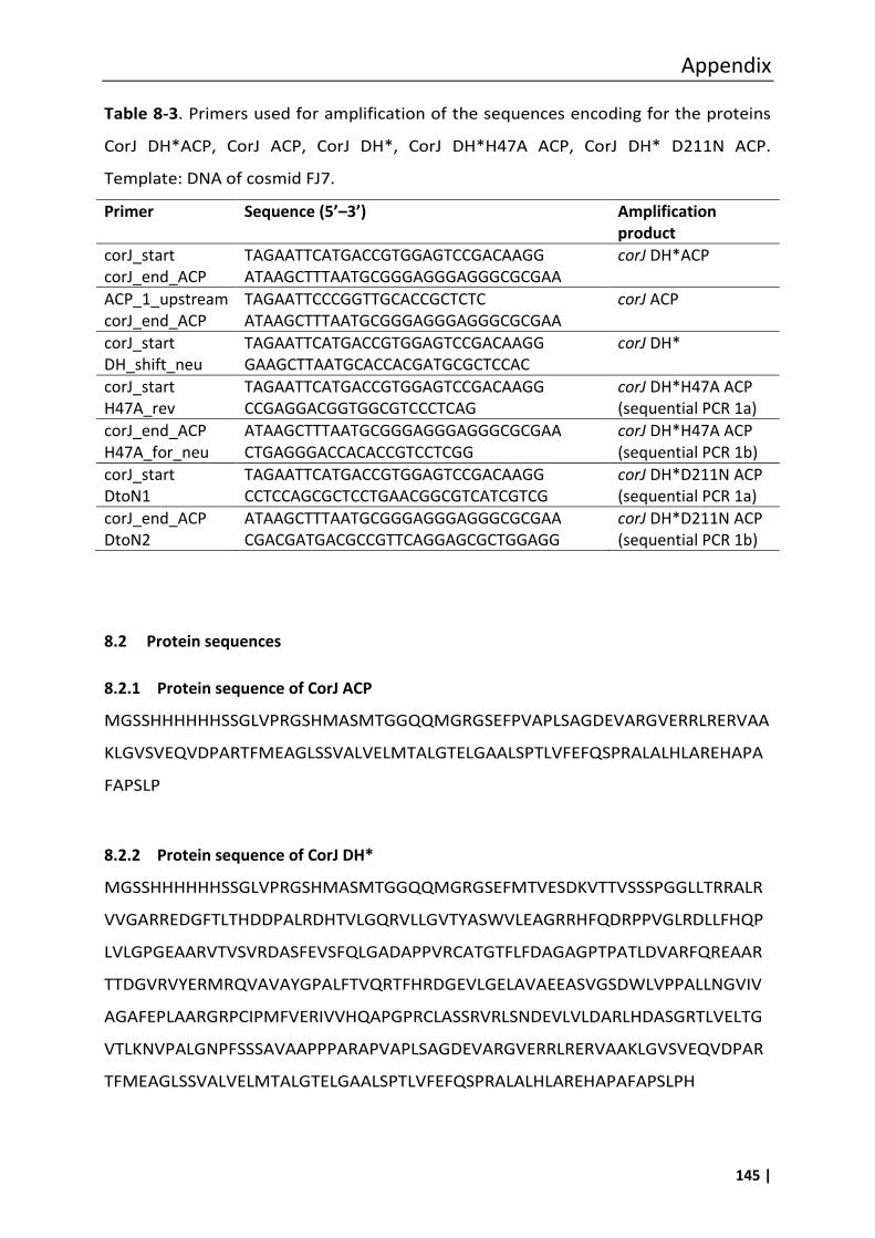
Appendix
145 |
Table 8-3. Primers used for amplification of the sequences encoding for the proteins
CorJ DH*ACP, CorJ ACP, CorJ DH*, CorJ DH*H47A ACP, CorJ DH* D211N ACP.
Template: DNA of cosmid FJ7.
Primer Sequence (5’–3’) Amplification product
corJ_start corJ_end_ACP
TAGAATTCATGACCGTGGAGTCCGACAAGG ATAAGCTTTAATGCGGGAGGGAGGGCGCGAA
corJ DH*ACP
ACP_1_upstream corJ_end_ACP
TAGAATTCCCGGTTGCACCGCTCTC ATAAGCTTTAATGCGGGAGGGAGGGCGCGAA
corJ ACP
corJ_start DH_shift_neu
TAGAATTCATGACCGTGGAGTCCGACAAGG GAAGCTTAATGCACCACGATGCGCTCCAC
corJ DH*
corJ_start H47A_rev
TAGAATTCATGACCGTGGAGTCCGACAAGG CCGAGGACGGTGGCGTCCCTCAG
corJ DH*H47A ACP (sequential PCR 1a)
corJ_end_ACP H47A_for_neu
ATAAGCTTTAATGCGGGAGGGAGGGCGCGAA CTGAGGGACCACACCGTCCTCGG
corJ DH*H47A ACP (sequential PCR 1b)
corJ_start DtoN1
TAGAATTCATGACCGTGGAGTCCGACAAGG CCTCCAGCGCTCCTGAACGGCGTCATCGTCG
corJ DH*D211N ACP (sequential PCR 1a)
corJ_end_ACP DtoN2
ATAAGCTTTAATGCGGGAGGGAGGGCGCGAA CGACGATGACGCCGTTCAGGAGCGCTGGAGG
corJ DH*D211N ACP (sequential PCR 1b)
8.2 Protein sequences
8.2.1 Protein sequence of CorJ ACP
MGSSHHHHHHSSGLVPRGSHMASMTGGQQMGRGSEFPVAPLSAGDEVARGVERRLRERVAA
KLGVSVEQVDPARTFMEAGLSSVALVELMTALGTELGAALSPTLVFEFQSPRALALHLAREHAPA
FAPSLP
8.2.2 Protein sequence of CorJ DH*
MGSSHHHHHHSSGLVPRGSHMASMTGGQQMGRGSEFMTVESDKVTTVSSSPGGLLTRRALR
VVGARREDGFTLTHDDPALRDHTVLGQRVLLGVTYASWVLEAGRRHFQDRPPVGLRDLLFHQP
LVLGPGEAARVTVSVRDASFEVSFQLGADAPPVRCATGTFLFDAGAGPTPATLDVARFQREAAR
TTDGVRVYERMRQVAVAYGPALFTVQRTFHRDGEVLGELAVAEEASVGSDWLVPPALLNGVIV
AGAFEPLAARGRPCIPMFVERIVVHQAPGPRCLASSRVRLSNDEVLVLDARLHDASGRTLVELTG
VTLKNVPALGNPFSSSAVAAPPPARAPVAPLSAGDEVARGVERRLRERVAAKLGVSVEQVDPAR
TFMEAGLSSVALVELMTALGTELGAALSPTLVFEFQSPRALALHLAREHAPAFAPSLPH
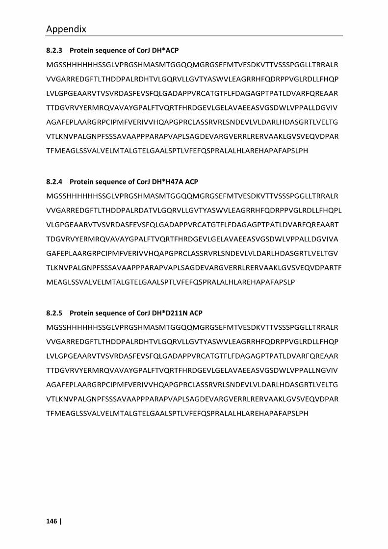
Appendix
146 |
8.2.3 Protein sequence of CorJ DH*ACP
MGSSHHHHHHSSGLVPRGSHMASMTGGQQMGRGSEFMTVESDKVTTVSSSPGGLLTRRALR
VVGARREDGFTLTHDDPALRDHTVLGQRVLLGVTYASWVLEAGRRHFQDRPPVGLRDLLFHQP
LVLGPGEAARVTVSVRDASFEVSFQLGADAPPVRCATGTFLFDAGAGPTPATLDVARFQREAAR
TTDGVRVYERMRQVAVAYGPALFTVQRTFHRDGEVLGELAVAEEASVGSDWLVPPALLDGVIV
AGAFEPLAARGRPCIPMFVERIVVHQAPGPRCLASSRVRLSNDEVLVLDARLHDASGRTLVELTG
VTLKNVPALGNPFSSSAVAAPPPARAPVAPLSAGDEVARGVERRLRERVAAKLGVSVEQVDPAR
TFMEAGLSSVALVELMTALGTELGAALSPTLVFEFQSPRALALHLAREHAPAFAPSLPH
8.2.4 Protein sequence of CorJ DH*H47A ACP
MGSSHHHHHHSSGLVPRGSHMASMTGGQQMGRGSEFMTVESDKVTTVSSSPGGLLTRRALR
VVGARREDGFTLTHDDPALRDATVLGQRVLLGVTYASWVLEAGRRHFQDRPPVGLRDLLFHQPL
VLGPGEAARVTVSVRDASFEVSFQLGADAPPVRCATGTFLFDAGAGPTPATLDVARFQREAART
TDGVRVYERMRQVAVAYGPALFTVQRTFHRDGEVLGELAVAEEASVGSDWLVPPALLDGVIVA
GAFEPLAARGRPCIPMFVERIVVHQAPGPRCLASSRVRLSNDEVLVLDARLHDASGRTLVELTGV
TLKNVPALGNPFSSSAVAAPPPARAPVAPLSAGDEVARGVERRLRERVAAKLGVSVEQVDPARTF
MEAGLSSVALVELMTALGTELGAALSPTLVFEFQSPRALALHLAREHAPAFAPSLP
8.2.5 Protein sequence of CorJ DH*D211N ACP
MGSSHHHHHHSSGLVPRGSHMASMTGGQQMGRGSEFMTVESDKVTTVSSSPGGLLTRRALR
VVGARREDGFTLTHDDPALRDHTVLGQRVLLGVTYASWVLEAGRRHFQDRPPVGLRDLLFHQP
LVLGPGEAARVTVSVRDASFEVSFQLGADAPPVRCATGTFLFDAGAGPTPATLDVARFQREAAR
TTDGVRVYERMRQVAVAYGPALFTVQRTFHRDGEVLGELAVAEEASVGSDWLVPPALLNGVIV
AGAFEPLAARGRPCIPMFVERIVVHQAPGPRCLASSRVRLSNDEVLVLDARLHDASGRTLVELTG
VTLKNVPALGNPFSSSAVAAPPPARAPVAPLSAGDEVARGVERRLRERVAAKLGVSVEQVDPAR
TFMEAGLSSVALVELMTALGTELGAALSPTLVFEFQSPRALALHLAREHAPAFAPSLPH
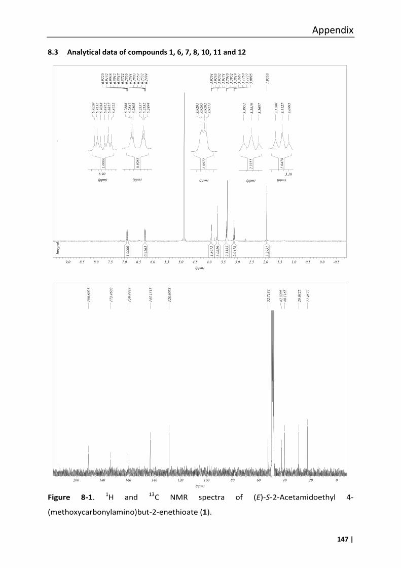
Appendix
147 |
8.3 Analytical data of compounds 1, 6, 7, 8, 10, 11 and 12
Figure 8-1. 1H and 13C NMR spectra of (E)-S-2-Acetamidoethyl 4-
(methoxycarbonylamino)but-2-enethioate (1).
2.3355
3.3952
3.3819
3.3687
(ppm)
2.0478
3.1260
3.1127
3.0995
(ppm)
3.10
1.0000
0.9263
1.8972
3.0629
2.3355
2.0478
3.2951
Integral
6.9220
6.9132
6.9038
6.8912
6.8817
6.8722
6.2866
6.2841
6.2803
6.2557
6.2532
6.2494
3.9291
3.9265
3.9202
3.9171
3.7040
3.3952
3.3819
3.3687
3.1260
3.1127
3.0995
1.9560
(ppm)
-0.50.00.51.01.52.02.53.03.54.04.55.05.56.06.57.07.58.08.59.0
1.8972
3.9291
3.9265
3.9202
3.9171
(ppm)
0.9263
6.2866
6.2841
6.2803
6.2557
6.2532
6.2494
(ppm)
1.0000
6.9220
6.9132
6.9038
6.8912
6.8817
6.8722
(ppm)
6.90
190.6025
173.4600
159.4449
143.1315
128.6073
52.7134
42.3203
40.1165
29.0325
22.4577
(ppm)
020406080100120140160180200
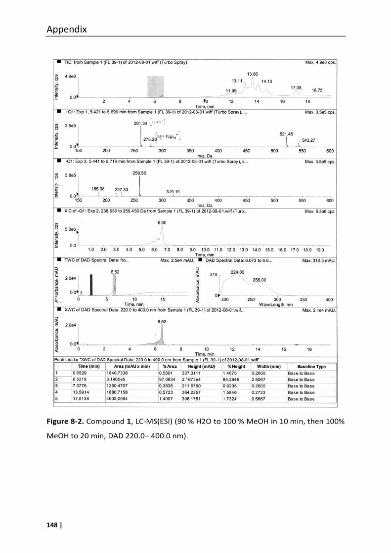
Appendix
148 |
Figure 8-2. Compound 1, LC-MS(ESI) (90 % H2O to 100 % MeOH in 10 min, then 100%
MeOH to 20 min, DAD 220.0– 400.0 nm).
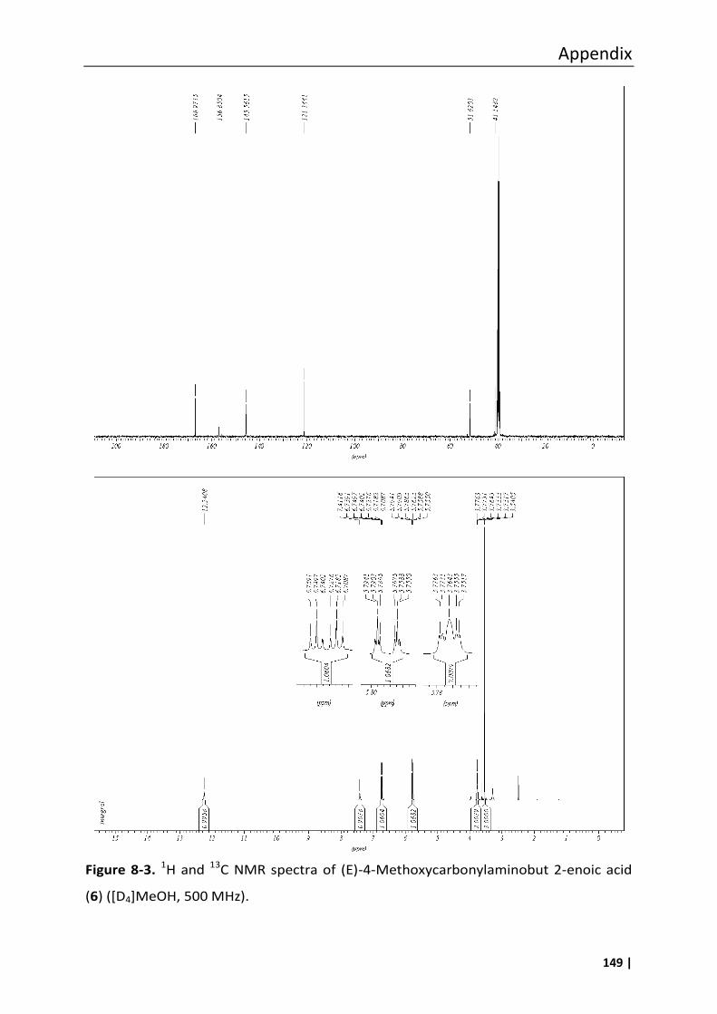
Appendix
149 |
Figure 8-3. 1H and 13C NMR spectra of (E)-4-Methoxycarbonylaminobut 2-enoic acid
(6) ([D4]MeOH, 500 MHz).
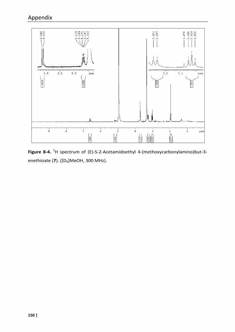
Appendix
150 |
Figure 8-4. 1H spectrum of (E)-S-2-Acetamidoethyl 4-(methoxycarbonylamino)but-3-
enethioate (7). ([D4]MeOH, 300 MHz).
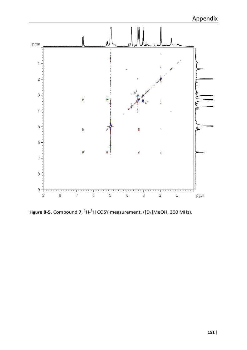
Appendix
151 |
Figure 8-5. Compound 7, 1H-1H COSY measurement. ([D4]MeOH, 300 MHz).
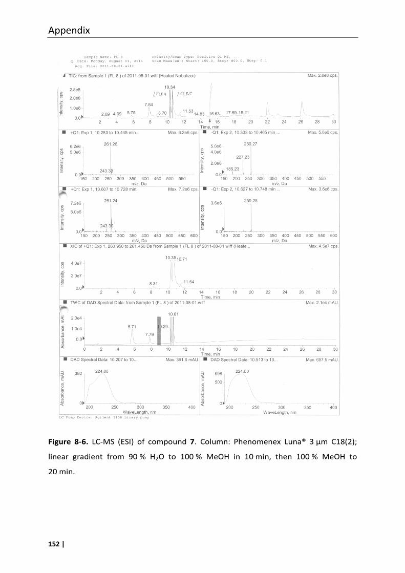
Appendix
152 |
Figure 8-6. LC-MS (ESI) of compound 7. Column: Phenomenex Luna® 3 µm C18(2);
linear gradient from 90 % H2O to 100 % MeOH in 10 min, then 100 % MeOH to
20 min.
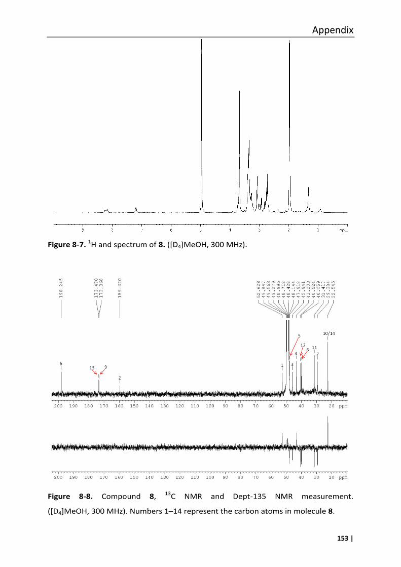
Appendix
153 |
Figure 8-7. 1H and spectrum of 8. ([D4]MeOH, 300 MHz).
Figure 8-8. Compound 8, 13C NMR and Dept-135 NMR measurement.
([D4]MeOH, 300 MHz). Numbers 1–14 represent the carbon atoms in molecule 8.
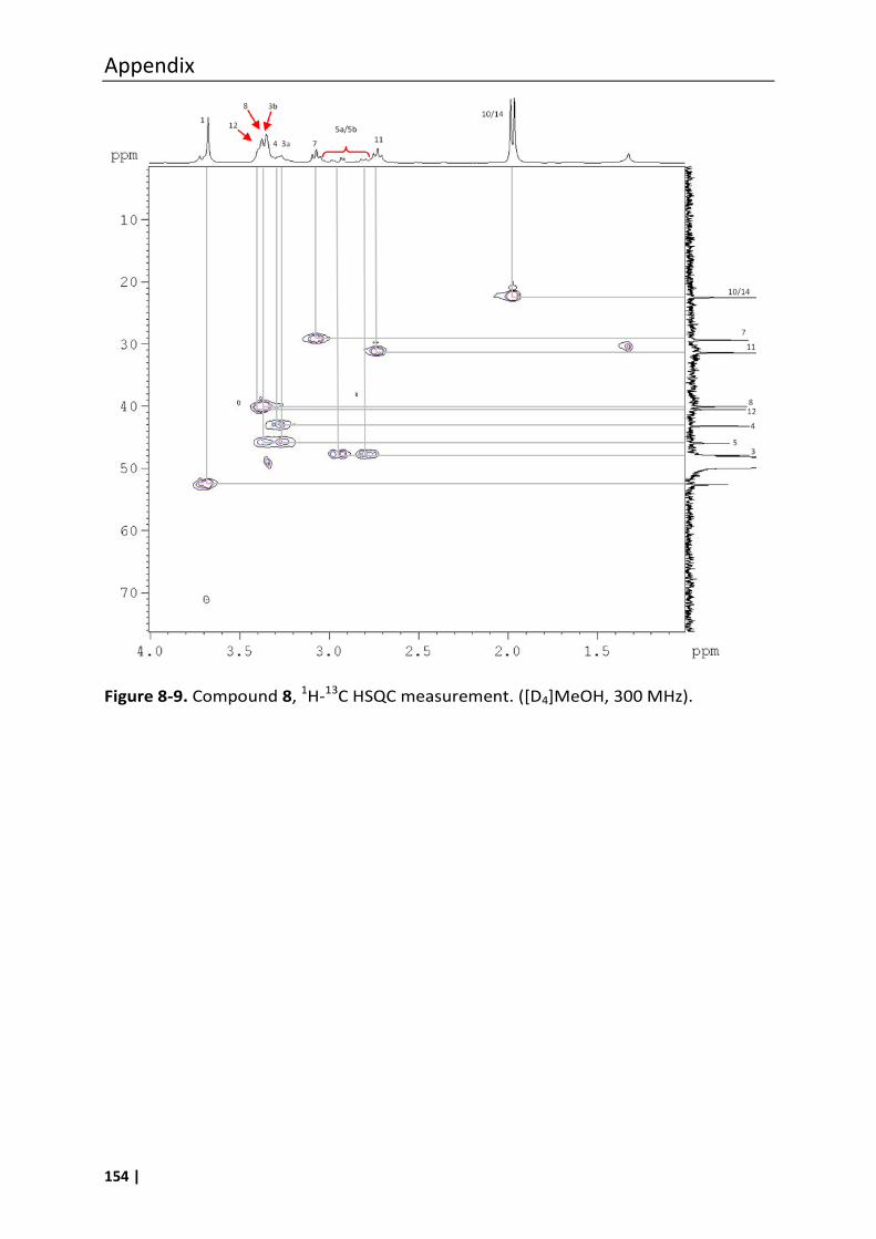
Appendix
154 |
Figure 8-9. Compound 8, 1H-13C HSQC measurement. ([D4]MeOH, 300 MHz).
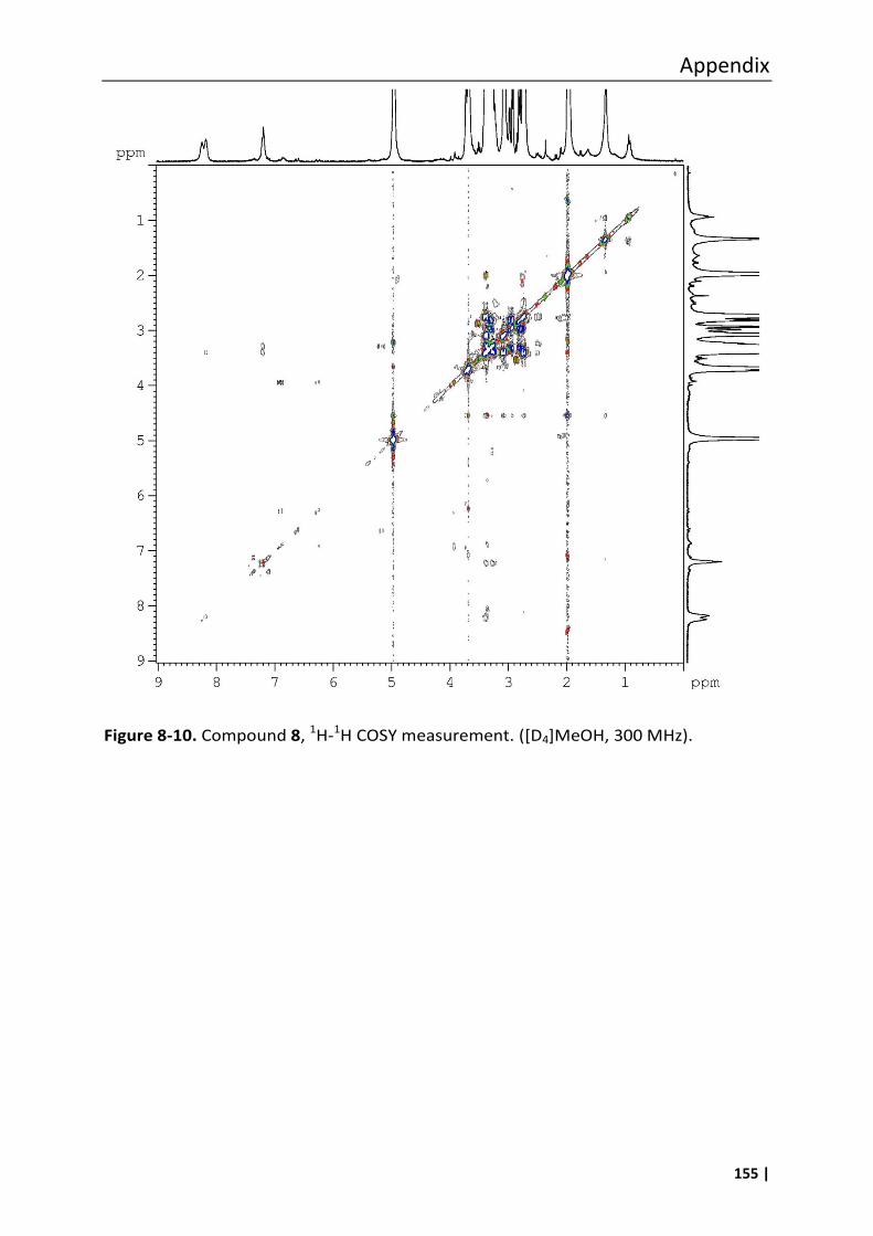
Appendix
155 |
Figure 8-10. Compound 8, 1H-1H COSY measurement. ([D4]MeOH, 300 MHz).
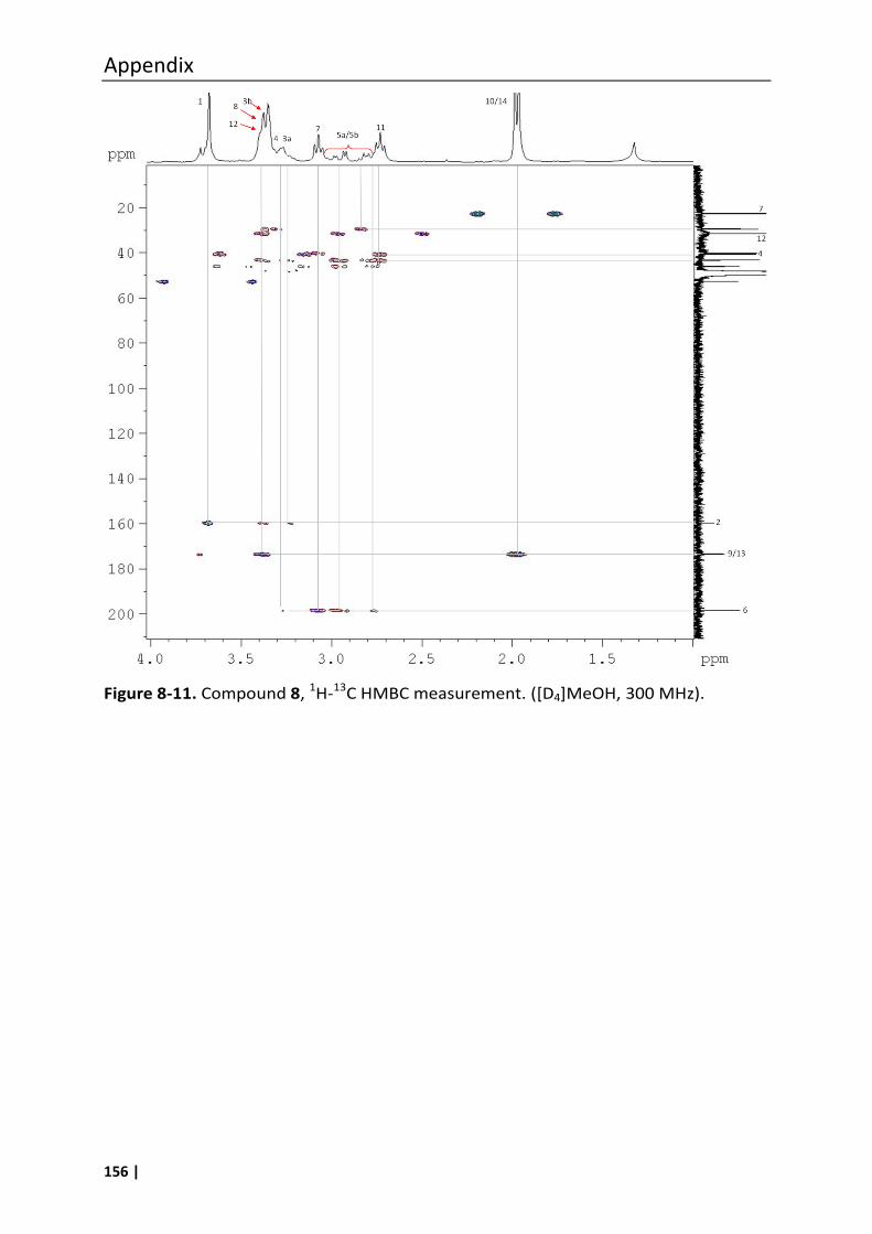
Appendix
156 |
Figure 8-11. Compound 8, 1H-13C HMBC measurement. ([D4]MeOH, 300 MHz).
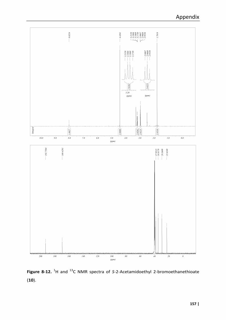
Appendix
157 |
Figure 8-12. 1H and 13C NMR spectra of S-2-Acetamidoethyl 2-bromoethanethioate
(10).
1.0027
2.0000
2.0262
2.0225
2.9539
Integral
8.0224
4.4261
3.2120
3.1988
3.1868
3.1730
2.9807
2.9668
2.9536
1.7818
(ppm)
0.01.02.03.04.05.06.07.08.09.010.0
2.0225
2.9807
2.9668
2.9536
(ppm)
2.0262
3.2120
3.1988
3.1868
3.1730
(ppm)
3.20
192.7504
169.4291
37.9515
34.8170
29.3609
22.6240
(ppm)
020406080100120140160180200
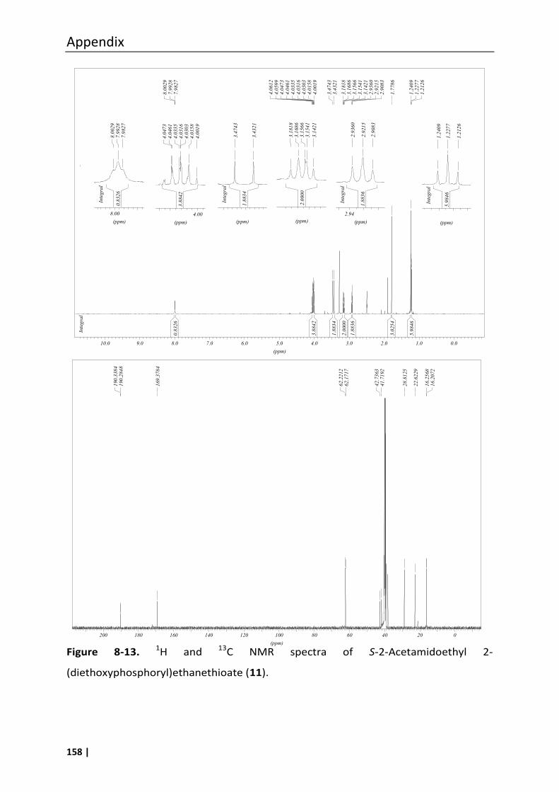
Appendix
158 |
Figure 8-13. 1H and 13C NMR spectra of S-2-Acetamidoethyl 2-
(diethoxyphosphoryl)ethanethioate (11).
2.0000
3.1818
3.1686
3.1566
3.1541
3.1421
(ppm)
3.8842
4.0473
4.0461
4.0335
4.0316
4.0303
4.0158
4.0019
(ppm)
4.00
0.8326
Integral
8.0029
7.9928
7.9827
(ppm)
8.00
5.9846
Integral
1.2409
1.2277
1.2126
(ppm)
1.8836
Integral
2.9360
2.9215
2.9083
(ppm)
2.94
0.8326
3.8842
1.8834
2.0000
1.8836
3.0254
5.9846
Integral
8.0029
7.9928
7.9827
4.0612
4.0599
4.0473
4.0461
4.0335
4.0316
4.0303
4.0158
4.0019
3.4743
3.4321
3.1818
3.1686
3.1566
3.1541
3.1421
2.9360
2.9215
2.9083
1.7786
1.2409
1.2277
1.2126
(ppm)
0.01.02.03.04.05.06.07.08.09.010.0
1.8834
Integral
3.4743
3.4321
(ppm)
190.3384
190.2848
169.3784
62.2212
62.1717
42.7363
41.7192
28.8125
22.6229
16.2568
16.2072
(ppm)
020406080100120140160180200
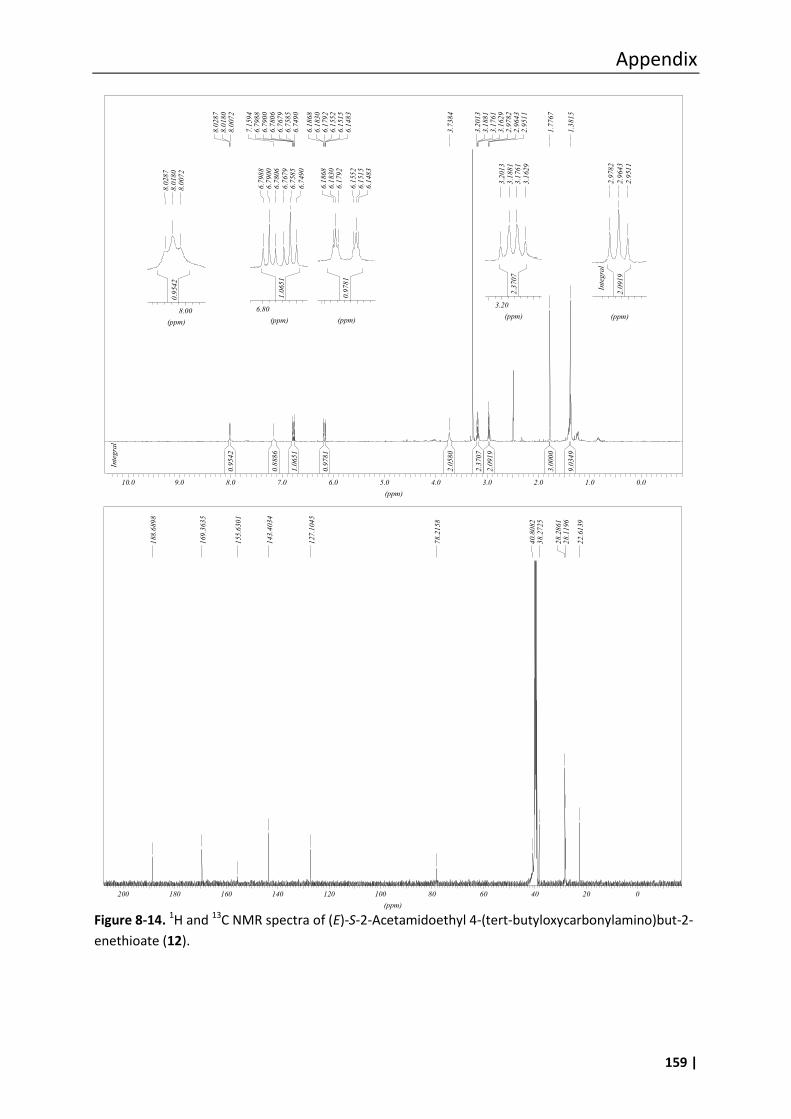
Appendix
159 |
Figure 8-14. 1H and 13C NMR spectra of (E)-S-2-Acetamidoethyl 4-(tert-butyloxycarbonylamino)but-2-
enethioate (12).
0.9542
0.8886
1.0651
0.9781
2.0580
2.3707
2.0919
3.0000
9.0349
Integral
8.0287
8.0180
8.0072
7.1594
6.7988
6.7900
6.7806
6.7679
6.7585
6.7490
6.1868
6.1830
6.1792
6.1552
6.1515
6.1483
3.7384
3.2013
3.1881
3.1761
3.1629
2.9782
2.9643
2.9511
1.7767
1.3815
(ppm)
0.01.02.03.04.05.06.07.08.09.010.0
2.0919
Integral
2.9782
2.9643
2.9511
(ppm)
2.3707
3.2013
3.1881
3.1761
3.1629
(ppm)
3.20
0.9781
6.1868
6.1830
6.1792
6.1552
6.1515
6.1483
(ppm)
1.0651
6.7988
6.7900
6.7806
6.7679
6.7585
6.7490
(ppm)
6.80
0.9542
8.0287
8.0180
8.0072
(ppm)
8.00
188.6898
169.3635
155.6301
143.4034
127.1045
78.2158
40.8082
38.2725
28.2861
28.1196
22.6139
(ppm)
020406080100120140160180200


