endothelial bmx tyrosine kinase activity is essential for myocardial hypertrophy and ... ·...
TRANSCRIPT
Endothelial Bmx tyrosine kinase activity is essential formyocardial hypertrophy and remodelingTanja Holopainena, Markus Räsänena, Andrey Anisimova, Tomi Tuomainenb, Wei Zhenga, Denis Tvorogova,Juha J. Hulmic, Leif C. Anderssond, Bruno Cennie, Pasi Tavib, Eero Mervaalaf, Riikka Kiveläa,1, and Kari Alitaloa,1,2
aWihuri Research Institute and Translational Cancer Biology Program, Biomedicum Helsinki, University of Helsinki, FI-00290 Helsinki, Finland; bDepartmentof Biotechnology and Molecular Medicine, A. I. Virtanen Institute for Molecular Sciences, University of Eastern Finland, FI-70211 Kuopio, Finland;cDepartment of Biology of Physical Activity, University of Jyväskylä, FI-40700 Jyväskylä, Finland; dDepartment of Pathology, Haartman Institute, University ofHelsinki, FI-00014 Helsinki, Finland; eNovartis Institutes for Biomedical Research, Basel CH-4002, Switzerland; and fDepartment of Pharmacology, Institute ofBiomedicine, Biomedicum Helsinki, University of Helsinki, FI-00014 Helsinki, Finland
Contributed by Kari Alitalo, September 8, 2015 (sent for review May 13, 2015; reviewed by Bernd Groner and Christian Kupatt)
Cardiac hypertrophy accompanies many forms of heart disease,including ischemic disease, hypertension, heart failure, and valvu-lar disease, and it is a strong predictor of increased cardiovascularmorbidity and mortality. Deletion of bone marrow kinase inchromosome X (Bmx), an arterial nonreceptor tyrosine kinase,has been shown to inhibit cardiac hypertrophy in mice. This find-ing raised the possibility of therapeutic use of Bmx tyrosine kinaseinhibitors, which we have addressed here by analyzing cardiachypertrophy in gene-targeted mice deficient in Bmx tyrosine ki-nase activity. We found that angiotensin II (Ang II)-induced car-diac hypertrophy is significantly reduced in mice deficient in Bmxand in mice with inactivated Bmx tyrosine kinase compared withWT mice. Genome-wide transcriptomic profiling showed thatBmx inactivation suppresses myocardial expression of genes re-lated to Ang II-induced inflammatory and extracellular matrixresponses whereas expression of RNAs encoding mitochondrialproteins after Ang II administration was maintained in Bmx-inac-tivated hearts. Very little or no Bmx mRNA was expressed inhuman cardiomyocytes whereas human cardiac endothelial cellsexpressed abundant amounts. Ang II stimulation of endothelialcells increased Bmx phosphorylation, and Bmx gene silencinginhibited downstream STAT3 signaling, which has been implicatedin cardiac hypertrophy. Furthermore, activation of the mechanistictarget of rapamycin complex 1 pathway by Ang II treatment wasdecreased in the Bmx-deficient hearts. Our results demonstratethat inhibition of the cross-talk between endothelial cells andcardiomyocytes by Bmx inactivation suppresses Ang II-inducedsignals for cardiac hypertrophy. These results suggest that theendothelial Bmx tyrosine kinase could provide a target to attenu-ate the development of cardiac hypertrophy.
Etk | endothelium | signaling | heart | cardiomyocyte
Heart failure is a continuously increasing global problem inaging populations. Despite some progress in treatment op-
tions, the prognosis of heart failure is worse than that for mostcancers (1). Cardiac hypertrophy due to exercise training is re-ferred to as physiological hypertrophy, in which the architectureand contractile function of the heart are maintained or enhanced,and it is reversible if the training is discontinued. Numerous dis-eases, such as myocardial infarction, aortic stenosis, hypertensionand metabolic stress, induce pathological hypertrophy, which isrelated to activation of maladaptive cellular events in the heartand progressively leads to heart failure (2). Understanding theunderlying regulatory processes in the development of patho-logical hypertrophy has provided effective therapeutic targets toattenuate disease progression, but there are currently no treat-ments to reverse cardiac hypertrophy.The Bmx (Etk) gene in the chromosome region Xp22.2 en-
codes a tyrosine kinase that was originally identified and clonedfrom hematopoietic cells (3). Striking new evidence indicatesthat Bmx phosphorylates a phosphotyrosine-primed motif medi-ating the activation of multiple receptor tyrosine kinases (4). Bmx
is highly expressed in the endocardium and in the endothelium oflarge arteries, starting between embryonic days 10.5–12.5 (5),indicating that Bmx is a signal transducer mainly in the arterialendothelium. However, Bmx deletion does not result in anyobvious developmental phenotype in mice (5), suggesting that ithas a redundant function during embryogenesis. In contrast,Bmx has been shown to be important in a variety of patholog-ical states, including tumor growth (6–8), and its overexpressionpromotes ischemia-induced arteriogenesis and inflammatoryangiogenesis (9).Previous reports have indicated that Bmx deficiency atten-
uates pressure overload-induced cardiac hypertrophy in re-sponse to thoracic aortic constriction or vascular endothelialgrowth factor-B (VEGF-B) overexpression (10, 11), but themechanisms of how this regulation occurs are as yet not known.We have here analyzed the molecular mechanisms of how Bmxregulates cardiac hypertrophy by using angiotensin II (Ang II)treatment that leads to cardiac hypertrophy and remodeling (12).Importantly, we show that pathological cardiac growth is sup-pressed in a mouse model where the Bmx kinase is inactivatedby a missense point mutation. Our findings provide therapeuticproof of principle indicating that the blocking of Bmx tyrosinekinase activity could be used to inhibit pathological cardiachypertrophy.
Significance
During the last decades, heart failure has developed into amajor burden in the western world, increasingly affectingmillions. Cardiac hypertrophy is an adaptive response to myo-cardial infarction or increased blood pressure, and it oftenleads to heart failure. Understanding the underlying regulatoryprocesses in the development of pathological hypertrophy isneeded for the development of effective therapies. Our resultsshow that the kinase activity of the endothelial bone marrowkinase in chromosome X (Bmx) protein is necessary for thedevelopment of pathological cardiac hypertrophy. This findingcould provide significant therapeutic applications when specificBmx kinase inhibitors become available in the clinics.
Author contributions: T.H., P.T., E.M., R.K., and K.A. designed research; T.H., M.R., A.A.,T.T., W.Z., D.T., J.J.H., and R.K. performed research; T.T., B.C., P.T., and E.M. contributednew reagents/analytic tools; T.H., M.R., J.J.H., L.C.A., E.M., R.K., and K.A. analyzed data;and T.H., R.K., and K.A. wrote the paper.
Reviewers: B.G., Georg Speyer Haus; and C.K., Klinikum Grosshadern Munich.
Conflict of interest statement: B.C. is an employee of Novartis.
Freely available online through the PNAS open access option.
Data deposition: The microarray data reported in this paper have been deposited in theGene Expression Omnibus (GEO) database, www.ncbi.nlm.nih.gov/geo (accession no.GSE47420).1R.K. and K.A. contributed equally to this work.2To whom correspondence should be addressed. Email: [email protected].
This article contains supporting information online at www.pnas.org/lookup/suppl/doi:10.1073/pnas.1517810112/-/DCSupplemental.
www.pnas.org/cgi/doi/10.1073/pnas.1517810112 PNAS | October 20, 2015 | vol. 112 | no. 42 | 13063–13068
MED
ICALSC
IENCE
S
Dow
nloa
ded
by g
uest
on
May
31,
202
0
ResultsBmx Tyrosine Kinase Activity Is Necessary for Ang II-Induced CardiacHypertrophy. Previous studies have shown that cardiac hypertro-phy in response to aortic coarctation or VEGF-B overexpressionis greatly attenuated in mice deficient of the Bmx protein (10,11). Because of the continuing development of increasinglyspecific Bmx tyrosine kinase inhibitors, it would be important toknow whether specific inactivation of the Bmx tyrosine kinaseactivity would be enough to inhibit cardiac hypertrophy. Thus,our aim was to determine whether attenuation of cardiac hy-pertrophy detected in Bmx knockout (KO) mice is dependent onthe tyrosine kinase activity of the Bmx protein. We analyzedcardiac growth upon Ang II administration to mice in which theBmx gene had been deleted, or replaced with a K421R mutantallele (equivalent to human mutation K445R) (13), renderingthe kinase catalytically inactive (referred to as TK mice) (14).To compare the effects of Bmx gene deletion or tyrosine ki-
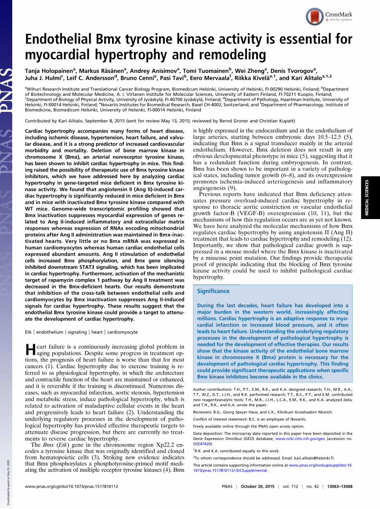
nase inactivation on Ang II-induced cardiac hypertrophy, we firstimplanted Ang II minipumps into Bmx KO and WT mice for2 wk. The WT mice developed cardiac hypertrophy, but, asexpected, the hypertrophic changes were significantly milder inKO mice than in WT mice (Fig. 1A). Immunostaining for dys-trophin demonstrated that the cardiomyocyte size was increasedin both groups after Ang II infusion; however, the increase inBmx KO mice was significantly smaller than in WT mice (Fig. 1B and C). Importantly, the Ang II-induced cardiac growth andcardiomyocyte size were reduced also in mice expressing thekinase-inactive Bmx allele (Fig. 1 E–G). The mRNA level ofskeletal α-actin, a marker for pathological cardiac hypertrophy,was significantly increased in WT mice, but not in Bmx KO miceor in TK mice (Fig. 1 D and H). Accordingly, after a prolongedAng II infusion for 6 wk, the extent of cardiac hypertrophy wasalso less in Bmx KO mice than in WT mice (Fig. S1). However,there were no significant differences in the blood pressure be-tween KO and WT mice before or 2 or 6 wk after Ang II infusionmeasured by the tail cuff method. Similar results were obtainedwhen a catheter was inserted into the carotid artery to monitorblood pressure more accurately. These results confirmed thatBmx deficiency does not affect blood pressure (Fig. S2). Takentogether; these data indicate that the development of cardiachypertrophy is dependent on Bmx tyrosine kinase activity andthat the difference in the cardiac Ang II response is not due todifferences in blood pressure.
The Bmx Tyrosine Kinase Is Important for Endothelial Cell–CardiomyocyteCross-Talk. A previous report has suggested that Bmx is expressedalso in cardiomyocytes in addition to its expression in the arterialendothelium, in endocardium, and in cells of the hematopoieticmyeloid lineage (15). Thus, we compared Bmx mRNA levels in
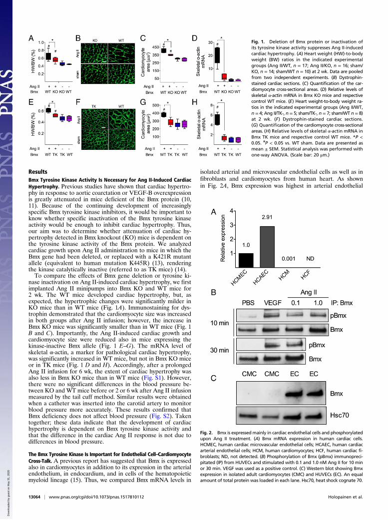
isolated arterial and microvascular endothelial cells as well as infibroblasts and cardiomyocytes from human heart. As shownin Fig. 2A, Bmx expression was highest in arterial endothelial
Fig. 1. Deletion of Bmx protein or inactivation ofits tyrosine kinase activity suppresses Ang II-inducedcardiac hypertrophy. (A) Heart weight (HW)-to-bodyweight (BW) ratios in the indicated experimentalgroups (Ang II/WT, n = 17; Ang II/KO, n = 16; sham/KO, n = 14; sham/WT n = 10) at 2 wk. Data are pooledfrom two independent experiments. (B) Dystrophin-stained cardiac sections. (C) Quantification of the car-diomyocyte cross-sectional areas. (D) Relative levels ofskeletal α-actin mRNA in Bmx KO mice and respectivecontrol WT mice. (E) Heart weight-to-body weight ra-tios in the indicated experimental groups (Ang II/WT,n = 4; Ang II/TK-, n = 5; sham/TK-, n = 7; sham/WT n = 8)at 2 wk. (F) Dystrophin-stained cardiac sections.(G) Quantification of the cardiomyocyte cross-sectionalareas. (H) Relative levels of skeletal α-actin mRNA inBmx TK mice and respective control WT mice. *P <0.05. #P < 0.05 vs. WT sham. Data are presented asmean ± SEM. Statistical analysis was performed withone-way ANOVA. (Scale bar: 20 μm.)
Fig. 2. Bmx is expressed mainly in cardiac endothelial cells and phosphorylatedupon Ang II treatment. (A) Bmx mRNA expression in human cardiac cells.HCMEC, human cardiac microvascular endothelial cells; HCAEC, human cardiacarterial endothelial cells; HCM, human cardiomyocytes; HCF, human cardiac fi-broblasts; ND, not detected. (B) Phosphorylation of Bmx (pBmx) immunopreci-pitated (IP) from HUVECs and stimulated with 0.1 and 1.0 nM Ang II for 10 minor 30 min. VEGF was used as a positive control. (C) Western blot showing Bmxexpression in isolated adult cardiomyocytes (CMC) and HUVECs (EC). An equalamount of total protein was loaded in each lane. Hsc70, heat shock cognate 70.
13064 | www.pnas.org/cgi/doi/10.1073/pnas.1517810112 Holopainen et al.
Dow
nloa
ded
by g
uest
on
May
31,
202
0
cells. The microvascular endothelial cells contained about 35% ofthis amount whereas expression in the cardiomyocytes was about0.1% of that found in microvascular endothelial cells, and noBmx RNA could be detected in cardiac fibroblasts. These resultsstrongly suggest that the effects of Bmx on cardiac hypertrophyare mediated via the endothelial cells of the coronary vasculature.Next, we analyzed whether Ang II can induce Bmx phosphor-
ylation in endothelial cells. Human umbilical venous endothelialcells (HUVECs) were stimulated with different concentrations ofAng II, and vascular endothelial growth factor (VEGF) was usedas a positive control. Similarly to VEGF, Ang II induced strongBmx phosphorylation in HUVECs after 10 min and 30 min ofstimulation (Fig. 2B). In contrast, Bmx was not detected in iso-lated adult mouse cardiomyocytes even after Ang II stimulation,further indicating that Bmx is specific for cardiac endothelialcells (Fig. 2C).
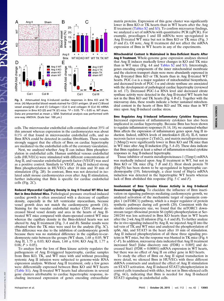
Reduced Myocardial Capillary Density in Ang II-Treated WT Mice butNot in Bmx-Deleted Mice. Pathological pressure overload-inducedcardiac growth is often associated with a decrease of capillarydensity, especially in the left ventricular myocardium, becausevessel growth does not match the cardiomyocyte growth (16).Staining for the vascular endothelial marker CD31 showed de-creased blood vessel density and area in the hearts of Ang II-treated WT mice compared with sham-operated control WT micewhereas the capillary density in the Bmx-deleted hearts was notaltered by Ang II treatment (Fig. 3 A and B). Similar results wereobtained when the TK mice were used for the analysis (Fig. 3C).This difference was due to the inhibition of cardiomyocyte growthbecause there was no statistical difference in the capillary-to-car-diomyocyte ratio between the groups (WT sham, 1.68 ± 0.05; WTAng II, 1.75 ± 0.03; KO sham, 1.60 ± 0.04; KO Ang II, 1.77 ±0.06; P > 0.05).To analyze how the loss of Bmx kinase activity regulates the
pathogenesis of Ang II-triggered hypertrophy, cardiac samplesfrom Bmx KO, TK, and WT mice with and without precedingsystemic Ang II infusion were subjected to genome-wide RNAexpression analysis. Without Ang II treatment, only a few geneswere differentially expressed between Bmx KO and WT hearts(Table S1). Ang II-treated WT hearts had alterations in severalgene clusters attributable to cardiac hypertrophic response, in-cluding increased expression of genes encoding extracellular
matrix proteins. Expression of this gene cluster was significantlylower in Bmx KO or TK hearts than in WT hearts after the AngII treatment (Tables S2 and S3). To confirm microarray findings,we analyzed a set of mRNAs with quantitative PCR (qPCR). Forexample, procollagen I and III mRNAs were up-regulated inAng II-treated WT mice but not in Bmx KO or TK mice (Fig. 3D and E). Of note, Ang II treatment did not affect the mRNAexpression of Bmx in WT hearts in any of the experiments.
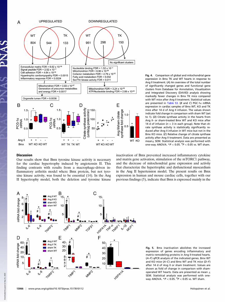
Mitochondrial Content Is Maintained in Bmx-Deficient Hearts AfterAng II Treatment. Whole-genome gene expression analyses showedthat Ang II induces markedly fewer changes in KO and TK micethan in WT mice (Fig. 4A and Tables S2 and S3). Interestingly,genes encoding components of the inner mitochondrial membraneand the electron transport chain were more abundantly expressed inAng II-treated Bmx KO or TK hearts than in Ang II-treated WThearts. PGC-1-α is a major regulator of mitochondrial biosynthesis,and decreased levels of PGC-1-α and citrate synthase are associatedwith the development of pathological cardiac hypertrophy (reviewedin ref. 17). Decreased PGC-1-α RNA level and decreased citratesynthase activity were detected in the Ang II-treated WT hearts butnot in the Bmx KO and TK hearts (Fig. 4 B–E). Together with themicroarray data, these results indicate a better sustained mitochon-drial content in the hearts of Bmx KO and TK mice than in WTmice upon Ang II-induced cardiac stress.
Bmx Regulates Ang II-Induced Inflammatory Cytokine Responses.Increased expression of inflammatory cytokines has also beenimplicated in cardiac hypertrophy. Because Bmx has been shownto regulate inflammatory responses (9, 14, 18), we examined whetherBmx affects the expression of inflammatory genes upon Ang II in-duction. Indeed, mRNA levels of interleukin-6 (IL-6), IL-8, tumornecrosis factor receptor-2 (Tnfr2), and matrix metalloproteinase2 (Mmp2) were significantly lower in Bmx KO and TK mice thanin WT mice after Ang II induction (Fig. 5 A–D). These data indicatethat Bmx regulates at least a subset of inflammation-related cytokineresponses in Ang II-induced hypertrophy.Tissue inhibitor of matrix metalloproteinases-1 (Timp1) mRNA
was markedly induced upon Ang II treatment in WT, but not inBmx KO or TK mice (Fig. 5 B and E). Heat shock protein 70(Hsp70) has been shown to be up-regulated in hypertrophic car-diomyopathy (19). Interestingly, a clear trend of Hsp1a mRNAinduction was detected in the hypertrophic WT hearts whereaslack of Bmx abolished this response (Fig. 5 C and F).
Involvement of Bmx Tyrosine Kinase Activity in Ang II-InducedDownstream Signaling. To elucidate the influence of Bmx inacti-vation on signaling pathways mediating cardiomyocyte growth, weanalyzed activation of the mechanistic target of rapamycin com-plex 1 (mTORC1) pathway, which is a major regulator of proteinsynthetic pathways during cell growth (20). Consistent with thesmaller cardiomyocyte size, we found that the mTORC1 down-stream target ribosomal protein S6 (rpS6) phosphorylation at Ser-240/244 was less activated in Bmx KO hearts than in WT heartsafter the 2-wk Ang II infusion (Fig. 6 A and B). To further analyzethe in vivo signaling induced by Ang II, we injected Ang II into thetail vein of TK and WT mice and analyzed the phosphorylation ofrpS6, Akt, and STAT3 in the heart after 10 min of stimulation.Ang II induced phosphorylation of all these three signaling pro-teins in WT mice, but the response was blunted in TK mice (Fig. 6C–F). In addition, microarray data indicated that Ang II treatmentincreased Stat3 [false discovery rate (FDR) = 0.005] and de-creased Stat1 (FDR = 0.00001) mRNA expression in WT hearts,but there was no effect of Ang II on either gene in KO hearts.To study the effect of Bmx on Ang II signal transduction in
more detail, we silenced Bmx in HUVECs with three differentshRNA constructs and analyzed the effect of Ang II stimulationon STAT3 activation. Ang II induced STAT3 phosphorylation incontrol cells transduced with shScr, but not in Bmx-silenced cells(Fig. 6G), indicating that Bmx is needed for Ang II-inducedSTAT3 signaling in endothelial cells.
Fig. 3. Attenuated Ang II-induced cardiac responses in Bmx KO and TKmice. (A) Myocardial blood vessels stained for CD31 antigen. (B and C) Bloodvessel area/grid. (D and E) Collagen I (Col I) and collagen III (Col III) mRNAexpression in Bmx KO (D) and TK (E) mice. *P < 0.05. #P < 0.05 vs. WT sham.Data are presented as mean ± SEM. Statistical analysis was performed withone-way ANOVA. (Scale bar: 100 μm.)
Holopainen et al. PNAS | October 20, 2015 | vol. 112 | no. 42 | 13065
MED
ICALSC
IENCE
S
Dow
nloa
ded
by g
uest
on
May
31,
202
0
DiscussionOur results show that Bmx tyrosine kinase activity is necessaryfor the cardiac hypertrophy induced by angiotensin II. Thisfinding contrasts with results from a macrophage-driven in-flammatory arthritis model where Bmx protein, but not tyro-sine kinase activity, was found to be essential (14). In the AngII hypertrophy model, both the deletion and tyrosine kinase
inactivation of Bmx prevented increased inflammatory cytokineand matrix gene activation, stimulation of the mTORC1 pathway,and the decrease of mitochondrial gene expression and activitythat characterize the hypertrophic and dysfunctional myocardiumin the Ang II hypertension model. The present results on Bmxexpression in human and mouse cardiac cells, together with ourprevious findings (5), indicate that Bmx is expressed mainly in the
Fig. 4. Comparison of global and mitochondrial geneexpression in Bmx TK and WT hearts in response toAng II treatment. (A) An overview of the total numberof significantly changed genes and functional geneclusters from Database for Annotation, Visualizationand Integrated Discovery (DAVID) analysis showingmarkedly fewer changes in Bmx TK mice comparedwith WT mice after Ang II treatment. Statistical valuesare presented in Table S3. (B and C) PGC-1α mRNAexpression in cardiac samples of Bmx WT, KO and TKmice after 14 d of Ang II infusion. The values shownindicate fold change in comparison with shamWT (setto 1). (D) Citrate synthase activity in the hearts fromAng II- or sham-treated Bmx WT and KO mice after14 d of infusion (n = 3 in each group). Note that cit-rate synthase activity is statistically significantly re-duced after Ang II infusion in WT mice but not in theBmx KO mice. (E) Relative change of citrate synthaseactivity after Ang II treatment. Data are presented asmean± SEM. Statistical analysis was performed withone-way ANOVA. *P < 0.05. #P < 0.05 vs. WT sham.
Fig. 5. Bmx inactivation abolishes the increasedexpression of genes encoding inflammatory andmatrix remodeling proteins in Ang II-treated hearts.(A–F) qPCR analysis of the indicated genes. Bmx WTand KO mice (A–C) and Bmx WT and TK mice (D–F)after 14 d of Ang II or sham treatment. Values areshown as fold of change in comparison with sham-operated WT hearts. Data are presented as mean ±SEM. Statistical analysis was performed with one-way ANOVA. *P < 0.05. #P < 0.05 vs. WT sham.
13066 | www.pnas.org/cgi/doi/10.1073/pnas.1517810112 Holopainen et al.
Dow
nloa
ded
by g
uest
on
May
31,
202
0
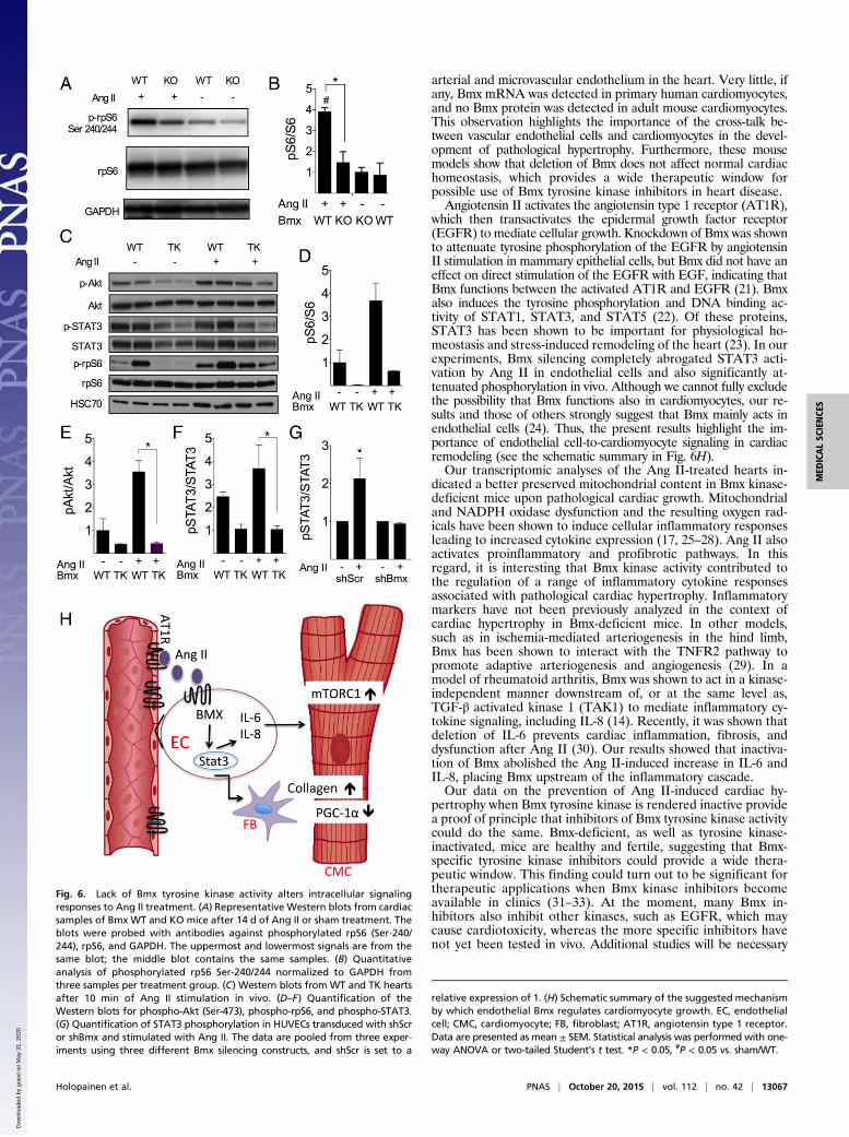
arterial and microvascular endothelium in the heart. Very little, ifany, Bmx mRNA was detected in primary human cardiomyocytes,and no Bmx protein was detected in adult mouse cardiomyocytes.This observation highlights the importance of the cross-talk be-tween vascular endothelial cells and cardiomyocytes in the devel-opment of pathological hypertrophy. Furthermore, these mousemodels show that deletion of Bmx does not affect normal cardiachomeostasis, which provides a wide therapeutic window forpossible use of Bmx tyrosine kinase inhibitors in heart disease.Angiotensin II activates the angiotensin type 1 receptor (AT1R),
which then transactivates the epidermal growth factor receptor(EGFR) to mediate cellular growth. Knockdown of Bmx was shownto attenuate tyrosine phosphorylation of the EGFR by angiotensinII stimulation in mammary epithelial cells, but Bmx did not have aneffect on direct stimulation of the EGFR with EGF, indicating thatBmx functions between the activated AT1R and EGFR (21). Bmxalso induces the tyrosine phosphorylation and DNA binding ac-tivity of STAT1, STAT3, and STAT5 (22). Of these proteins,STAT3 has been shown to be important for physiological ho-meostasis and stress-induced remodeling of the heart (23). In ourexperiments, Bmx silencing completely abrogated STAT3 acti-vation by Ang II in endothelial cells and also significantly at-tenuated phosphorylation in vivo. Although we cannot fully excludethe possibility that Bmx functions also in cardiomyocytes, our re-sults and those of others strongly suggest that Bmx mainly acts inendothelial cells (24). Thus, the present results highlight the im-portance of endothelial cell-to-cardiomyocyte signaling in cardiacremodeling (see the schematic summary in Fig. 6H).Our transcriptomic analyses of the Ang II-treated hearts in-
dicated a better preserved mitochondrial content in Bmx kinase-deficient mice upon pathological cardiac growth. Mitochondrialand NADPH oxidase dysfunction and the resulting oxygen rad-icals have been shown to induce cellular inflammatory responsesleading to increased cytokine expression (17, 25–28). Ang II alsoactivates proinflammatory and profibrotic pathways. In thisregard, it is interesting that Bmx kinase activity contributed tothe regulation of a range of inflammatory cytokine responsesassociated with pathological cardiac hypertrophy. Inflammatorymarkers have not been previously analyzed in the context ofcardiac hypertrophy in Bmx-deficient mice. In other models,such as in ischemia-mediated arteriogenesis in the hind limb,Bmx has been shown to interact with the TNFR2 pathway topromote adaptive arteriogenesis and angiogenesis (29). In amodel of rheumatoid arthritis, Bmx was shown to act in a kinase-independent manner downstream of, or at the same level as,TGF-β activated kinase 1 (TAK1) to mediate inflammatory cy-tokine signaling, including IL-8 (14). Recently, it was shown thatdeletion of IL-6 prevents cardiac inflammation, fibrosis, anddysfunction after Ang II (30). Our results showed that inactiva-tion of Bmx abolished the Ang II-induced increase in IL-6 andIL-8, placing Bmx upstream of the inflammatory cascade.Our data on the prevention of Ang II-induced cardiac hy-
pertrophy when Bmx tyrosine kinase is rendered inactive providea proof of principle that inhibitors of Bmx tyrosine kinase activitycould do the same. Bmx-deficient, as well as tyrosine kinase-inactivated, mice are healthy and fertile, suggesting that Bmx-specific tyrosine kinase inhibitors could provide a wide thera-peutic window. This finding could turn out to be significant fortherapeutic applications when Bmx kinase inhibitors becomeavailable in clinics (31–33). At the moment, many Bmx in-hibitors also inhibit other kinases, such as EGFR, which maycause cardiotoxicity, whereas the more specific inhibitors havenot yet been tested in vivo. Additional studies will be necessary
Fig. 6. Lack of Bmx tyrosine kinase activity alters intracellular signalingresponses to Ang II treatment. (A) Representative Western blots from cardiacsamples of Bmx WT and KO mice after 14 d of Ang II or sham treatment. Theblots were probed with antibodies against phosphorylated rpS6 (Ser-240/244), rpS6, and GAPDH. The uppermost and lowermost signals are from thesame blot; the middle blot contains the same samples. (B) Quantitativeanalysis of phosphorylated rpS6 Ser-240/244 normalized to GAPDH fromthree samples per treatment group. (C) Western blots from WT and TK heartsafter 10 min of Ang II stimulation in vivo. (D–F) Quantification of theWestern blots for phospho-Akt (Ser-473), phospho-rpS6, and phospho-STAT3.(G) Quantification of STAT3 phosphorylation in HUVECs transduced with shScror shBmx and stimulated with Ang II. The data are pooled from three exper-iments using three different Bmx silencing constructs, and shScr is set to a
relative expression of 1. (H) Schematic summary of the suggested mechanismby which endothelial Bmx regulates cardiomyocyte growth. EC, endothelialcell; CMC, cardiomyocyte; FB, fibroblast; AT1R, angiotensin type 1 receptor.Data are presented as mean ± SEM. Statistical analysis was performed with one-way ANOVA or two-tailed Student’s t test. *P < 0.05, #P < 0.05 vs. sham/WT.
Holopainen et al. PNAS | October 20, 2015 | vol. 112 | no. 42 | 13067
MED
ICALSC
IENCE
S
Dow
nloa
ded
by g
uest
on
May
31,
202
0
to explore the possible benefits of modulating Bmx activity asa therapeutic target.
Materials and MethodsA detailed description of all materials and methods can be found in SI Ma-terials and Methods.
Ang II-Induced Cardiac Hypertrophy. Bmx KO mice (described in ref. 5),backcrossed to the C57BL/6J background at least eight times, and age- andgender-matched WT C57BL/6J mice were used in the present studies. Ang IIinfusion at 0.1 mg·kg−1·h−1 for 14 d was induced by s.c. implantation ofosmotic minipumps (Alzet model 1002; Durect Corporation). Sham-operatedKO mice (n = 14) and WT mice (n = 10) were used as controls. At 2 or 6 wk,the mice were weighed and killed, and then the hearts were excised,weighed, and processed for further analysis as described in SI Materials andMethods. We also studied mice with the kinase-deficient Bmx K421R mu-tation (equivalent to the human K445R mutation) (13) in the BALB/c back-ground, referred to here as Bmx TK mice (14).
The National Animal Board for Animal Experiments at the Provincial StateOffice of Southern Finland approved all animal experiments, which wereperformed in accordance with Finnish legislation regarding the humane careand use of laboratory animals.
Ang II–Bmx Signal Transduction, RT-qPCR, and Western Blot Analysis. AngII–Bmx signal transduction, RT-qPCR, and Western blot analysis are detailedin SI Materials and Methods. Primers used are listed in Table S4.
Whole-Genome Microarray and Data Analysis. RNA samples were analyzedwith genome-wide Illumina MouseWG-6 v2 microarrays (Illumina Inc.). De-tailed microarray data analysis is described in SI Materials and Methods. The
data have been deposited in the Gene Expression Omnibus (GEO) data re-pository with accession number GSE47420.
Immunohistochemical Analyses. Five- to 7-μm frozen sections were fixedwith acetone, immunostained, and analyzed as detailed in SI Materialsand Methods.
Statistical Analysis. Values are indicated as mean ± SEM in the figures. Sta-tistical analysis of multiple groups was performed with one-way ANOVA,followed by Tukey’s post hoc test for groups with equal variances and byGames–Howell’s post hoc test for groups with unequal variances. Statisticalanalysis of two groups was performed with unpaired t test. All statisticaltests were two-tailed. Differences were considered statistically significant atP < 0.05.
ACKNOWLEDGMENTS. We thank Dr. Thomas M. Vondriska for helpfulcomments and Dr. Ulrich Pohl, Dr. Seppo Kaijalainen, and Dr. Jaana Rysä forthe help with experiments. Katja Palonen, Miia Taavitsainen, Salli Antila, TanjaLaakkonen, Katja Salo, Kirsi Lintula, Päivi Leinikka, Tapio Tainola, and theBiomedicum Imaging Unit are acknowledged for excellent technical assistance.This study was supported by the Antti and Jenny Wihuri Foundation, Academyof Finland Grants 262976 (to K.A.), 131711 (to R.K.), and 267637 (to P.T.),European Research Council (TX-FACTORS) Grant ERC-2010-AdG-268804,the Sigrid Juselius Foundation, the Finnish Foundation for CardiovascularResearch, the Leducq Foundation, Biocenter Finland, and the Helsinki Uni-versity Central Hospital. T.H. and R.K. have been personally supported by theFinnish Foundation for Cardiovascular Research, and T.H. has also been sup-ported by Finska Läkaresällskapet, the Instrumentarium Science Foundation,the Maud Kuistila Memorial Foundation, the Paulo Foundation, the IdaMontin Foundation, and the Finnish Cultural Foundation. The research lead-ing to these results has received funding from the People Programme (MarieCurie Actions) of European Union Seventh Framework Programme FP7/2007-2013 under Research Executive Agency Grant 317250.
1. Braunwald E (2015) The war against heart failure: The Lancet lecture. Lancet385(9970):812–824.
2. Oka T, Akazawa H, Naito AT, Komuro I (2014) Angiogenesis and cardiac hypertrophy:Maintenance of cardiac function and causative roles in heart failure. Circ Res 114(3):565–571.
3. Tamagnone L, et al. (1994) BMX, a novel nonreceptor tyrosine kinase gene of the BTK/ITK/TEC/TXK family located in chromosome Xp22.2. Oncogene 9(12):3683–3688.
4. Chen S, et al. (2013) Tyrosine kinase BMX phosphorylates phosphotyrosine-primed motifmediating the activation of multiple receptor tyrosine kinases. Sci Signal 6(277):ra40.
5. Rajantie I, et al. (2001) Bmx tyrosine kinase has a redundant function downstream ofangiopoietin and vascular endothelial growth factor receptors in arterial endothe-lium. Mol Cell Biol 21(14):4647–4655.
6. Holopainen T, et al. (2012) Deletion of the endothelial Bmx tyrosine kinase decreasestumor angiogenesis and growth. Cancer Res 72(14):3512–3521.
7. Guryanova OA, et al. (2011) Nonreceptor tyrosine kinase BMX maintains self-renewaland tumorigenic potential of glioblastoma stem cells by activating STAT3. Cancer Cell19(4):498–511.
8. Cohen I, et al. (2010) Etk/Bmx regulates proteinase-activated-receptor1 (PAR1) inbreast cancer invasion: Signaling partners, hierarchy and physiological significance.PLoS One 5(6):e11135.
9. Paavonen K, et al. (2004) Bmx tyrosine kinase transgene induces skin hyperplasia, in-flammatory angiogenesis, and accelerated wound healing.Mol Biol Cell 15(9):4226–4233.
10. Mitchell-Jordan SA, et al. (2008) Loss of Bmx nonreceptor tyrosine kinase preventspressure overload-induced cardiac hypertrophy. Circ Res 103(12):1359–1362.
11. Bry M, et al. (2010) Vascular endothelial growth factor-B acts as a coronary growthfactor in transgenic rats without inducing angiogenesis, vascular leak, or in-flammation. Circulation 122(17):1725–1733.
12. Yamazaki T, Komuro I, Yazaki Y (1999) Role of the renin-angiotensin system in car-diac hypertrophy. Am J Cardiol 83(12A):53H–57H.
13. Ekman N, et al. (2000) The Bmx tyrosine kinase is activated by IL-3 and G-CSF in a PI-3Kdependent manner. Oncogene 19(36):4151–4158.
14. Gottar-Guillier M, et al. (2011) The tyrosine kinase BMX is an essential mediatorof inflammatory arthritis in a kinase-independent manner. J Immunol 186(10):6014–6023.
15. Willey CD, et al. (2008) STAT3 activation in pressure-overloaded feline myocardium:Role for integrins and the tyrosine kinase BMX. Int J Biol Sci 4(3):184–199.
16. Shiojima I, et al. (2005) Disruption of coordinated cardiac hypertrophy and angio-genesis contributes to the transition to heart failure. J Clin Invest 115(8):2108–2118.
17. Rosca MG, Tandler B, Hoppel CL (2013) Mitochondria in cardiac hypertrophy andheart failure. J Mol Cell Cardiol 55:31–41.
18. Pan S, et al. (2002) Etk/Bmx as a tumor necrosis factor receptor type 2-specific kinase:Role in endothelial cell migration and angiogenesis. Mol Cell Biol 22(21):7512–7523.
19. Kee HJ, et al. (2008) Activation of histone deacetylase 2 by inducible heat shockprotein 70 in cardiac hypertrophy. Circ Res 103(11):1259–1269.
20. Ma XM, Blenis J (2009) Molecular mechanisms of mTOR-mediated translational con-trol. Nat Rev Mol Cell Biol 10(5):307–318.
21. George AJ, et al. (2013) A functional siRNA screen identifies genes modulating an-giotensin II-mediated EGFR transactivation. J Cell Sci 126(Pt 23):5377–5390.
22. Saharinen P, et al. (1997) The Bmx tyrosine kinase induces activation of the Statsignaling pathway, which is specifically inhibited by protein kinase Cdelta. Blood90(11):4341–4353.
23. Haghikia A, Ricke-Hoch M, Stapel B, Gorst I, Hilfiker-Kleiner D (2014) STAT3, a keyregulator of cell-to-cell communication in the heart. Cardiovasc Res 102(2):281–289.
24. Noels H, et al. (2014) Deficiency of endothelial CXCR4 reduces reendothelializationand enhances neointimal hyperplasia after vascular injury in atherosclerosis-pronemice. Arterioscler Thromb Vasc Biol 34(6):1209–1220.
25. Abel ED, Doenst T (2011) Mitochondrial adaptations to physiological vs. pathologicalcardiac hypertrophy. Cardiovasc Res 90(2):234–242.
26. Maejima Y, Kuroda J, Matsushima S, Ago T, Sadoshima J (2011) Regulation of myo-cardial growth and death by NADPH oxidase. J Mol Cell Cardiol 50(3):408–416.
27. Lassègue B, San Martín A, Griendling KK (2012) Biochemistry, physiology, and path-ophysiology of NADPH oxidases in the cardiovascular system. Circ Res 110(10):1364–1390.
28. Burgoyne JR, Mongue-Din H, Eaton P, Shah AM (2012) Redox signaling in cardiacphysiology and pathology. Circ Res 111(8):1091–1106.
29. Luo Y, et al. (2010) Endothelial-specific transgenesis of TNFR2 promotes adaptivearteriogenesis and angiogenesis. Arterioscler Thromb Vasc Biol 30(7):1307–1314.
30. González GE, et al. (2015) Deletion of interleukin-6 prevents cardiac inflammation,fibrosis and dysfunction without affecting blood pressure in angiotensin II-high salt-induced hypertension. J Hypertens 33(1):144–152.
31. Hur W, et al. (2008) Clinical stage EGFR inhibitors irreversibly alkylate Bmx kinase.Bioorg Med Chem Lett 18(22):5916–5919.
32. Jarboe JS, Dutta S, Velu SE, Willey CD (2013) Mini-review: Bmx kinase inhibitors forcancer therapy. Recent Patents Anticancer Drug Discov 8(3):228–238.
33. Liu F, et al. (2013) Discovery of a selective irreversible BMX inhibitor for prostatecancer. ACS Chem Biol 8(7):1423–1428.
34. O’Connell T, Ni Y (2002) Isolation of Adult Mouse Cardiac Myocytes from One Heart.AfCS Procedure Protocol ID PP00000125. Available at www.signaling-gateway.org/data/cgi-bin/ProtocolFile.cgi?pid=PP00000125. Accessed September 22, 2015.
35. Schefe JH, Lehmann KE, Buschmann IR, Unger T, Funke-Kaiser H (2006) Quantitativereal-time RT-PCR data analysis: Current concepts and the novel “gene expression’s CTdifference” formula. J Mol Med (Berl) 84(11):901–910.
36. HuangW, Sherman BT, Lempicki RA (2009) Bioinformatics enrichment tools: Paths towardthe comprehensive functional analysis of large gene lists. Nucleic Acids Res 37(1):1–13.
37. Huang W, Sherman BT, Lempicki RA (2009) Systematic and integrative analysis oflarge gene lists using DAVID bioinformatics resources. Nat Protoc 4(1):44–57.
38. Subramanian A, et al. (2005) Gene set enrichment analysis: A knowledge-based ap-proach for interpreting genome-wide expression profiles. Proc Natl Acad Sci USA102(43):15545–15550.
39. Mootha VK, et al. (2003) PGC-1alpha-responsive genes involved in oxidative phos-phorylation are coordinately downregulated in human diabetes. Nat Genet 34(3):267–273.
13068 | www.pnas.org/cgi/doi/10.1073/pnas.1517810112 Holopainen et al.
Dow
nloa
ded
by g
uest
on
May
31,
202
0