new ultrasound techniques for lymph node evaluation
TRANSCRIPT
New ultrasound techniques for lymph node evaluation
Xin-Wu Cui, Christian Jenssen, Adrian Saftoiu, Andre Ignee, Christoph F Dietrich
Xin-Wu Cui, Andre Ignee, Christoph F Dietrich, Med. Klinik 2, Caritas-Krankenhaus, 97980 Bad Mergentheim, GermanyChristian Jenssen, Klinik für Innere Medizin, Krankenhaus Märkisch Oderland, 15334 Strausberg, GermanyAdrian Saftoiu, Department of Gastroenterology, Research Cen-ter of Gastroenterology and Hepatology, University of Medicine and Pharmacy Craiova, 200349 Craiova, RomaniaAuthor contributions: Cui XW, Jenssen C and Dietrich CF es-tablished the design and conception of the paper; Cui XW, Jens-sen C, Saftoiu A, Ignee A and Dietrich CF analyzed the literature data; Dietrich CF provided the first draft of the manuscript; Cui XW, Jenssen C, Saftoiu A, Ignee A and Dietrich CF were criti-cally discussed and revised the intellectual content of the manu-script; Jenssen C, Ignee A and Dietrich CF provided figures; all authors discussed the statement and conclusions and approved the final version to be published.Correspondence to: Christoph F Dietrich, Professor, Head of Med. Klinik 2, Caritas-Krankenhaus, Uhlandstraße 7, 97980 Bad Mergentheim, Germany. [email protected] Telephone: +49-7931-582201 Fax: +49-7931-582290Received: March 4, 2013 Revised: April 4, 2013Accepted: May 7, 2013Published online: August 14, 2013
Abstract Conventional ultrasound (US) is the recommended im-aging method for lymph node (LN) diseases with the advantages of high resolution, real time evaluation and relative low costs. Current indications of transcutane-ous ultrasound and endoscopic ultrasound include the detection and characterization of lymph nodes and the guidance for LN biopsy. Recent advances in US tech-nology, such as contrast enhanced ultrasound (CEUS), contrast enhanced endoscopic ultrasound (CE-EUS), and real time elastography show potential to improve the accuracy of US for the differential diagnosis of be-nign and malignant lymph nodes. In addition, CEUS and CE-EUS have been also used for the guidance of fine needle aspiration and assessment of treatment response. Complementary to size criteria, CEUS could also be used to evaluate response of tumor angio-genesis to anti-angiogenic therapies. In this paper we
review current literature regarding evaluation of lymph-adenopathy by new and innovative US techniques.
© 2013 Baishideng. All rights reserved.
Key words: Lymph nodes; Ultrasound; Endoscopic ul-trasound; Lymph node metastasis; Lymphoma
Core tip: The differentiation of malignant from benign lymph nodes by ultrasound, computed tomography and magnetic resonance imaging traditionally relies mainly on size measurements and topographic distribution. However, sensitivity and specificity in the differentiation of benign and malignant lymph nodes are disappoint-ing using only size parameters. The presented paper is intended to discuss, comment and illustrate the clinical important work-up of lymphadenopathy with respect of recently introduced imaging techniques including con-trast enhanced ultrasound and elastography.
Cui XW, Jenssen C, Saftoiu A, Ignee A, Dietrich CF. New ultra-sound techniques for lymph node evaluation. World J Gastroen-terol 2013; 19(30): 4850-4860 Available from: URL: http://www.wjgnet.com/1007-9327/full/v19/i30/4850.htm DOI: http://dx.doi.org/10.3748/wjg.v19.i30.4850
INTRODUCTORY CONSIDERATIONSThe differentiation of malignant from benign lymph nodes by ultrasound (US), computed tomography (CT) and magnetic resonance imaging (MRI) traditionally relies mainly on size measurements and topographic distribu-tion[1-3]. However, sensitivity and specificity in the differ-entiation of benign and malignant lymph nodes are disap-pointing using only size parameters. Reasons for the low accuracy include that malignant lymph node infiltration occurs in up to 30% in lymph nodes of less than 5 mm which has been shown for lung, esophageal, gastric, pan-creatic and rectal carcinoma[4-10]. The evaluation of shape and border often adds no or only little more information
REVIEW
Online Submissions: http://www.wjgnet.com/esps/[email protected]:10.3748/wjg.v19.i30.4850
4850 August 14, 2013|Volume 19|Issue 30|WJG|www.wjgnet.com
World J Gastroenterol 2013 August 14; 19(30): 4850-4860 ISSN 1007-9327 (print) ISSN 2219-2840 (online)
© 2013 Baishideng. All rights reserved.
to exclude malignancy[11,12]. New imaging methods should be able to delineate the early and circumscribed malignant infiltration and to improve ultrasound guided biopsy.
Colour Doppler ultrasound (CDI) adds value for the differentiation of malignant from normal or reactive nodes by displaying the macrovessel architecture. Normal LNs generally show hilar predominant normal vascular-ity. Inflammatory lymph nodes are typically more vascu-larised without changes of the predominant hilar vessel architecture. In contrast metastatic lymph nodes present peripheral or mixed vascularity and loss of hilar type of vascularisation[13].
Contrast enhanced CDI has improved the viusalisa-tion of macrovessels (angioarchitecture) but does not allow evaluation of microvessels[14]. Demonstration of malignant neovascularisation, e.g., vessels penetrating the LN capsule, has been used as the characteristic feature of lymph node metastases.
Spectral Doppler ultrasound contributes to differentia-tion of malignant and benign solid neoplasia[15]. Likewise, normal and inflammatory lymph nodes show lower vascu-lar resistance [resistive index (RI)] as compared to malig-nant lymph nodes[16] but overall results are disappointing.
Although Doppler ultrasound techniques have ex-tended the opportunities for the differentiation of ma-lignant from benign lymph nodes by displaying changes of macrovascularity and the vascular resistance[13,17,18], they do not improve lymph node detection rate and vascularity is often not detected in small lymph nodes[19]. Therefore, Doppler techniques and contrast enhanced Doppler techniques in general have not significantly improved the diagnostic work up of lymphadenopathy. There is a need for new imaging techniques for better characterisation of lymph nodes with the opportunity to assess also the internal microvessel architecture of lymph nodes and tissue elasticity for detection of early circumscribed malignant infiltration.
In the presented paper we discuss current knowledge about recent advances in ultrasound technology for im-proved lymph node evaluation.
CONTRAST ENHANCED ULTRASOUND Contrast enhanced ultrasound (CEUS) is the applica-tion of ultrasound contrast agents (UCA) to traditional sonography. The currently used UCA are microbubbles stabilized by a shell which has a high degree echogenic-ity. Since their physical size is just 1-4 micrometres in diameter (equal to or smaller than red blood cells), UCA allow depiction of both the macrovasculature and the microvasculature[20]. CEUS has been introduced more than ten years ago and guidelines have been published for the liver[20,21] and non-liver indications[22]. Currently 4.8 mL SonoVue® is recommended for imaging superficial LNs with a high frequency probe and for imaging the mediastinal and abdominal LNs with a high frequency endoscopic probe in CE-EUS.
CEUS techniques provide information on vascularisa-tion and perfusion patterns, and exploit the differences in blood flow characteristics between normal and pathologi-cal tissue but knowledge about lymph node evaluation is limited[22]. CEUS could be helpful by identifying changes in vascular architecture of macro- and micro-vessels and avascular areas as signs of malignant infiltration.
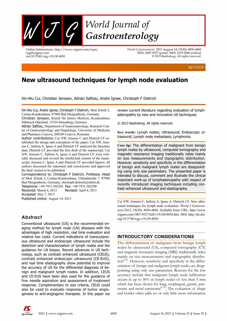
CarcinomaCarcinoma infiltration causes the development of patho-logical vessels (neoangiogenesis) and, therefore, a change of the perfusion pattern with heterogeneous enhance-ment due to the presence of caliber changes of the neo-plastic vessels and arteriovenous shunts[23-27]. Focal hy-poenhancement may result from the partial insufficiency of blood-supply due to overpressure in the LN caused by the neoplastic infiltration. Malignant lymph nodes not only have a greater number of peripheral vessels, but also longer contrast enhancement duration than benign lymph nodes[28]. Destructive avascular necroses are an impor-tant imaging sign for malignant infiltration (Figures 1-3). Avascular areas are detected by the lack of contrast agent uptake in the necrotic zones and the peripherally located pronounced hyperenhancement (rim enhancement)[29,30]. The contrast enhancement pattern of focal cortical
4851 August 14, 2013|Volume 19|Issue 30|WJG|www.wjgnet.com
Cui XW et al . Lymph node evaluation
Figure 1 Lymph node infiltration, carcinoma. A: With lymph node (LN) specific contrast agents malignant infiltration can be delineated (LKmg) as focal hypoen-hancement in the upper part of this perihepatic LN. The lower part (LKnx) shows normal (physiological) enhancement; B: With SonoVue®. Necrotic (non-enhancing, arrows) areas can be detected within this perihepatic lymph node. Necrotic areas are typically for carcinoma infiltration and tuberculosis. IVC: Inferior vena cava; Ao: Aorta; DHC: Common bile duct.
DHC
LKmgLKnx
IVCAo
A B
thickening has been also identified as an important sign to differentiate benign and malignant lymphadenopathy. In benign lymph nodes contrast enhancement within the cortex is homogeneous, whereas in malignant lymph the cortical thickening is less well vascularized than the adja-cent normal lymph node parenchyma[24].
In conclusion, criteria for carcinomatous lymph node infiltration on CEUS are centripetal inhomogeneous en-hancement and perfusion defects.
Lymphoma It is essential to consider lymphoma separately because some of its features are different from other LN dis-ease[13,31]. The very few studies published so far showed that in lymphoma contrast enhancement patterns are highly variable. The most often observed pattern is in-tense homogeneous enhancement, which is not different from reactive inflammatory lymph nodes[25,31] (Figure 4).
In conclusion, there is evidence that the vascular pat-tern of lymphomatous lymph node infiltration resembles that of non-malignant nodes.
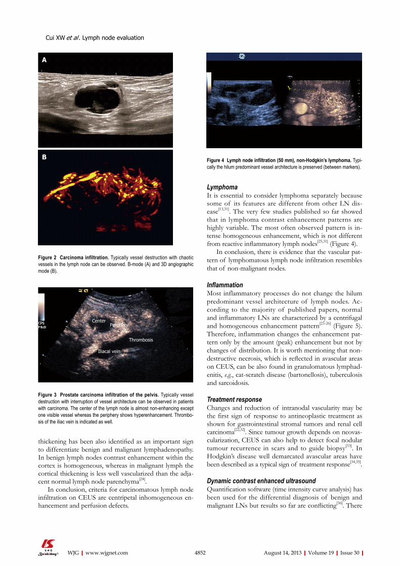
InflammationMost inflammatory processes do not change the hilum predominant vessel architecture of lymph nodes. Ac-cording to the majority of published papers, normal and inflammatory LNs are characterized by a centrifugal and homogeneous enhancement pattern[23-26] (Figure 5). Therefore, inflammation changes the enhancement pat-tern only by the amount (peak) enhancement but not by changes of distribution. It is worth mentioning that non-destructive necrosis, which is reflected in avascular areas on CEUS, can be also found in granulomatous lymphad-enitis, e.g., cat-scratch disease (bartonellosis), tuberculosis and sarcoidosis.
Treatment responseChanges and reduction of intranodal vascularity may be the first sign of response to antineoplastic treatment as shown for gastrointestinal stromal tumors and renal cell carcinoma[22,32]. Since tumour growth depends on neovas-cularization, CEUS can also help to detect focal nodular tumour recurrence in scars and to guide biopsy[33]. In Hodgkin’s disease well demarcated avascular areas have been described as a typical sign of treatment response[34,35].
Dynamic contrast enhanced ultrasoundQuantification software (time intensity curve analysis) has been used for the differential diagnosis of benign and malignant LNs but results so far are conflicting[36]. There
4852 August 14, 2013|Volume 19|Issue 30|WJG|www.wjgnet.com
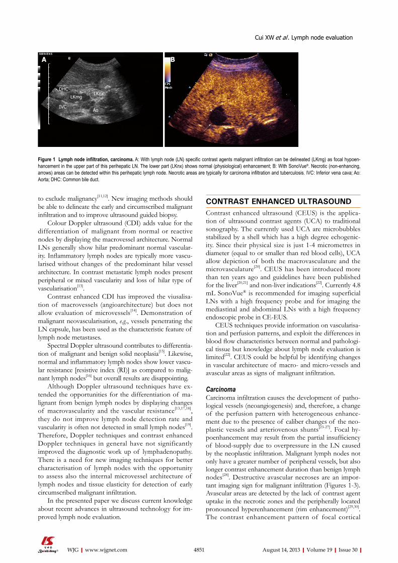
Figure 2 Carcinoma infiltration. Typically vessel destruction with chaotic vessels in the lymph node can be observed. B-mode (A) and 3D angiographic mode (B).
Figure 3 Prostate carcinoma infiltration of the pelvis. Typically vessel destruction with interruption of vessel architecture can be observed in patients with carcinoma. The center of the lymph node is almost non-enhancing except one visible vessel whereas the periphery shows hyperenhancement. Thrombo-sis of the iliac vein is indicated as well.
Figure 4 Lymph node infiltration (50 mm), non-Hodgkin’s lymphoma. Typi-cally the hilum predominant vessel architecture is preserved (between markers).
A
B
CenterPerphery
Thrombosis
Iliacal vein
Cui XW et al . Lymph node evaluation
4853 August 14, 2013|Volume 19|Issue 30|WJG|www.wjgnet.com
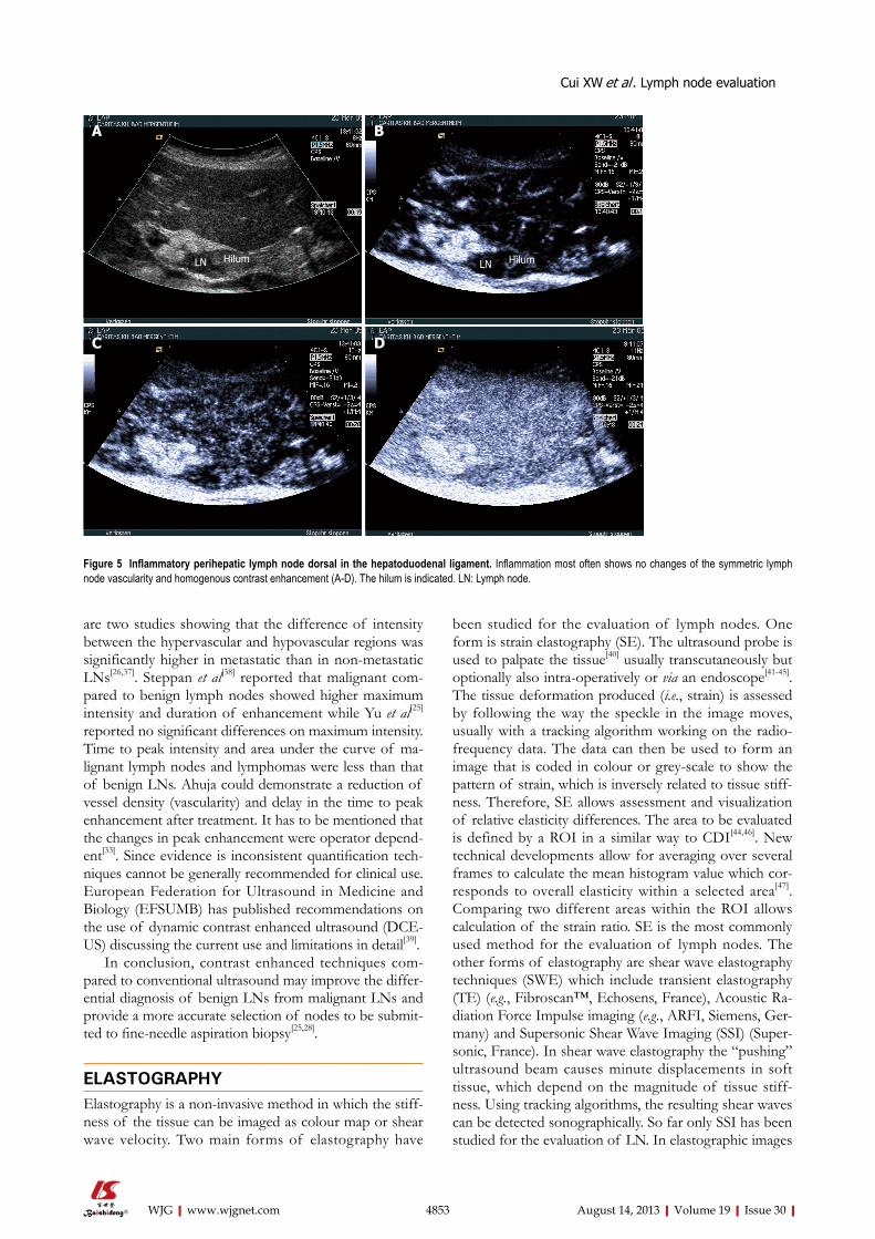
been studied for the evaluation of lymph nodes. One form is strain elastography (SE). The ultrasound probe is used to palpate the tissue[40] usually transcutaneously but optionally also intra-operatively or via an endoscope[41-45]. The tissue deformation produced (i.e., strain) is assessed by following the way the speckle in the image moves, usually with a tracking algorithm working on the radio-frequency data. The data can then be used to form an image that is coded in colour or grey-scale to show the pattern of strain, which is inversely related to tissue stiff-ness. Therefore, SE allows assessment and visualization of relative elasticity differences. The area to be evaluated is defined by a ROI in a similar way to CDI[44,46]. New technical developments allow for averaging over several frames to calculate the mean histogram value which cor-responds to overall elasticity within a selected area[47]. Comparing two different areas within the ROI allows calculation of the strain ratio. SE is the most commonly used method for the evaluation of lymph nodes. The other forms of elastography are shear wave elastography techniques (SWE) which include transient elastography (TE) (e.g., Fibroscan™, Echosens, France), Acoustic Ra-diation Force Impulse imaging (e.g., ARFI, Siemens, Ger-many) and Supersonic Shear Wave Imaging (SSI) (Super-sonic, France). In shear wave elastography the “pushing” ultrasound beam causes minute displacements in soft tissue, which depend on the magnitude of tissue stiff-ness. Using tracking algorithms, the resulting shear waves can be detected sonographically. So far only SSI has been studied for the evaluation of LN. In elastographic images
are two studies showing that the difference of intensity between the hypervascular and hypovascular regions was significantly higher in metastatic than in non-metastatic LNs[26,37]. Steppan et al[38] reported that malignant com-pared to benign lymph nodes showed higher maximum intensity and duration of enhancement while Yu et al[25] reported no significant differences on maximum intensity. Time to peak intensity and area under the curve of ma-lignant lymph nodes and lymphomas were less than that of benign LNs. Ahuja could demonstrate a reduction of vessel density (vascularity) and delay in the time to peak enhancement after treatment. It has to be mentioned that the changes in peak enhancement were operator depend-ent[33]. Since evidence is inconsistent quantification tech-niques cannot be generally recommended for clinical use. European Federation for Ultrasound in Medicine and Biology (EFSUMB) has published recommendations on the use of dynamic contrast enhanced ultrasound (DCE-US) discussing the current use and limitations in detail[39].
In conclusion, contrast enhanced techniques com-pared to conventional ultrasound may improve the differ-ential diagnosis of benign LNs from malignant LNs and provide a more accurate selection of nodes to be submit-ted to fine-needle aspiration biopsy[25,28].
ELASTOGRAPHYElastography is a non-invasive method in which the stiff-ness of the tissue can be imaged as colour map or shear wave velocity. Two main forms of elastography have
Figure 5 Inflammatory perihepatic lymph node dorsal in the hepatoduodenal ligament. Inflammation most often shows no changes of the symmetric lymph node vascularity and homogenous contrast enhancement (A-D). The hilum is indicated. LN: Lymph node.
A B
C D
LN Hilum LN Hilum
Cui XW et al . Lymph node evaluation
4854 August 14, 2013|Volume 19|Issue 30|WJG|www.wjgnet.com
of normal lymph nodes the nodal cortex is significantly harder than the medulla and the hilum[48,49].
EFSUMB has prepared recommendations on the use of elastography. In two sets of papers the techniques are explained in more detail[50,51].
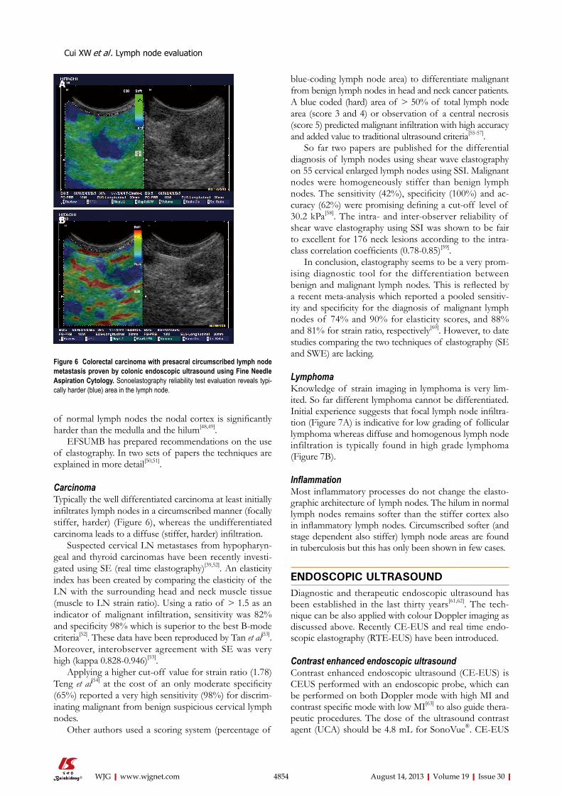
CarcinomaTypically the well differentiated carcinoma at least initially infiltrates lymph nodes in a circumscribed manner (focally stiffer, harder) (Figure 6), whereas the undifferentiated carcinoma leads to a diffuse (stiffer, harder) infiltration.
Suspected cervical LN metastases from hypopharyn-geal and thyroid carcinomas have been recently investi-gated using SE (real time elastography)[39,52]. An elasticity index has been created by comparing the elasticity of the LN with the surrounding head and neck muscle tissue (muscle to LN strain ratio). Using a ratio of > 1.5 as an indicator of malignant infiltration, sensitivity was 82% and specificity 98% which is superior to the best B-mode criteria[52]. These data have been reproduced by Tan et al[53]. Moreover, interobserver agreement with SE was very high (kappa 0.828-0.946)[53].
Applying a higher cut-off value for strain ratio (1.78) Teng et al[54] at the cost of an only moderate specificity (65%) reported a very high sensitivity (98%) for discrim-inating malignant from benign suspicious cervical lymph nodes.
Other authors used a scoring system (percentage of
blue-coding lymph node area) to differentiate malignant from benign lymph nodes in head and neck cancer patients. A blue coded (hard) area of > 50% of total lymph node area (score 3 and 4) or observation of a central necrosis (score 5) predicted malignant infiltration with high accuracy and added value to traditional ultrasound criteria[55-57].
So far two papers are published for the differential diagnosis of lymph nodes using shear wave elastography on 55 cervical enlarged lymph nodes using SSI. Malignant nodes were homogeneously stiffer than benign lymph nodes. The sensitivity (42%), specificity (100%) and ac-curacy (62%) were promising defining a cut-off level of 30.2 kPa[58]. The intra- and inter-observer reliability of shear wave elastography using SSI was shown to be fair to excellent for 176 neck lesions according to the intra-class correlation coefficients (0.78-0.85)[59].
In conclusion, elastography seems to be a very prom-ising diagnostic tool for the differentiation between benign and malignant lymph nodes. This is reflected by a recent meta-analysis which reported a pooled sensitiv-ity and specificity for the diagnosis of malignant lymph nodes of 74% and 90% for elasticity scores, and 88% and 81% for strain ratio, respectively[60]. However, to date studies comparing the two techniques of elastography (SE and SWE) are lacking.
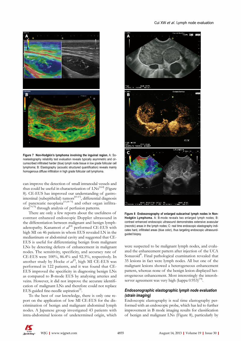
Lymphoma Knowledge of strain imaging in lymphoma is very lim-ited. So far different lymphoma cannot be differentiated. Initial experience suggests that focal lymph node infiltra-tion (Figure 7A) is indicative for low grading of follicular lymphoma whereas diffuse and homogenous lymph node infiltration is typically found in high grade lymphoma (Figure 7B).
InflammationMost inflammatory processes do not change the elasto-graphic architecture of lymph nodes. The hilum in normal lymph nodes remains softer than the stiffer cortex also in inflammatory lymph nodes. Circumscribed softer (and stage dependent also stiffer) lymph node areas are found in tuberculosis but this has only been shown in few cases.
ENDOSCOPIC ULTRASOUNDDiagnostic and therapeutic endoscopic ultrasound has been established in the last thirty years[61,62]. The tech-nique can be also applied with colour Doppler imaging as discussed above. Recently CE-EUS and real time endo-scopic elastography (RTE-EUS) have been introduced.
Contrast enhanced endoscopic ultrasoundContrast enhanced endoscopic ultrasound (CE-EUS) is CEUS performed with an endoscopic probe, which can be performed on both Doppler mode with high MI and contrast specific mode with low MI[63] to also guide thera-peutic procedures. The dose of the ultrasound contrast agent (UCA) should be 4.8 mL for SonoVue®. CE-EUS
Figure 6 Colorectal carcinoma with presacral circumscribed lymph node metastasis proven by colonic endoscopic ultrasound using Fine Needle Aspiration Cytology. Sonoelastography reliability test evaluation reveals typi-cally harder (blue) area in the lymph node.
A
B
Cui XW et al . Lymph node evaluation
4855 August 14, 2013|Volume 19|Issue 30|WJG|www.wjgnet.com
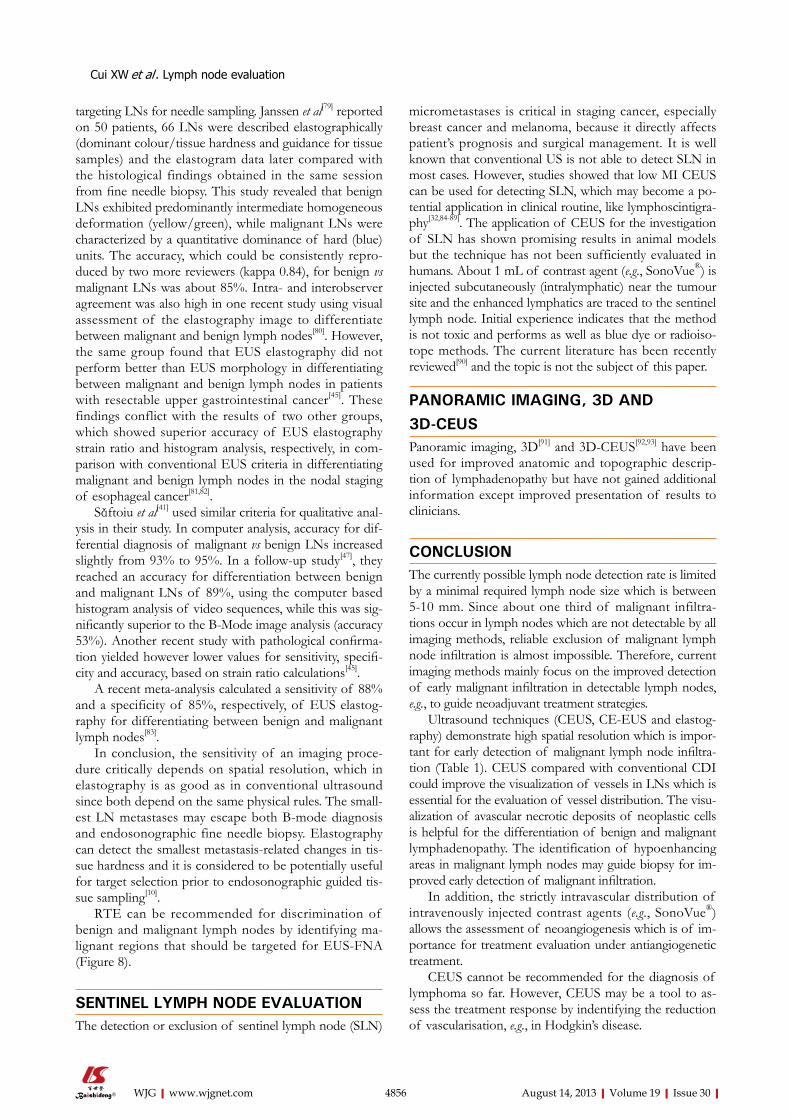
can improve the detection of small intranodal vessels and thus could be useful in characterization of LNs[3,64] (Figure 8). CE-EUS has improved our understanding of gastro-intestinal (subepithelial) tumors[65-67], differential diagnosis of pancreatic neoplasia[15,68-75] and other organ infiltra-tion[74,75] through analysis of perfusion patterns.
There are only a few reports about the usefulness of contrast enhanced endoscopic Doppler ultrasound in the differentiation between malignant and benign lymph-adenopathy. Kanamori et al[64] performed CE-EUS with high MI on 46 patients in whom EUS revealed LN in the mediastinum or abdominal cavity and suggested that CE-EUS is useful for differentiating benign from malignant LNs by detecting defects of enhancement in malignant nodes. The sensitivity, specificity, and accuracy rate of CE-EUS were 100%, 86.4% and 92.3%, respectively. In another study by Hocke et al[3], high MI CE-EUS was performed in 122 patients, and it was found that CE-EUS improved the specificity in diagnosing benign LNs as compared to B-mode EUS by analysing arteries and veins. However, it did not improve the accurate identifi-cation of malignant LNs and therefore could not replace EUS-guided fine-needle aspiration[3].
To the best of our knowledge, there is only one re-port on the application of low MI CE-EUS for the dis-crimination of benign and malignant abdominal lymph nodes. A Japanese group investigated 43 patients with intra-abdominal lesions of undetermined origin, which
were suspected to be malignant lymph nodes, and evalu-ated the enhancement pattern after injection of the UCA Sonazoid®. Final pathological examination revealed that 35 lesions in fact were lymph nodes. All but one of the malignant lesions showed a heterogeneous enhancement pattern, whereas none of the benign lesion displayed het-erogeneous enhancement. Most interestingly the interob-server agreement was very high (kappa 0.953)[78].
Endosonographic elastographic lymph node evaluation (strain imaging)Endoscopic elastography is real time elastography per-formed with an endoscopic probe, which has led to further improvement in B mode imaging results for classification of benign and malignant LNs (Figure 8), particularly by
Figure 8 Endosonography of enlarged subcarinal lymph nodes in Non-Hodgkin Lymphoma. A: B-mode reveals two enlarged lymph nodes; B: contrast enhanced endoscopic ultrasound demonstrates extensive avascular (necrotic) areas in the lymph nodes; C: real time endoscopic elastography indi-cates hard, infiltrated areas (blue color), thus targeting endoscopic ultrasound-guided biopsy.
A
B
C
Cui XW et al . Lymph node evaluation
Figure 7 Non-Hodgkin’s lymphoma involving the inguinal region. A: So-noelastography reliability test evaluation reveals typically asymmetric and cir-cumscribed infiltrated harder (blue) lymph node tissue in low grade follicular cell lymphoma; B: Elastography (acoustic structured quantification) reveals mainly homogenous diffuse infiltration in high grade follicular cell lymphoma.
A
B
4856 August 14, 2013|Volume 19|Issue 30|WJG|www.wjgnet.com
targeting LNs for needle sampling. Janssen et al[79] reported on 50 patients, 66 LNs were described elastographically (dominant colour/tissue hardness and guidance for tissue samples) and the elastogram data later compared with the histological findings obtained in the same session from fine needle biopsy. This study revealed that benign LNs exhibited predominantly intermediate homogeneous deformation (yellow/green), while malignant LNs were characterized by a quantitative dominance of hard (blue) units. The accuracy, which could be consistently repro-duced by two more reviewers (kappa 0.84), for benign vs malignant LNs was about 85%. Intra- and interobserver agreement was also high in one recent study using visual assessment of the elastography image to differentiate between malignant and benign lymph nodes[80]. However, the same group found that EUS elastography did not perform better than EUS morphology in differentiating between malignant and benign lymph nodes in patients with resectable upper gastrointestinal cancer[45]. These findings conflict with the results of two other groups, which showed superior accuracy of EUS elastography strain ratio and histogram analysis, respectively, in com-parison with conventional EUS criteria in differentiating malignant and benign lymph nodes in the nodal staging of esophageal cancer[81,82].
Sǎftoiu et al[41] used similar criteria for qualitative anal-ysis in their study. In computer analysis, accuracy for dif-ferential diagnosis of malignant vs benign LNs increased slightly from 93% to 95%. In a follow-up study[47], they reached an accuracy for differentiation between benign and malignant LNs of 89%, using the computer based histogram analysis of video sequences, while this was sig-nificantly superior to the B-Mode image analysis (accuracy 53%). Another recent study with pathological confirma-tion yielded however lower values for sensitivity, specifi-city and accuracy, based on strain ratio calculations[45].
A recent meta-analysis calculated a sensitivity of 88% and a specificity of 85%, respectively, of EUS elastog-raphy for differentiating between benign and malignant lymph nodes[83].
In conclusion, the sensitivity of an imaging proce-dure critically depends on spatial resolution, which in elastography is as good as in conventional ultrasound since both depend on the same physical rules. The small-est LN metastases may escape both B-mode diagnosis and endosonographic fine needle biopsy. Elastography can detect the smallest metastasis-related changes in tis-sue hardness and it is considered to be potentially useful for target selection prior to endosonographic guided tis-sue sampling[10].
RTE can be recommended for discrimination of benign and malignant lymph nodes by identifying ma-lignant regions that should be targeted for EUS-FNA (Figure 8).
SENTINEL LYMPH NODE EVALUATION The detection or exclusion of sentinel lymph node (SLN)
micrometastases is critical in staging cancer, especially breast cancer and melanoma, because it directly affects patient’s prognosis and surgical management. It is well known that conventional US is not able to detect SLN in most cases. However, studies showed that low MI CEUS can be used for detecting SLN, which may become a po-tential application in clinical routine, like lymphoscintigra-phy[32,84-89]. The application of CEUS for the investigation of SLN has shown promising results in animal models but the technique has not been sufficiently evaluated in humans. About 1 mL of contrast agent (e.g., SonoVue®) is injected subcutaneously (intralymphatic) near the tumour site and the enhanced lymphatics are traced to the sentinel lymph node. Initial experience indicates that the method is not toxic and performs as well as blue dye or radioiso-tope methods. The current literature has been recently reviewed[90] and the topic is not the subject of this paper.
PANORAMIC IMAGING, 3D AND 3D-CEUSPanoramic imaging, 3D[91] and 3D-CEUS[92,93] have been used for improved anatomic and topographic descrip-tion of lymphadenopathy but have not gained additional information except improved presentation of results to clinicians.
CONCLUSIONThe currently possible lymph node detection rate is limited by a minimal required lymph node size which is between 5-10 mm. Since about one third of malignant infiltra-tions occur in lymph nodes which are not detectable by all imaging methods, reliable exclusion of malignant lymph node infiltration is almost impossible. Therefore, current imaging methods mainly focus on the improved detection of early malignant infiltration in detectable lymph nodes, e.g., to guide neoadjuvant treatment strategies.
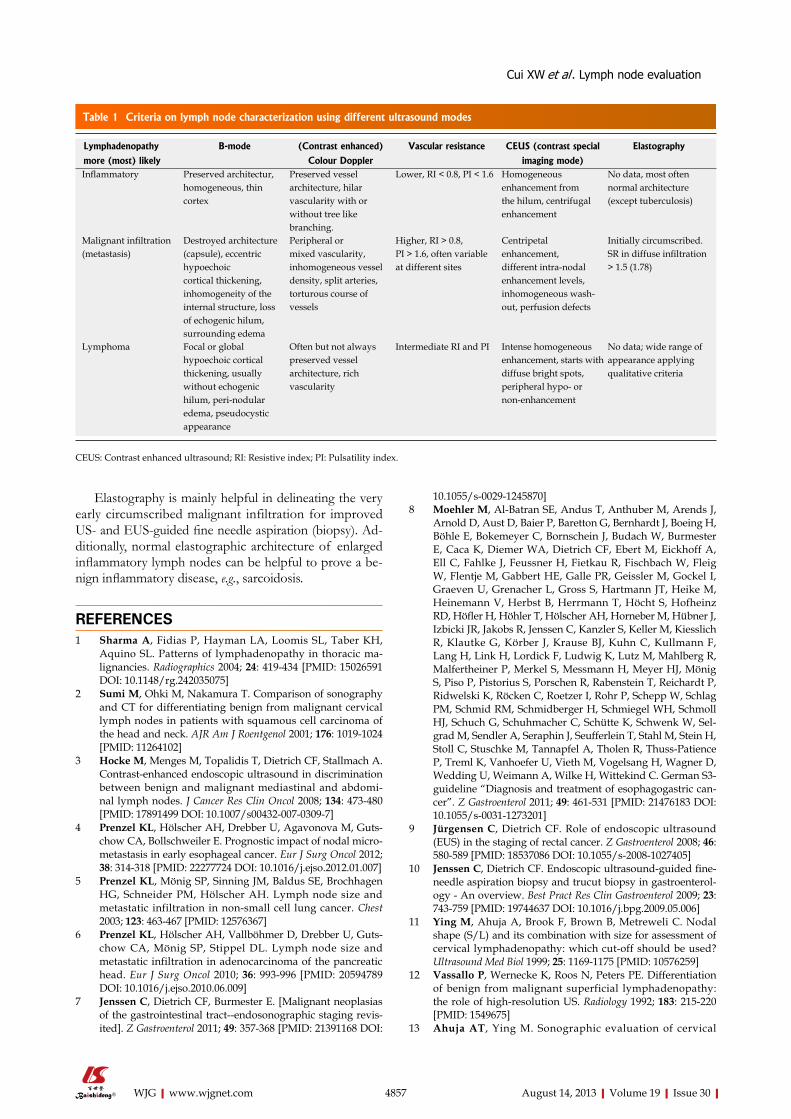
Ultrasound techniques (CEUS, CE-EUS and elastog-raphy) demonstrate high spatial resolution which is impor-tant for early detection of malignant lymph node infiltra-tion (Table 1). CEUS compared with conventional CDI could improve the visualization of vessels in LNs which is essential for the evaluation of vessel distribution. The visu-alization of avascular necrotic deposits of neoplastic cells is helpful for the differentiation of benign and malignant lymphadenopathy. The identification of hypoenhancing areas in malignant lymph nodes may guide biopsy for im-proved early detection of malignant infiltration.
In addition, the strictly intravascular distribution of intravenously injected contrast agents (e.g., SonoVue®) allows the assessment of neoangiogenesis which is of im-portance for treatment evaluation under antiangiogenetic treatment.
CEUS cannot be recommended for the diagnosis of lymphoma so far. However, CEUS may be a tool to as-sess the treatment response by indentifying the reduction of vascularisation, e.g., in Hodgkin’s disease.
Cui XW et al . Lymph node evaluation
4857 August 14, 2013|Volume 19|Issue 30|WJG|www.wjgnet.com
Elastography is mainly helpful in delineating the very early circumscribed malignant infiltration for improved US- and EUS-guided fine needle aspiration (biopsy). Ad-ditionally, normal elastographic architecture of enlarged inflammatory lymph nodes can be helpful to prove a be-nign inflammatory disease, e.g., sarcoidosis.
REFERENCES1 Sharma A, Fidias P, Hayman LA, Loomis SL, Taber KH,
Aquino SL. Patterns of lymphadenopathy in thoracic ma-lignancies. Radiographics 2004; 24: 419-434 [PMID: 15026591 DOI: 10.1148/rg.242035075]
2 Sumi M, Ohki M, Nakamura T. Comparison of sonography and CT for differentiating benign from malignant cervical lymph nodes in patients with squamous cell carcinoma of the head and neck. AJR Am J Roentgenol 2001; 176: 1019-1024 [PMID: 11264102]
3 Hocke M, Menges M, Topalidis T, Dietrich CF, Stallmach A. Contrast-enhanced endoscopic ultrasound in discrimination between benign and malignant mediastinal and abdomi-nal lymph nodes. J Cancer Res Clin Oncol 2008; 134: 473-480 [PMID: 17891499 DOI: 10.1007/s00432-007-0309-7]
4 Prenzel KL, Hölscher AH, Drebber U, Agavonova M, Guts-chow CA, Bollschweiler E. Prognostic impact of nodal micro-metastasis in early esophageal cancer. Eur J Surg Oncol 2012; 38: 314-318 [PMID: 22277724 DOI: 10.1016/j.ejso.2012.01.007]
5 Prenzel KL, Mönig SP, Sinning JM, Baldus SE, Brochhagen HG, Schneider PM, Hölscher AH. Lymph node size and metastatic infiltration in non-small cell lung cancer. Chest 2003; 123: 463-467 [PMID: 12576367]
6 Prenzel KL, Hölscher AH, Vallböhmer D, Drebber U, Guts-chow CA, Mönig SP, Stippel DL. Lymph node size and metastatic infiltration in adenocarcinoma of the pancreatic head. Eur J Surg Oncol 2010; 36: 993-996 [PMID: 20594789 DOI: 10.1016/j.ejso.2010.06.009]
7 Jenssen C, Dietrich CF, Burmester E. [Malignant neoplasias of the gastrointestinal tract--endosonographic staging revis-ited]. Z Gastroenterol 2011; 49: 357-368 [PMID: 21391168 DOI:
10.1055/s-0029-1245870]8 Moehler M, Al-Batran SE, Andus T, Anthuber M, Arends J,
Arnold D, Aust D, Baier P, Baretton G, Bernhardt J, Boeing H, Böhle E, Bokemeyer C, Bornschein J, Budach W, Burmester E, Caca K, Diemer WA, Dietrich CF, Ebert M, Eickhoff A, Ell C, Fahlke J, Feussner H, Fietkau R, Fischbach W, Fleig W, Flentje M, Gabbert HE, Galle PR, Geissler M, Gockel I, Graeven U, Grenacher L, Gross S, Hartmann JT, Heike M, Heinemann V, Herbst B, Herrmann T, Höcht S, Hofheinz RD, Höfler H, Höhler T, Hölscher AH, Horneber M, Hübner J, Izbicki JR, Jakobs R, Jenssen C, Kanzler S, Keller M, Kiesslich R, Klautke G, Körber J, Krause BJ, Kuhn C, Kullmann F, Lang H, Link H, Lordick F, Ludwig K, Lutz M, Mahlberg R, Malfertheiner P, Merkel S, Messmann H, Meyer HJ, Mönig S, Piso P, Pistorius S, Porschen R, Rabenstein T, Reichardt P, Ridwelski K, Röcken C, Roetzer I, Rohr P, Schepp W, Schlag PM, Schmid RM, Schmidberger H, Schmiegel WH, Schmoll HJ, Schuch G, Schuhmacher C, Schütte K, Schwenk W, Sel-grad M, Sendler A, Seraphin J, Seufferlein T, Stahl M, Stein H, Stoll C, Stuschke M, Tannapfel A, Tholen R, Thuss-Patience P, Treml K, Vanhoefer U, Vieth M, Vogelsang H, Wagner D, Wedding U, Weimann A, Wilke H, Wittekind C. German S3-guideline “Diagnosis and treatment of esophagogastric can-cer”. Z Gastroenterol 2011; 49: 461-531 [PMID: 21476183 DOI: 10.1055/s-0031-1273201]
9 Jürgensen C, Dietrich CF. Role of endoscopic ultrasound (EUS) in the staging of rectal cancer. Z Gastroenterol 2008; 46: 580-589 [PMID: 18537086 DOI: 10.1055/s-2008-1027405]
10 Jenssen C, Dietrich CF. Endoscopic ultrasound-guided fine-needle aspiration biopsy and trucut biopsy in gastroenterol-ogy - An overview. Best Pract Res Clin Gastroenterol 2009; 23: 743-759 [PMID: 19744637 DOI: 10.1016/j.bpg.2009.05.006]
11 Ying M, Ahuja A, Brook F, Brown B, Metreweli C. Nodal shape (S/L) and its combination with size for assessment of cervical lymphadenopathy: which cut-off should be used? Ultrasound Med Biol 1999; 25: 1169-1175 [PMID: 10576259]
12 Vassallo P, Wernecke K, Roos N, Peters PE. Differentiation of benign from malignant superficial lymphadenopathy: the role of high-resolution US. Radiology 1992; 183: 215-220 [PMID: 1549675]
13 Ahuja AT, Ying M. Sonographic evaluation of cervical
P- Reviewers Bener A S- Editor Wen LL L- Editor Cant MR E- Editor Li JY
P- Reviewers Bener A S- Editor Song XX L- Editor Stewart GJ E- Editor Li JY
Cui XW et al . Lymph node evaluation
Lymphadenopathy B-mode (Contrast enhanced) Vascular resistance CEUS (contrast special Elastography
more (most) likely Colour Doppler imaging mode) Inflammatory Preserved architectur,
homogeneous, thin cortex
Preserved vessel architecture, hilar vascularity with or without tree like branching.
Lower, RI < 0.8, PI < 1.6 Homogeneous enhancement from the hilum, centrifugal enhancement
No data, most often normal architecture (except tuberculosis)
Malignant infiltration (metastasis)
Destroyed architecture (capsule), eccentric hypoechoic cortical thickening, inhomogeneity of the internal structure, loss of echogenic hilum, surrounding edema
Peripheral or mixed vascularity, inhomogeneous vessel density, split arteries, torturous course of vessels
Higher, RI > 0.8, PI > 1.6, often variableat different sites
Centripetal enhancement, different intra-nodal enhancement levels, inhomogeneous wash-out, perfusion defects
Initially circumscribed. SR in diffuse infiltration > 1.5 (1.78)
Lymphoma Focal or global hypoechoic cortical thickening, usually without echogenic hilum, peri-nodular edema, pseudocystic appearance
Often but not always preserved vessel architecture, rich vascularity
Intermediate RI and PI Intense homogeneous enhancement, starts with diffuse bright spots, peripheral hypo- or non-enhancement
No data; wide range of appearance applying qualitative criteria
Table 1 Criteria on lymph node characterization using different ultrasound modes
CEUS: Contrast enhanced ultrasound; RI: Resistive index; PI: Pulsatility index.
4858 August 14, 2013|Volume 19|Issue 30|WJG|www.wjgnet.com
lymph nodes. AJR Am J Roentgenol 2005; 184: 1691-1699 [PMID: 15855141]
14 Schmid-Wendtner MH, Partscht K, Korting HC, Volke-nandt M. Improved differentiation of benign and malignant lymphadenopathy in patients with cutaneous melanoma by contrast-enhanced color Doppler sonography. Arch Dermatol 2002; 138: 491-497 [PMID: 11939811]
15 Hocke M, Schulze E, Gottschalk P, Topalidis T, Dietrich CF. Contrast-enhanced endoscopic ultrasound in discrimination between focal pancreatitis and pancreatic cancer. World J Gastroenterol 2006; 12: 246-250 [PMID: 16482625]
16 Ying M, Ahuja A. Sonography of neck lymph nodes. Part I: normal lymph nodes. Clin Radiol 2003; 58: 351-358 [PMID: 12727162]
17 Ahuja A, Ying M. Sonographic evaluation of cervical lymph-adenopathy: is power Doppler sonography routinely indicat-ed? Ultrasound Med Biol 2003; 29: 353-359 [PMID: 12706185]
18 Tschammler A, Heuser B, Ott G, Schmitt S, Hahn D. Patho-logical angioarchitecture in lymph nodes: underlying histo-pathologic findings. Ultrasound Med Biol 2000; 26: 1089-1097 [PMID: 11053743]
19 Moritz JD, Ludwig A, Oestmann JW. Contrast-enhanced color Doppler sonography for evaluation of enlarged cervi-cal lymph nodes in head and neck tumors. AJR Am J Roent-genol 2000; 174: 1279-1284 [PMID: 10789776]
20 Claudon M, Dietrich CF, Choi BI, Cosgrove DO, Kudo M, Nolsøe CP, Piscaglia F, Wilson SR, Barr RG, Chammas MC, Chaubal NG, Chen MH, Clevert DA, Correas JM, Ding H, Forsberg F, Fowlkes JB, Gibson RN, Goldberg BB, Lassau N, Leen EL, Mattrey RF, Moriyasu F, Solbiati L, Weskott HP, Xu HX. Guidelines and good clinical practice recommenda-tions for contrast enhanced ultrasound (CEUS) in the liver--update 2012: a WFUMB-EFSUMB initiative in cooperation with representatives of AFSUMB, AIUM, ASUM, FLAUS and ICUS. Ultraschall Med 2013; 34: 11-29 [PMID: 23129518 DOI: 10.1055/s-0032-1325499]
21 Claudon M, Dietrich CF, Choi BI, Cosgrove DO, Kudo M, Nolsøe CP, Piscaglia F, Wilson SR, Barr RG, Chammas MC, Chaubal NG, Chen MH, Clevert DA, Correas JM, Ding H, Forsberg F, Fowlkes JB, Gibson RN, Goldberg BB, Lassau N, Leen EL, Mattrey RF, Moriyasu F, Solbiati L, Weskott HP, Xu HX. Guidelines and good clinical practice recommenda-tions for Contrast Enhanced Ultrasound (CEUS) in the liver - update 2012: A WFUMB-EFSUMB initiative in cooperation with representatives of AFSUMB, AIUM, ASUM, FLAUS and ICUS. Ultrasound Med Biol 2013; 39: 187-210 [PMID: 23137926 DOI: 10.1016/j.ultrasmedbio.2012.09.002]
22 Piscaglia F, Nolsøe C, Dietrich CF, Cosgrove DO, Gilja OH, Bachmann Nielsen M, Albrecht T, Barozzi L, Bertolotto M, Catalano O, Claudon M, Clevert DA, Correas JM, D’Onofrio M, Drudi FM, Eyding J, Giovannini M, Hocke M, Ignee A, Jung EM, Klauser AS, Lassau N, Leen E, Mathis G, Saftoiu A, Seidel G, Sidhu PS, ter Haar G, Timmerman D, Weskott HP. The EFSUMB Guidelines and Recommendations on the Clin-ical Practice of Contrast Enhanced Ultrasound (CEUS): up-date 2011 on non-hepatic applications. Ultraschall Med 2012; 33: 33-59 [PMID: 21874631 DOI: 10.1055/s-0031-1281676]
23 Rubaltelli L, Khadivi Y, Tregnaghi A, Stramare R, Ferro F, Borsato S, Fiocco U, Adami F, Rossi CR. Evaluation of lymph node perfusion using continuous mode harmonic ultraso-nography with a second-generation contrast agent. J Ultra-sound Med 2004; 23: 829-836 [PMID: 15244307]
24 Rubaltelli L, Beltrame V, Tregnaghi A, Scagliori E, Frigo AC, Stramare R. Contrast-enhanced ultrasound for characterizing lymph nodes with focal cortical thickening in patients with cutaneous melanoma. AJR Am J Roentgenol 2011; 196: W8-12 [PMID: 21178038 DOI: 10.2214/AJR.10.4711]
25 Yu M, Liu Q, Song HP, Han ZH, Su HL, He GB, Zhou XD. Clinical application of contrast-enhanced ultrasonography in diagnosis of superficial lymphadenopathy. J Ultrasound Med
2010; 29: 735-740 [PMID: 20427785]26 Ouyang Q, Chen L, Zhao H, Xu R, Lin Q. Detecting metasta-
sis of lymph nodes and predicting aggressiveness in patients with breast carcinomas. J Ultrasound Med 2010; 29: 343-352 [PMID: 20194931]
27 Wan CF, Du J, Fang H, Li FH, Zhu JS, Liu Q. Enhance-ment patterns and parameters of breast cancers at contrast-enhanced US: correlation with prognostic factors. Radiology 2012; 262: 450-459 [PMID: 22282183 DOI: 10.1148/radi-ol.11110789]
28 Yang WT, Metreweli C, Lam PK, Chang J. Benign and ma-lignant breast masses and axillary nodes: evaluation with echo-enhanced color power Doppler US. Radiology 2001; 220: 795-802 [PMID: 11526284]
29 Sakaguchi T, Yamashita Y, Katahira K, Nishimura R, Baba Y, Arakawa A, Takahashi M, Yumoto E, Shinohara M. Differ-ential diagnosis of small round cervical lymph nodes: com-parison of power Doppler US with contrast-enhanced CT and pathologic results. Radiat Med 2001; 19: 119-125 [PMID: 11467378]
30 King AD, Tse GM, Ahuja AT, Yuen EH, Vlantis AC, To EW, van Hasselt AC. Necrosis in metastatic neck nodes: diagnos-tic accuracy of CT, MR imaging, and US. Radiology 2004; 230: 720-726 [PMID: 14990838 DOI: 10.1148/radiol.2303030157]
31 Nakase K, Yamamoto K, Hiasa A, Tawara I, Yamaguchi M, Shiku H. Contrast-enhanced ultrasound examination of lymph nodes in different types of lymphoma. Cancer Detect Prev 2006; 30: 188-191 [PMID: 16632242 DOI: 10.1016/j.cdp.2006.03.005]
32 Berho M, Oviedo M, Stone E, Chen C, Nogueras J, Weiss E, Sands D, Wexner S. The correlation between tumour regression grade and lymph node status after chemoradia-tion in rectal cancer. Colorectal Dis 2009; 11: 254-258 [PMID: 18513188 DOI: 10.1111/j.1463-1318.2008.01597.x]
33 Ahuja AT, Ying M, Ho SY, Antonio G, Lee YP, King AD, Wong KT. Ultrasound of malignant cervical lymph nodes. Cancer Imaging 2008; 8: 48-56 [PMID: 18390388 DOI: 10.1102/1470-7330.2008.0006]
34 Eich HT, Müller RP, Engenhart-Cabillic R, Lukas P, Schmid-berger H, Staar S, Willich N. Involved-node radiotherapy in early-stage Hodgkin’s lymphoma. Definition and guidelines of the German Hodgkin Study Group (GHSG). Strahlenther Onkol 2008; 184: 406-410 [PMID: 18956517]
35 Engert A, Eichenauer DA, Dreyling M. Hodgkin’s lym-phoma: ESMO clinical recommendations for diagnosis, treat-ment and follow-up. Ann Oncol 2009; 20 Suppl 4: 108-109 [PMID: 19454425 DOI: 10.1093/annonc/mdp144]
36 Ignee A, Jedrejczyk M, Schuessler G, Jakubowski W, Dietrich CF. Quantitative contrast enhanced ultrasound of the liver for time intensity curves-Reliability and potential sources of errors. Eur J Radiol 2010; 73: 153-158 [PMID: 19157739 DOI: 10.1016/j.ejrad.2008.10.016]
37 Rubaltelli L, Corradin S, Dorigo A, Tregnaghi A, Adami F, Rossi CR, Stramare R. Automated quantitative evaluation of lymph node perfusion on contrast-enhanced sonography. AJR Am J Roentgenol 2007; 188: 977-983 [PMID: 17377033 DOI: 10.2214/AJR.06.0562]
38 Steppan I, Reimer D, Müller-Holzner E, Marth C, Aigner F, Frauscher F, Frede T, Zeimet AG. Breast cancer in women: evaluation of benign and malignant axillary lymph nodes with contrast-enhanced ultrasound. Ultraschall Med 2010; 31: 63-67 [PMID: 20094979 DOI: 10.1055/s-0028-1109847]
39 Dietrich CF, Averkiou MA, Correas JM, Lassau N, Leen E, Piscaglia F. An EFSUMB introduction into Dynamic Con-trast-Enhanced Ultrasound (DCE-US) for quantification of tumour perfusion. Ultraschall Med 2012; 33: 344-351 [PMID: 22843433 DOI: 10.1055/s-0032-1313026]
40 Dietrich CF. Elastography, the new dimension in ultraso-nography. Praxis (Bern 1994) 2011; 100: 1533-1542 [PMID: 22161880 DOI: 10.1024/1661-8157/a000735]
Cui XW et al . Lymph node evaluation
4859 August 14, 2013|Volume 19|Issue 30|WJG|www.wjgnet.com
41 Săftoiu A, Vilmann P, Hassan H, Gorunescu F. Analysis of endoscopic ultrasound elastography used for characterisa-tion and differentiation of benign and malignant lymph nodes. Ultraschall Med 2006; 27: 535-542 [PMID: 17160759 DOI: 10.1055/s-2006-927117]
42 Janssen J. [(E)US elastography: current status and perspec-tives]. Z Gastroenterol 2008; 46: 572-579 [PMID: 18537085 DOI: 10.1055/s-2008-1027379]
43 Giovannini M, Thomas B, Erwan B, Christian P, Fabrice C, Benjamin E, Geneviève M, Paolo A, Pierre D, Robert Y, Walter S, Hanz S, Carl S, Christoph D, Pierre E, Jean-Luc VL, Jacques D, Peter V, Andrian S. Endoscopic ultrasound elastography for evaluation of lymph nodes and pancreatic masses: a multicenter study. World J Gastroenterol 2009; 15: 1587-1593 [PMID: 19340900]
44 Dietrich CF. Elastography Applications. Endo heute 2011; 24: 177-212
45 Larsen MH, Fristrup C, Hansen TP, Hovendal CP, Mortensen MB. Endoscopic ultrasound, endoscopic sonoelastography, and strain ratio evaluation of lymph nodes with histology as gold standard. Endoscopy 2012; 44: 759-766 [PMID: 22752891 DOI: 10.1055/s-0032-1309817]
46 Bachmann-Nielsen M, Săftoiu A.[Elastography - true or false?. Ultraschall Med 2011; 32: 5-7 [PMID: 21305435 DOI: 10.1055/s-0029-1246008]
47 Săftoiu A, Vilmann P, Ciurea T, Popescu GL, Iordache A, Hassan H, Gorunescu F, Iordache S. Dynamic analysis of EUS used for the differentiation of benign and malignant lymph nodes. Gastrointest Endosc 2007; 66: 291-300 [PMID: 17643702 DOI: 10.1016/j.gie.2006.12.039]
48 Wojcinski S, Dupont J, Schmidt W, Cassel M, Hillemanns P. Real-time ultrasound elastography in 180 axillary lymph nodes: elasticity distribution in healthy lymph nodes and prediction of breast cancer metastases. BMC Med Imaging 2012; 12: 35 [PMID: 23253859 DOI: 10.1186/1471-2342-12-35]
49 Wing-Han Yuen Q, Zheng YP, Huang YP, He JF, Chung-Wai Cheung J, Ying M. In-vitro Strain and Modulus Mea-surements in Porcine Cervical Lymph Nodes. Open Biomed Eng J 2011; 5: 39-46 [PMID: 21643424 DOI: 10.2174/1874120701105010039]
50 Bamber J, C1 Bamber J, Cosgrove D, Dietrich CF, Fromageau J, Bojunga J, Calliada F, Cantisani V, Correas JM, D'Onofrio M, Drakonaki EE, Fink M, Friedrich-Rust M, Gilja OH, Havre RF, Jenssen C, Klauser AS, Ohlinger R, Saftoiu A, Schaefer F, Sporea I, Piscaglia F. EFSUMB guidelines and recommenda-tions on the clinical use of ultrasound elastography. Part 1: Basic principles and technology. Ultraschall Med 2013; 34: 169-184 [PMID: 23558397 DOI: 10.1055/s-0033-1335205]
51 Cosgrove D, Piscaglia F, Bamber J, Bojunga J, Correas JM, Gilja OH, Klauser AS, Sporea I, Calliada F, Cantisani V, D'Onofrio M, Drakonaki EE, Fink M, Friedrich-Rust M, Fromageau J, Havre RF, Jenssen C, Ohlinger R, Săftoiu A, Schaefer F, Dietrich CF. EFSUMB guidelines and recommen-dations on the clinical use of ultrasound elastography. Part 2: Clinical applications. Ultraschall Med 2013; 34: 238-253 [PMID: 23605169 DOI: 10.1055/s-0033-1335375]
52 Lyshchik A, Higashi T, Asato R, Tanaka S, Ito J, Hiraoka M, Insana MF, Brill AB, Saga T, Togashi K. Cervical lymph node metastases: diagnosis at sonoelastography--initial ex-perience. Radiology 2007; 243: 258-267 [PMID: 17293571 DOI: 10.1148/radiol.2431052032]
53 Tan R, Xiao Y, He Q. Ultrasound elastography: Its poten-tial role in assessment of cervical lymphadenopathy. Acad Radiol 2010; 17: 849-855 [PMID: 20540909 DOI: 10.1016/j.acra.2010.03.014]
54 Teng DK, Wang H, Lin YQ, Sui GQ, Guo F, Sun LN. Value of ultrasound elastography in assessment of enlarged cervi-cal lymph nodes. Asian Pac J Cancer Prev 2012; 13: 2081-2085 [PMID: 22901174]
55 Ishibashi N, Yamagata K, Sasaki H, Seto K, Shinya Y, Ito
H, Shinozuka K, Yanagawa T, Onizawa K, Bukawa H. Real-time tissue elastography for the diagnosis of lymph node metastasis in oral squamous cell carcinoma. Ultrasound Med Biol 2012; 38: 389-395 [PMID: 22266228 DOI: 10.1016/j.ultrasmedbio.2011.12.004]
56 Choi JJ, Kang BJ, Kim SH, Lee JH, Jeong SH, Yim HW, Song BJ, Jung SS. Role of sonographic elastography in the differ-ential diagnosis of axillary lymph nodes in breast cancer. J Ultrasound Med 2011; 30: 429-436 [PMID: 21460142]
57 Taylor K, O’Keeffe S, Britton PD, Wallis MG, Treece GM, Housden J, Parashar D, Bond S, Sinnatamby R. Ultrasound elastography as an adjuvant to conventional ultrasound in the preoperative assessment of axillary lymph nodes in suspected breast cancer: a pilot study. Clin Radiol 2011; 66: 1064-1071 [PMID: 21835398 DOI: 10.1016/j.crad.2011.05.015]
58 Bhatia KS, Cho CC, Tong CS, Yuen EH, Ahuja AT. Shear wave elasticity imaging of cervical lymph nodes. Ultrasound Med Biol 2012; 38: 195-201 [PMID: 22178167 DOI: 10.1016/j.ultrasmedbio.2011.10.024]
59 Bhatia K, Tong CS, Cho CC, Yuen EH, Lee J, Ahuja AT. Reliability of shear wave ultrasound elastography for neck lesions identified in routine clinical practice. Ultraschall Med 2012; 33: 463-468 [PMID: 23070932 DOI: 10.1055/s-0032-1325330]
60 Ying L, Hou Y, Zheng HM, Lin X, Xie ZL, Hu YP. Real-time elastography for the differentiation of benign and malignant superficial lymph nodes: a meta-analysis. Eur J Radiol 2012; 81: 2576-2584 [PMID: 22138121 DOI: 10.1016/j.ejrad.2011.10.026]
61 Dietrich CF, Jenssen C. Evidence based endoscopic ultra-sound. Z Gastroenterol 2011; 49: 599-621 [PMID: 21544753 DOI: 10.1055/s-0029-1246021]
62 Dietrich CF, Hocke M, Jenssen C. Interventional endosonog-raphy. Ultraschall Med 2011; 32: 8-22, quiz 23-25 [PMID: 21305436 DOI: 10.1055/s-0029-1246017]
63 Dietrich CF. Contrast-enhanced low mechanical index endo-scopic ultrasound (CELMI-EUS). Endoscopy 2009; 41 Suppl 2: E43-E44 [PMID: 19288418 DOI: 10.1055/s-0028-1119491]
64 Kanamori A, Hirooka Y, Itoh A, Hashimoto S, Kawashima H, Hara K, Uchida H, Goto J, Ohmiya N, Niwa Y, Goto H. Usefulness of contrast-enhanced endoscopic ultrasonogra-phy in the differentiation between malignant and benign lymphadenopathy. Am J Gastroenterol 2006; 101: 45-51 [PMID: 16405532 DOI: 10.1111/j.1572-0241.2006.00394.x]
65 Sakamoto H, Kitano M, Matsui S, Kamata K, Komaki T, Imai H, Dote K, Kudo M. Estimation of malignant potential of GI stromal tumors by contrast-enhanced harmonic EUS (with videos). Gastrointest Endosc 2011; 73: 227-237 [PMID: 21295636 DOI: 10.1016/j.gie.2010.10.011]
66 Kannengiesser K, Mahlke R, Petersen F, Peters A, Ross M, Kucharzik T, Maaser C. Contrast-enhanced harmonic endo-scopic ultrasound is able to discriminate benign submucosal lesions from gastrointestinal stromal tumors. Scand J Gastro-enterol 2012; 47: 1515-1520 [PMID: 23148660 DOI: 10.3109/00365521.2012.729082]
67 Dietrich CF, Jenssen C, Hocke M, Cui XW, Woenckhaus M, Ignee A. Imaging of gastrointestinal stromal tumours with modern ultrasound techniques - a pictorial essay. Z Gas-troenterol 2012; 50: 457-467 [PMID: 22581701 DOI: 10.1055/s-0031-1282076]
68 Gong TT, Hu DM, Zhu Q. Contrast-enhanced EUS for differ-ential diagnosis of pancreatic mass lesions: a meta-analysis. Gastrointest Endosc 2012; 76: 301-309 [PMID: 22703697 DOI: 10.1016/j.gie.2012.02.051]
69 Napoleon B, Alvarez-Sanchez MV, Gincoul R, Pujol B, Le-fort C, Lepilliez V, Labadie M, Souquet JC, Queneau PE, Scoazec JY, Chayvialle JA, Ponchon T. Contrast-enhanced harmonic endoscopic ultrasound in solid lesions of the pan-creas: results of a pilot study. Endoscopy 2010; 42: 564-570 [PMID: 20593334 DOI: 10.1055/s-0030-1255537]
Cui XW et al . Lymph node evaluation
4860 August 14, 2013|Volume 19|Issue 30|WJG|www.wjgnet.com
70 Kitano M, Kudo M, Yamao K, Takagi T, Sakamoto H, Ko-maki T, Kamata K, Imai H, Chiba Y, Okada M, Murakami T, Takeyama Y. Characterization of small solid tumors in the pancreas: the value of contrast-enhanced harmonic endo-scopic ultrasonography. Am J Gastroenterol 2012; 107: 303-310 [PMID: 22008892 DOI: 10.1038/ajg.2011.354]
71 Reddy NK, Ioncică AM, Săftoiu A, Vilmann P, Bhutani MS. Contrast-enhanced endoscopic ultrasonography. World J Gastroenterol 2011; 17: 42-48 [PMID: 21218082 DOI: 10.3748/wjg.v17.i1.42]
72 Săftoiu A, Vilmann P, Gorunescu F, Janssen J, Hocke M, Larsen M, Iglesias-Garcia J, Arcidiacono P, Will U, Giovanni-ni M, Dietrich C, Havre R, Gheorghe C, McKay C, Gheonea DI, Ciurea T. Accuracy of endoscopic ultrasound elastogra-phy used for differential diagnosis of focal pancreatic mass-es: a multicenter study. Endoscopy 2011; 43: 596-603 [PMID: 21437851 DOI: 10.1055/s-0030-1256314]
73 Săftoiu A, Dietrich CF, Vilmann P. Contrast-enhanced har-monic endoscopic ultrasound. Endoscopy 2012; 44: 612-617 [PMID: 22528674 DOI: 10.1055/s-0032-1308909]
74 Hocke M, Ignee A, Topalidis T, Stallmach A, Dietrich CF. Contrast-enhanced endosonographic Doppler spectrum analysis is helpful in discrimination between focal chronic pancreatitis and pancreatic cancer. Pancreas 2007; 35: 286-288 [PMID: 17895854 DOI: 10.1097/MPA.0b013e318093f964]
75 Dietrich CF, Ignee A, Braden B, Barreiros AP, Ott M, Hocke M. Improved differentiation of pancreatic tumors using contrast-enhanced endoscopic ultrasound. Clin Gastroenterol Hepatol 2008; 6: 590-597.e1 [PMID: 18455699 DOI: 10.1016/j.cgh.2008.02.030]
76 Park CH, Chung MJ, Oh TG, Park JY, Bang S, Park SW, Kim H, Hwang HK, Lee WJ, Song SY. Differential diagnosis be-tween gallbladder adenomas and cholesterol polyps on con-trast-enhanced harmonic endoscopic ultrasonography. Surg Endosc 2013; 27: 1414-1421 [PMID: 23233003 DOI: 10.1007/s00464-012-2620-x]
77 Romagnuolo J, Hoffman B, Vela S, Hawes R, Vignesh S. Ac-curacy of contrast-enhanced harmonic EUS with a second-generation perflutren lipid microsphere contrast agent (with video). Gastrointest Endosc 2011; 73: 52-63 [PMID: 21184870 DOI: 10.1016/j.gie.2010.09.014]
78 Xia Y, Kitano M, Kudo M, Imai H, Kamata K, Sakamoto H, Komaki T. Characterization of intra-abdominal lesions of undetermined origin by contrast-enhanced harmonic EUS (with videos). Gastrointest Endosc 2010; 72: 637-642 [PMID: 20646696 DOI: 10.1016/j.gie.2010.04.013]
79 Janssen J, Dietrich CF, Will U, Greiner L. Endosonographic elastography in the diagnosis of mediastinal lymph nodes. Endoscopy 2007; 39: 952-957 [PMID: 18008203 DOI: 10.1055/s-2007-966946]
80 Larsen MH, Fristrup CW, Mortensen MB. Intra- and interob-server agreement of endoscopic sonoelastography in the evaluation of lymph nodes. Ultraschall Med 2011; 32 Suppl 2: E45-E50 [PMID: 22194049 DOI: 10.1055/s-0031-1273493]
81 Paterson S, Duthie F, Stanley AJ. Endoscopic ultrasound-guided elastography in the nodal staging of oesophageal cancer. World J Gastroenterol 2012; 18: 889-895 [PMID:
22408347 DOI: 10.3748/wjg.v18.i9.889]82 Knabe M, Günter E, Ell C, Pech O. Can EUS elastography
improve lymph node staging in esophageal cancer? Surg Endosc 2013; 27: 1196-1202 [PMID: 23093233 DOI: 10.1007/s00464-012-2575-y]
83 Xu W, Shi J, Zeng X, Li X, Xie WF, Guo J, Lin Y. EUS elas-tography for the differentiation of benign and malignant lymph nodes: a meta-analysis. Gastrointest Endosc 2011; 74: 1001-1009; quiz 1115.e1-4 [PMID: 22032315 DOI: 10.1016/j.gie.2011.07.026]
84 Omoto K, Matsunaga H, Take N, Hozumi Y, Takehara M, Omoto Y, Shiozawa M, Mizunuma H, Harashima H, Tani-guchi N, Kawano M. Sentinel node detection method using contrast-enhanced ultrasonography with sonazoid in breast cancer: preliminary clinical study. Ultrasound Med Biol 2009; 35: 1249-1256 [PMID: 19520493 DOI: 10.1016/j.ultrasmedbio.2009.02.004]
85 De Giorgi V, Gori A, Grazzini M, Rossari S, Marino G, D’Elia G, Crocetti E, Roselli G, Innocenti P, Dini M, Lotti T. Contrast-enhanced ultrasound: a filter role in AJCC stage I/II melanoma patients. Oncology 2010; 79: 370-375 [PMID: 21430406 DOI: 10.1159/000323494]
86 Sever A, Jones S, Cox K, Weeks J, Mills P, Jones P. Preopera-tive localization of sentinel lymph nodes using intradermal microbubbles and contrast-enhanced ultrasonography in pa-tients with breast cancer. Br J Surg 2009; 96: 1295-1299 [PMID: 19847869 DOI: 10.1002/bjs.6725]
87 Sever AR, Mills P, Weeks J, Jones SE, Fish D, Jones PA, Mali W. Preoperative needle biopsy of sentinel lymph nodes using intradermal microbubbles and contrast-enhanced ultrasound in patients with breast cancer. AJR Am J Roentgenol 2012; 199: 465-470 [PMID: 22826414 DOI: 10.2214/AJR.11.7702]
88 Sever AR, Mills P, Jones SE, Mali W, Jones PA. Sentinel node identification using microbubbles and contrast-enhanced ul-trasonography. Clin Radiol 2012; 67: 687-694 [PMID: 22226568 DOI: 10.1016/j.crad.2011.11.009]
89 Sever AR, Mills P, Jones SE, Cox K, Weeks J, Fish D, Jones PA. Preoperative sentinel node identification with ultra-sound using microbubbles in patients with breast cancer. AJR Am J Roentgenol 2011; 196: 251-256 [PMID: 21257873 DOI: 10.2214/AJR.10.4865]
90 Cui XW, Ignee A, Nielsen MB, Schreiber-Dietrich D, De Molo C, Pirri C, Jedrzejczyk M, Dietrich CF. Contrast en-hanced ultrasound of sentinel lymph nodes. J Ultrason 2013; 13: 73-81
91 Bialek EJ, Jakubowski W, Szczepanik AB, Maryniak RK, Bil-ski R, Prochorec-Sobieszek M, Serafin-Krol M. 3D ultrasound examination of the superficial lymph nodes--does it provide additional information? Ultraschall Med 2006; 27: 467-472 [PMID: 17033947 DOI: 10.1055/s-2006-927064]
92 Dietrich CF. 3D real time contrast enhanced ultrasonography,a new technique. Rofo 2002; 174: 160-163 [PMID: 11898076 DOI: 10.1055/s-2002-20102]
93 Hocke M, Dietrich CF. New technology--combined use of 3D contrast enhanced endoscopic ultrasound techniques. Ultraschall Med 2011; 32: 317-318 [PMID: 21667410 DOI: 10.1055/s-0031-1274695]
P- Reviewers Levent D, Korpanty G S- Editor Song XX L- Editor A E- Editor Li JY
Cui XW et al . Lymph node evaluation

Baishideng Publishing Group Co., Limited © 2013 Baishideng. All rights reserved.
Published by Baishideng Publishing Group Co., LimitedFlat C, 23/F., Lucky Plaza,
315-321 Lockhart Road, Wan Chai, Hong Kong, ChinaFax: +852-65557188
Telephone: +852-31779906E-mail: [email protected]
http://www.wjgnet.com
I S S N 1 0 0 7 - 9 3 2 7
9 7 7 1 0 07 9 3 2 0 45
3 0