protective effect of calcium nanophosphate and cpp-acp
TRANSCRIPT
Cariology
Braz Oral Res., (São Paulo) 2013 Nov-Dec;27(6):463-70 463
Fabíola Galbiatti de Carvalho(a)
Veruska Lima Moura Brasil(b)
Tiago João da Silva Filho(b)
Hugo Lemes Carlo(b)
Rogério Lacerda dos Santos(a) Bruno Alessandro Silva Guedes de Lima(c)
(a) Department of Biological Science, Division of Dentistry, Universidade Federal de Campina Grande - UFCG, Patos, PB, Brazil.
(b) Department of Operative Dentistry, Centro de Ciências da Saúde, Universidade Federal da Paraíba - UFPB, João Pessoa, PB, Brazil.
(c) Department of Mechanical Technology, Laboratório de Solidificação Rápida, Universidade Federal da Paraíba - UFPB, João Pessoa, PB, Brazil.
Corresponding Author: Fabíola Galbiatti de Carvalho Email: [email protected]
Protective effect of calcium nanophosphate and CPP-ACP agents on enamel erosion
Abstract: The aim of this study was to assess the effect of differ-ent remineralizing agents on enamel microhardness (KHN) and sur-face topography after an erosive challenge. Forty-eight human enamel specimens (4 × 4 mm) were randomly assigned to 4 groups: control (no treatment), fluoride varnish, calcium nanophosphate paste and ca-sein phosphopeptide–amorphous calcium phosphate paste (CPP-ACP). Both pastes were applied for 5 minutes, and fluoride varnish, for 24 h. Four daily erosive cycles of 5 minutes of immersion in a cola drink and 2 h in artificial saliva were conducted for 5 days. KHN readings were performed at baseline and after 5 days. The percentage of enamel hardness change (%KHN) was obtained after erosion. The surface to-pography was evaluated by atomic force microscopy (AFM). The data were tested using ANOVA, Tukey’s and paired-T tests (p < 0.05). Af-ter an erosive challenge, there was no statistically significant difference between the control (96.8 ± 11.4 KHN / 72.4 ± 3.0 %KHN) and the varnish (91.7 ± 14.1 KHN / 73.4 ± 5.5 %KHN) groups. The nano-phosphate group showed lower enamel hardness loss (187.2 ± 27.9 / 49.0 ± 7.9 %KHN), compared with the CPP-ACP group (141.8 ± 16.5 / 60.6 ± 4.0 %KHN), and both were statistically different from the var-nish and the control groups. AFM images showed a rough surface for the control and the varnish groups, a non-homogeneous layer with globular irregularities for CPP-ACP, and a thick homogeneous layer for the nano-phosphate group. None of the agents provided protection against the de-velopment of erosion; however, nanophosphate paste was able to reduce enamel surface softening after the erosive challenge.
Descriptors: Tooth Erosion; Fluorides; Microscopy, Atomic Force.
IntroductionDental erosion is defined as the loss of tooth substance due to chemi-
cal processes not involving bacteria.1 Although erosive lesions have a multifactorial etiology, the increasing consumption of acidic food and soft drinks has become an important factor driving their development.2-4
Many strategies have been developed for the prevention and treatment of erosion, and fluoride is the main agent used to enhance enamel rem-ineralization.5,6 However, to control mineral loss caused by erosion, high concentrations and frequency seem to be needed, because the protective effect of fluoride against demineralization depends on the pH level, F
Declaration of Interests: The authors certify that they have no commercial or associative interest that represents a conflict of interest in connection with the manuscript.
Submitted: Feb 20, 2013 Accepted for publication: Aug 12, 2013 Last revision: Aug 26, 2013
http://dx.doi.org/10.1590/S1806-83242013000600004
Protective effect of calcium nanophosphate and CPP-ACP agents on enamel erosion
464 Braz Oral Res., (São Paulo) 2013 Nov-Dec;27(6):463-70
concentration, type of F salt and presence of mineral ions (Ca and P) in the saliva and in the agents.5-7 So-dium fluoride varnishes have been used due to their ability to adhere to the tooth surface and their high fluoride concentration, which increases the forma-tion of calcium fluoride (CaF2) deposits that act as fluoride reservoirs.4-6
In recent years, other agents for inhibiting erosion have been investigated, such as casein phosphopep-tides with amorphous calcium phosphate complex (CPP-ACP).8-10 CPP-ACP complex may increase the level of calcium and inorganic phosphate ions at the tooth surface, thereby permitting immediate enamel surface remineralization.8-10 Additionally, there have been advancements in nanotechnological develop-ments of products for the remineralization of enam-el, such as nano-hydroxyapatite (HA).11 However, HA has been studied only as a biomimetic material to remineralize enamel carious lesions,11 whereas its effect on eroded enamel has not been investigated.
Calcium nanophosphate organized in the crystal-line form of HA was recently developed as a paste. Calcium nanophosphate crystals are smaller than 100 nm, leading to improved bioactivity of the prod-uct, resulting from the increase in surface area and wettability of HA nanoparticles (manufacturer’s in-formation). Calcium, phosphate and fluoride ions are released and organized in fluorapatite and CaF2 on demineralized tooth surfaces. Based on these mecha-nisms, it would be interesting to evaluate the effect of this bioactive material on erosion prevention, in comparison with other remineralizing agents.
The aim of this study was to investigate the in vi-tro effect of fluoride varnish, calcium nanophosphate HA paste and CPP-ACP paste on preventing the de-velopment of enamel erosion. The null hypothesis tested was that there would be no difference between the effects of fluoride varnish and of remineralizing pastes on enamel microhardness and surface topog-raphy after an in vitro erosive challenge.
MethodologySpecimen preparation
After obtaining approval from the Universidade Federal de Campina Grande - UFCG Research Eth-ics Committee (11/2011), forty sound human third
molars were selected for this study. The teeth were stored in 0.1% thymol at 4°C and used within 1 month after extraction. Two enamel specimens (4 × 4 × 2 mm) were obtained from the buccal and lingual surfaces of each tooth, using a water-cooled low-speed diamond saw (Isomet; Buehler Ltd., Lake Bluff, USA). Next, they were embedded in acrylic res-in (Vipi Flash, Pirassununga, Brazil), and the enamel surfaces were ground flat with SiC paper discs (400, 600 and 1200 grades) and polished with 1 µm alu-mina suspension (Erios Corp., São Paulo, Brazil). Af-terwards, the baseline hardness of the enamel surface was determined and specimens with a KHN between 300 and 380 were selected12,13 and randomly divided into 4 groups (n = 12), according to the agent ap-plied: • control, no treatment; •fluoride varnish (Duraphat, Colgate, São Paulo,
Brazil); • calcium nanophosphate paste (Desensibilize
Nano P, FGM Produtos Odontológicos, Joinville, Brazil) and
•CPP-ACP paste (MI Paste Plus, GC America Inc., Alsip, USA; Table 1).
Treatment Before exposure to acid, 0.3 mL of each agent
was inserted into an insulin syringe (BD Ultra-fine, Franklin Lakes, USA) to standardize the volume of product applied to the enamel surface. The vol-ume of 20 µL of all agents was sufficient to cover the surface of the specimens. Thus, according to each group, 20 µL of the agent was applied to the
Table 1 - Composition of remineralizing agents.
Agent Composition (batch number)
MI Paste Plus(GC America Inc.)
Water, glycerol, CPP-ACP, D-sorbitol, CMC propylene glycol, silicone and titanium dioxide, xylitol, phosphoric acid, flavor, sodium saccharin, ethyl propyl butyl p-hydroxybenzoate, 900 ppm sodium fluoride (090813M)
Desensibilize Nano P (FGM)
Calcium nanophosphate organized in crystalline form of hydroxyapatite, potassium nitrate, water, surfactant, tensoactive, flavor, 9000 ppm sodium fluoride (170610)
Duraphat(Colgate)
Alcohol, natural resins, wax, saccharine, flavor, 22600 ppm sodium fluoride (P008203LA)
Carvalho FG, Brasil VLM, Silva Filho TJ, Carlo HL, Santos RL, Lima BASG
465Braz Oral Res., (São Paulo) 2013 Nov-Dec;27(6):463-70
readout, at least 50 µm apart, and their average rep-resented the specimen KHN value. In addition, the percentage of hardness loss (%KHN change) was calculated using the following formula:
% = 100 (KHN(I) – KHN(F)) / KHN(I),
where KHN(I) is the average of the initial hard-ness measurements and KHN(F) is the average of the final hardness values.12
Atomic force microscopyAFM is an important tool for obtaining a source
of new structural information.19 Three randomized specimens of each group were analyzed using atom-ic force microscopy (AFM; SPM-9600, Shimadzu Corp., Kyoto, Japan). Each specimen was fixed to the microscope holder on a stub (2 × 3 mm). The block surface morphology was probed in ‘‘contact mode.’’ Imaging was performed with a standard ge-ometry silicon nitride Micro-Cantilever (OMCL-TR, Olympus, Tokyo, Japan) and probed with 0.15 N/m constant elastic and 24 kHz resonant frequency. Im-ages 30 µm x 30 µm with resolution of 512 x 512 pixels and an operating point of 1.5 V were collected at a very low scan rate to obtain details of the enam-el structure and to avoid damaging the tip.
Statistical analysisData analysis was performed with the Graph-
Pad Instat version 2.0 (GraphPad software, La Jol-la, USA) at a level of significance of α = 0.05. Since all the variables tested satisfied the assumptions of equality and normal distribution, one-way ANOVA and Tukey’s test were carried out for statistical com-parisons of enamel hardness change percentages. The paired t-test was used to compare the enamel hardness before and after the erosive challenge in the same group.
ResultsTable 2 shows the following KHN values for all
groups: • initial, •after erosion, and • %KHN change.
specimens, in accordance with the manufacturer’s instructions: 1. Varnish was applied and specimens were stored
in artificial saliva. After 24 h, cotton tips were immersed in deionized water and applied to the surface to soften the varnish. The varnish was removed with a scalpel blade,12 taking care to avoid touching the surface;
2. Desensibilize Nano P paste was rubbed on with a microbrush for 10 s. After this, the paste was kept in contact with the enamel for 5 minutes and removed with deionized water;
3. MI Paste Plus was applied and kept in contact with the enamel for 5 minutes and then removed with deionized water. Paste application time was standardized at 5 minutes.
Erosive challenge and treatmentSpecimens were immersed in a cola drink (Coca-
Cola, Jundiaí, Brazil; pH 2.3), using separate con-tainers (10 mL/specimen), at room temperature, for 5 minutes 4 times/day.14 The specimens were rinsed thoroughly with deionized water and immersed in artificial saliva, pH 7.0 (10 mL/block) at room tem-perature for 2 h, between erosive challenges14 and overnight. The artificial saliva was made according to the Moretto et al.14 study. This erosive challenge was repeated for 5 days. The cola drink and arti-ficial saliva were changed after every cycle. During the demineralization cycles, the specimens were kept in hermetically sealed containers to prevent the loss of gas, which can increase the pH.15 The enamel specimens were exposed to one of the pastes imme-diately after each erosive challenge.14 The fluoride varnish was applied only once, just before the ero-sive challenge, to simulate a clinical situation.12,16,17
Hardness assessmentMicrohardness is the most useful method of as-
sessing enamel softening caused by erosion.18 Enam-el hardness (KHN) was measured with a Knoop microhardness diamond applied under a 25 g load for 10 s (HMV II; Shimadzu Corporation, Kyoto, Japan) at baseline and after 5 days of erosive chal-lenge. All readouts were performed by the same examiner. Five indentations were made in each

Protective effect of calcium nanophosphate and CPP-ACP agents on enamel erosion
466 Braz Oral Res., (São Paulo) 2013 Nov-Dec;27(6):463-70
No statistically significant differences were ob-served among the groups for initial KHN values (p = 0.87). After erosion, all groups showed lower KHN values (p < 0.05), in comparison with the ini-tial values.
After the erosive challenge, there was no statisti-cally significant difference between the control and varnish groups (p = 0.79), which showed the high-est KHN loss values. Furthermore, the control and varnish groups showed a statistically significant dif-ference in the remineralizing pastes used (p < 0.05). The nanophosphate group showed a lower KHN loss value, compared with the CPP-ACP group, after the erosive challenge (p < 0.05). There was a significant amount of protection against deminer-alization produced by nanophosphate paste, rep-resented by the highest KHN value after erosion, compared with the CPP-ACP, the varnish and the control groups (p < 0.001). The varnish and control groups showed no statistically significant difference with regard to %KHN change (p = 0.75).
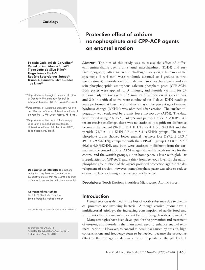
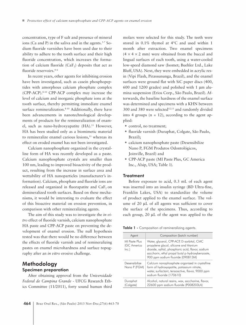
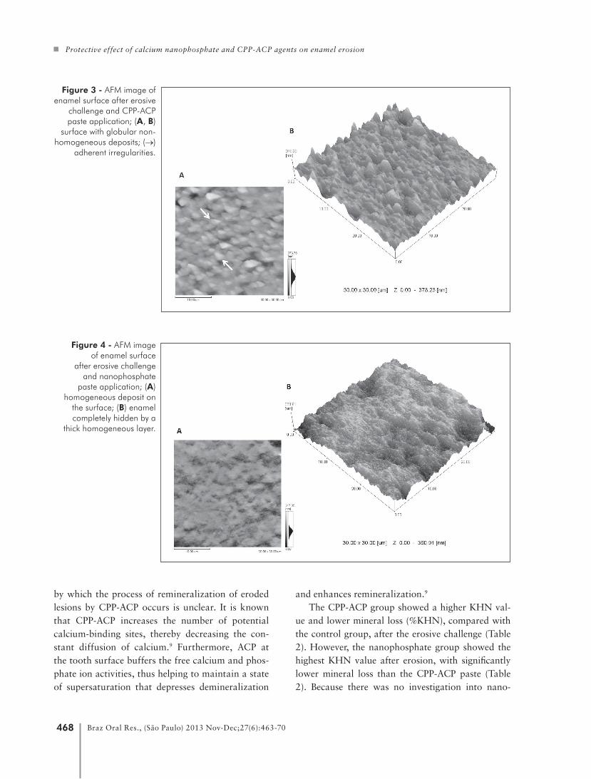
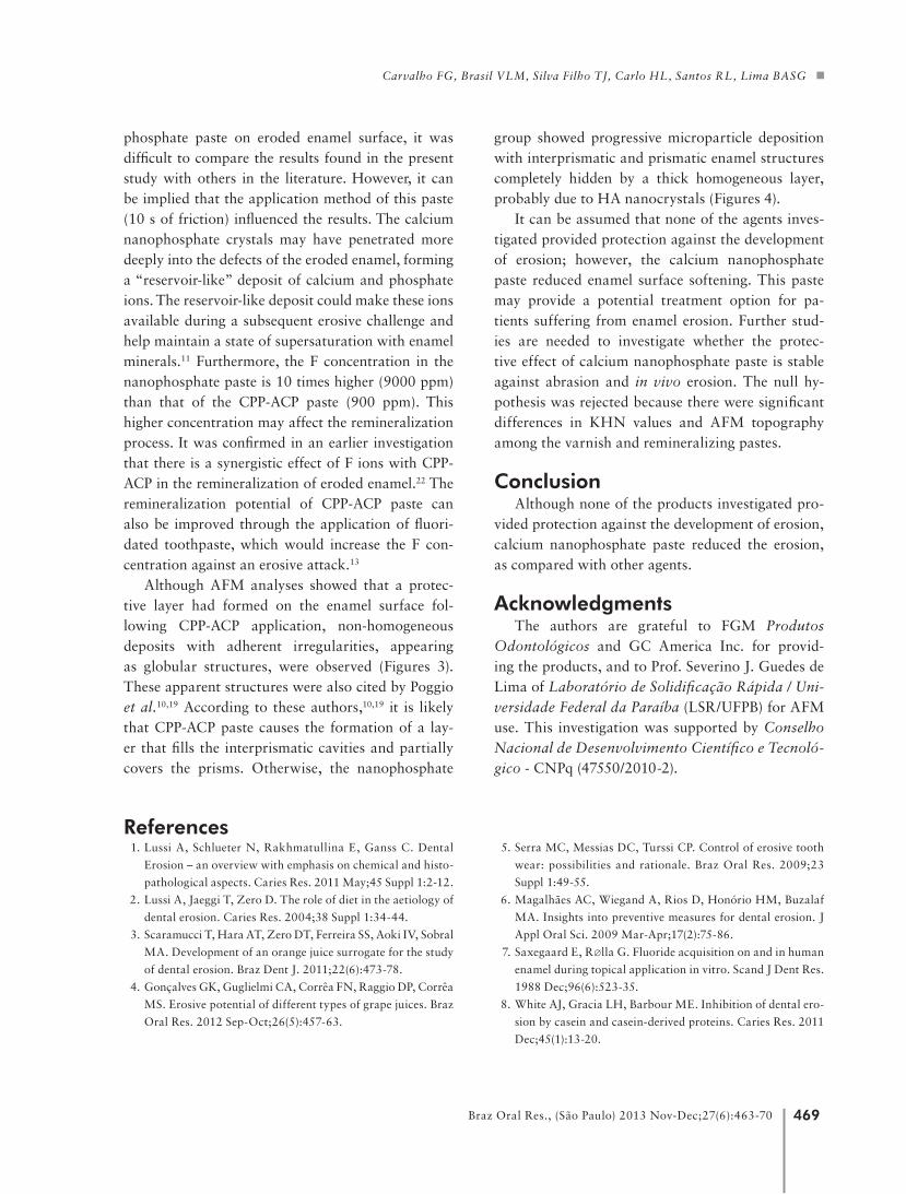
Figures 1, 2, 3 and 4 show the AFM images after applying the remineralizing agent and erosive chal-lenge. The control and varnish groups showed simi-lar topography. Figures 1 and 2 showed areas of de-mineralized enamel with a honeycomb-like structure and a rough surface without a protective layer (Fig-ure 1B and 2B). The treatment with the two pastes caused different surface topographies. The CPP-ACP group showed a surface interspersed with globu-lar deposits (Figure 3). The nanophosphate group showed progressive microparticle deposition, and the interprismatic and prismatic enamel structures appeared to be completely hidden by a thick homo-geneous layer (Figures 4).
DiscussionThe present study is an innovative investigation
about the potential for remineralization of eroded enamel with a new agent containing calcium nano-phosphate organized in HA crystals. The micro-hardness test and AFM were used. The standard-ization of baseline enamel KHN (Table 2) made it possible to establish the %KHN change among the groups after treatment.
Formation of the CaF2-like layer by fluoride (F) agents on eroded dental surfaces partially re-duces enamel mineral loss by subsequent ero-sion.14 High-concentrated F applications have been considered very effective in reducing the enamel erosion.6,20 The agents investigated contain ap-proximately 22600 ppm F (varnish), 9000 ppm F (nanophosphate paste) and 900 ppm F (CPP-ACP paste), according to the manufacturers’ informa-tion. Although the varnish contained the highest F concentration among the agents tested, according to our protocol, it showed no protective effect against enamel erosion. This could be observed by the sta-tistically lower remineralization rates (represented by a loss in KHN = higher %KHN change), as com-pared with the CPP-ACP and nanophosphate groups (Table 2). Furthermore, no statistically significant difference was found for KHN values and surface topography (Figures 1 and 2) between the varnish and control groups (Table 2). Figures 1A and 2A showed that the enamel demineralization caused by the cola beverage involved mainly the inner area of the prism, and created a honeycomb-like structure, as described by Poggio et al.10
The varnish was removed after 24 h to simulate in vivo conditions (a single professional application
Agent/treatment Initial KHN KHN after erosion %KNH change
Control 351.3 ± 10.7 a*,A** 96.8 ± 11.4 a,B 72.4 ± 3.0 a*
Fluoride varnish 354.4 ± 24.6 a,A 91.7 ± 14.1 a,B 73.4 ± 5.5 a
Nanophosphate 367.1 ± 6.5 a,A 187.2 ± 27.9 b,B 49.0 ± 7.9 b
CPP-ACP 360.4 ± 10.1 a,A 141.8 ± 16.5 c,B 60.6 ± 4.0 c
* Same lowercase letters indicate that there was no significant difference among the agents in each treatment and in the %KNH change among agents (one-way ANOVA and Tukey’s test, p > 0.05). ** Same uppercase letters indicate that there was no significant difference between initial and post-erosion values of each agent (paired-T test, p > 0.05).
Table 2 - Change in surface hardness (KHN) of enamel after
erosion by a cola beverage, expressed in mean ±standard deviation.
Carvalho FG, Brasil VLM, Silva Filho TJ, Carlo HL, Santos RL, Lima BASG
467Braz Oral Res., (São Paulo) 2013 Nov-Dec;27(6):463-70
was made every 7 days), as used in other in vitro studies.12,16,17 The fluoride varnish may have reacted chemically with the enamel during the first 24 h, but this reaction was not enough to reduce the enamel loss caused by the erosive attack. Figure 2B shows a rough and eroded surface with no formation of a protective layer for the varnish group, visualized as a honeycomb-like demineralized enamel structure (Figure 2A). In some studies, which showed the pro-
tective effect of fluoride varnish against dental ero-sion,21 the varnishes were not completely removed during the experimental period, and this mechani-cal protection may have played a role in the protec-tive effect evidenced.
Apart from fluoride, the CPP-ACP complex showed an increase in enamel microhardness and a decrease in enamel surface roughness after ero-sion by cola drinks.9,10,13 However, the mechanism
Figure 1 - AFM image of enamel surface after erosive
challenge and application of no agent; (A) (→)
honeycomb-like structure of enamel; (B) rough surface
with no protection layer.
Figure 2 - AFM image of enamel surface after erosive challenge and
varnish application; (A) (→) honeycomb-like structure of
enamel; (B) rough surface with no protection layer.
Protective effect of calcium nanophosphate and CPP-ACP agents on enamel erosion
468 Braz Oral Res., (São Paulo) 2013 Nov-Dec;27(6):463-70
by which the process of remineralization of eroded lesions by CPP-ACP occurs is unclear. It is known that CPP-ACP increases the number of potential calcium-binding sites, thereby decreasing the con-stant diffusion of calcium.9 Furthermore, ACP at the tooth surface buffers the free calcium and phos-phate ion activities, thus helping to maintain a state of supersaturation that depresses demineralization
and enhances remineralization.9
The CPP-ACP group showed a higher KHN val-ue and lower mineral loss (%KHN), compared with the control group, after the erosive challenge (Table 2). However, the nanophosphate group showed the highest KHN value after erosion, with significantly lower mineral loss than the CPP-ACP paste (Table 2). Because there was no investigation into nano-
Figure 4 - AFM image of enamel surface
after erosive challenge and nanophosphate
paste application; (A) homogeneous deposit on
the surface; (B) enamel completely hidden by a
thick homogeneous layer.
Figure 3 - AFM image of enamel surface after erosive
challenge and CPP-ACP paste application; (A, B)
surface with globular non-homogeneous deposits; (→)
adherent irregularities.
Carvalho FG, Brasil VLM, Silva Filho TJ, Carlo HL, Santos RL, Lima BASG
469Braz Oral Res., (São Paulo) 2013 Nov-Dec;27(6):463-70
phosphate paste on eroded enamel surface, it was difficult to compare the results found in the present study with others in the literature. However, it can be implied that the application method of this paste (10 s of friction) influenced the results. The calcium nanophosphate crystals may have penetrated more deeply into the defects of the eroded enamel, forming a “reservoir-like” deposit of calcium and phosphate ions. The reservoir-like deposit could make these ions available during a subsequent erosive challenge and help maintain a state of supersaturation with enamel minerals.11 Furthermore, the F concentration in the nanophosphate paste is 10 times higher (9000 ppm) than that of the CPP-ACP paste (900 ppm). This higher concentration may affect the remineralization process. It was confirmed in an earlier investigation that there is a synergistic effect of F ions with CPP-ACP in the remineralization of eroded enamel.22 The remineralization potential of CPP-ACP paste can also be improved through the application of fluori-dated toothpaste, which would increase the F con-centration against an erosive attack.13
Although AFM analyses showed that a protec-tive layer had formed on the enamel surface fol-lowing CPP-ACP application, non-homogeneous deposits with adherent irregularities, appearing as globular structures, were observed (Figures 3). These apparent structures were also cited by Poggio et al.10,19 According to these authors,10,19 it is likely that CPP-ACP paste causes the formation of a lay-er that fills the interprismatic cavities and partially covers the prisms. Otherwise, the nanophosphate
group showed progressive microparticle deposition with interprismatic and prismatic enamel structures completely hidden by a thick homogeneous layer, probably due to HA nanocrystals (Figures 4).
It can be assumed that none of the agents inves-tigated provided protection against the development of erosion; however, the calcium nanophosphate paste reduced enamel surface softening. This paste may provide a potential treatment option for pa-tients suffering from enamel erosion. Further stud-ies are needed to investigate whether the protec-tive effect of calcium nanophosphate paste is stable against abrasion and in vivo erosion. The null hy-pothesis was rejected because there were significant differences in KHN values and AFM topography among the varnish and remineralizing pastes.
ConclusionAlthough none of the products investigated pro-
vided protection against the development of erosion, calcium nanophosphate paste reduced the erosion, as compared with other agents.
Acknowledgments The authors are grateful to FGM Produtos
Odontológicos and GC America Inc. for provid-ing the products, and to Prof. Severino J. Guedes de Lima of Laboratório de Solidificação Rápida / Uni-versidade Federal da Paraíba (LSR/UFPB) for AFM use. This investigation was supported by Conselho Nacional de Desenvolvimento Científico e Tecnoló-gico - CNPq (47550/2010-2).
References1. Lussi A, Schlueter N, Rakhmatullina E, Ganss C. Dental
Erosion – an overview with emphasis on chemical and histo-
pathological aspects. Caries Res. 2011 May;45 Suppl 1:2-12.
2. Lussi A, Jaeggi T, Zero D. The role of diet in the aetiology of
dental erosion. Caries Res. 2004;38 Suppl 1:34-44.
3. Scaramucci T, Hara AT, Zero DT, Ferreira SS, Aoki IV, Sobral
MA. Development of an orange juice surrogate for the study
of dental erosion. Braz Dent J. 2011;22(6):473-78.
4. Gonçalves GK, Guglielmi CA, Corrêa FN, Raggio DP, Corrêa
MS. Erosive potential of different types of grape juices. Braz
Oral Res. 2012 Sep-Oct;26(5):457-63.
5. Serra MC, Messias DC, Turssi CP. Control of erosive tooth
wear: possibilities and rationale. Braz Oral Res. 2009;23
Suppl 1:49-55.
6. Magalhães AC, Wiegand A, Rios D, Honório HM, Buzalaf
MA. Insights into preventive measures for dental erosion. J
Appl Oral Sci. 2009 Mar-Apr;17(2):75-86.
7. Saxegaard E, R∅lla G. Fluoride acquisition on and in human
enamel during topical application in vitro. Scand J Dent Res.
1988 Dec;96(6):523-35.
8. White AJ, Gracia LH, Barbour ME. Inhibition of dental ero-
sion by casein and casein-derived proteins. Caries Res. 2011
Dec;45(1):13-20.
Protective effect of calcium nanophosphate and CPP-ACP agents on enamel erosion
470 Braz Oral Res., (São Paulo) 2013 Nov-Dec;27(6):463-70
9. Ranjitkar S, Kaidonis JA, Richards LC, Townsend GC. The
effect of CPP-ACP on enamel wear under severe erosive condi-
tions. Arch Oral Biol. 2009 Jun;54(6):527-32.
10. Poggio C, Lombardini M, Dagna A, Chiesa M, Bianchi S. Protec-
tive effect on enamel demineralization of a CPP-ACP paste: an
AFM in vitro study. J Dent. 2009 Dec;37(12):949-54.
11. Huang S, Gao S, Cheng L, Yu H. Remineralization potential
of nano-hydroxyapatite on initial enamel lesions: an in vitro
study. Caries Res. 2011 Sep;45(5):460-68.
12. Murakami C, Bönecker M, Corrêa MS, Mendes FM, Ro-
drigues CR. Effect of fluoride varnish and gel on dental ero-
sion in primary and permanent teeth. Arch Oral Biol. 2009
Nov;54(11):997-1001.
13. Tantbirojn D, Huang A, Ericson MD, Poolthong S. Change in
surface hardness of enamel by a cola drink and a CPP–ACP
paste. J Dent. 2008 Jan;36(1):74-9.
14. Moretto MJ, Magalhães AC, Sassaki KT, Delbem AC, Mar-
tinhon CC. Effect of different fluoride concentrations of ex-
perimental dentifrices on enamel erosion and abrasion. Caries
Res. 2010 Apr;44(2):135-40.
15. Parry J, Shaw L, Arnaud M, Smith A. Investigation of mineral
waters and soft drinks in relation to dental erosion. J Oral
Rehabil. 2001 Aug;28(8):766-72.
16. Magalhães AC, Kato MT, Rios D, Wiegand A, Attin T, Bu-
zalaf MA. The effect of an experimental 4% TIF4 varnish
compared to NAF varnishes and 4% TIF4 solution on dental
erosion in vitro. Caries Res 2008 Jun;42(4):269-74.
17. Magalhães AC, Stancari FH, Rios D, Buzalaf MA. Effect of an
experimental 4% titanium tetrafluoride varnish on dental ero-
sion by a soft drink. J Dent. 2007 Nov;35(11):858-61.
18. Shellis RP, Ganss C, Ren Y, Zero DT, Lussi A. Methodology
and models in erosion research: discussion and conclusions.
Caries Res. 2011;45 Suppl 1:69-77.
19. Poggio C, Lombardini M, Colombo M, Bianchi S. Im-
pact of two toothpastes on repairing enamel erosion pro-
duced by a soft drink: an AFM in vitro study. J Dent. 2010
Nov;38(11):868-74.
20. Magalhães AC, Rios D, Honório HM, Delbem AC, Buzalaf
MA. Effect of 4% titanium tetrafluoride solution on the ero-
sion of permanent and deciduous human enamel: an in situ/
ex vivo study. J Appl Oral Sci. 2009 Jan-Feb;17(1):56-60.
21. Sorvari R, Meurman JH, Alakuijala P, Frank RM. Effect
of fluoride varnish and solution on enamel erosion in vitro.
Caries Res.1994;28(4):227-32.
22. Srinivasan N, Kavitha M, Loganathan SC. Comparison of the
remineralization potential of CPP-ACP and CPP-ACP with
900 ppm fluoride on eroded human enamel: an in situ study.
Arch Oral Biol. 2010 Jul;55(7):541-44.