regulation of nitrogen fixation in klebsiella pneumoniae
TRANSCRIPT
Regulation of nitrogen fixation in Klebsiella pneumoniae:
The role of Fnr in oxygen signal-transduction
Dissertation
Zur Erlangung des Doktorgrades
der Mathematisch-Naturwissenschaftlichen Fakultäten
der Georg-August Universität zu Göttingen
vorgelegt von
Roman Grabbe aus Helmarshausen
Göttingen 2002
Die vorliegende Arbeit wurde am Lehrstuhl für Allgemeine Mikrobiologie im Institut für
Mikrobiologie und Genetik der Georg-August Universität Göttingen angefertigt.
Finanziert wurde diese Arbeit aus Mitteln der Deutschen Forschungsgemeinschaft im
Schwerpunkt „Regulatorische Netzwerke in Bakterien“ und dem Fonds der chemischen
Industrie.
Teile dieser Arbeit wurden veröffentlicht in:
Grabbe, R., Kuhn, A., and Schmitz, R.A. 2001a. Cloning, sequencing and characterization
of Fnr from Klebsiella pneumoniae. Antonie Van Leeuwenhoek 79: 319-326.
Grabbe, R., Klopprogge, K., and Schmitz, R.A. 2001b. Fnr is Required for NifL-dependent
oxygen control of nif gene expression in Klebsiella pneumoniae. J. Bacteriol. 183:
1385-1393.
Klopprogge, K., Grabbe, R., Hoppert, M., and Schmitz, R.A. 2002. Membrane association
of Klebsiella pneumoniae NifL is affected by molecular oxygen and combined
nitrogen. Arch. Microbiol. 177(3): 223-34.
Grabbe, R., and Schmitz, R.A. 2002. Oxygen Control of nif Gene Expression in Klebsiella
pneumoniae is dependent on NifL reduction at the cytoplasmic membrane by electrons
derived from the reduced quinone pool. (submitted)
Außerdem gingen folgende Veröffentlichungen aus der Arbeit hervor:
Ehlers, C, Grabbe R, Veit K, Schmitz RA 2002.Characterization of GlnK1 from
Methanosarcina mazei strain Go1: complementation of an Escherichia coli glnK
mutant strain by GlnK1. J. Bacteriol. 184(4): 1028-40.
D7
Referent: Prof. Dr. G. Gottschalk
Korreferent: PD. Dr. R.A. Schmitz-Streit
Tag der mündlichen Prüfung: 20.06.02

Acknowledgements
Especially I would like to thank PD Dr. Ruth Schmitz for supervising and supporting this
thesis. It has not always been easy to work with you because of your high demands on the
people in the lab. But finally, I do believe that I will never learn more about science than I
did under your inspiring guidance.
I thank Prof. Dr. G. Gottschalk for generous support and helpful discussions.
Special thanks to the former and current members of lab. 214: Anita, Anne, Christian,
Claudia, Daniela, Edna, Jessica, Julia, Jutta, Kai, Katharina, Katja, Korinna, Melanie. It has
been a pleasure working in such a friendly atmosphere. I will always remember that when
looking back.
I like to thank all the people at the Institute for Microbiology and Genetics, Göttingen,
especially the members of LII, G2L, the workshop and Mr. Hellwig.
Additionally, I want to thank my family and friends for giving me help and support.
Finally, I like to thank Tanja, just for being there.
Table of contents Summary 1 Chapter 1:
Introduction 3 NifL modulates NifA transcriptional activity by direct protein protein interaction 4 Nitrogen signal transduction 5 NifL response to molecular oxygen 6
Chapter 2:
Cloning, sequencing and characterization of Fnr from Klebsiella pneumoniae 8 Abstract 8 Introduction 8 Materials & Methods 9 Results & Discussion 15 Summary 19
Chapter 3: Fnr is required for NifL dependent oxygen control of nif gene expression in Klebsiella pneumoniae 21 Abstract 21 Introduction 22 Materials & Methods 23 Results 30 Discussion 41
Chapter 4: Membrane association of Klebsiella pneumoniae NifL is effected by molecular oxygen and combined nitrogen 45 Abstract 45 Introduction 45 Materials & Methods 47 Results 51 Discussion 63
Chapter 5: Oxygen control of nif gene expression in Klebsiella pneumoniae is dependent on NifL reduction at the cytoplasmic membrane by electrons derived from tne reduced quinone pool 67 Abstract 67 Introduction 68 Materials & Methods 70 Results 75 Discussion 88
Conclusions 93
Current working model 96 Further studies 98
References 99 Curriculum vitae 114
Summary 1
Summary • In the free-living diazotroph Klebsiella pneumoniae, a member of the γ-subgroup of
Proteobacteria, nitrogen fixation (nif) genes are under the control of the nifLA operon, the
products of which regulate transcription of the nif operons. NifA activates nif gene
transcription by alternative RNA polymerase, σ54-holoenzyme; the negative regulator
NifL modulates activity of NifA in response to molecular oxygen and combined nitrogen.
Transcriptionally coupled synthesis, immunological studies and complex analysis of both
regulators indicate that NifL-mediated inhibition of NifA depends on direct protein-
protein interaction.
• The negative regulator NifL is a flavoprotein, which modulates NifA activity depending
on the redox state of its N-terminally bound FAD-cofactor. Thus, oxygen might be sensed
directly by the redox-sensitive cofactor of NifL or by a global oxygen sensor, for example
Fnr (fumarate nitrate reductase regulator), which transduces the oxygen signal towards the
NifL-bound cofactor.
• The fnr gene of K. pneumoniae was cloned, sequenced and biochemically analyzed. The
analysis of the deduced amino acid sequence revealed 98 % similarity to the Escherichia
coli Fnr protein. The conserved cystein residues, which establish the oxygen-sensing
[4Fe-4S]-cluster, are located in the N-terminal domain of the K. pneumoniae Fnr as it is
kown for the E. coli protein. Biochemical analysis of the glutathionS-transferase (GST)
fusion protein Fnr-GST expressed and purified under aerobic or anaerobic conditions,
revealed decreased amounts of iron and acid-labile sulphur in the aerobic protein
compared to the anaerobic protein. This indicates that K. pneumoniae Fnr sesnes oxygen
based on an oxygen-sensitive iron-sulphur cluster.
• Studying the oxygen dependent regulation of nif induction in fnr mutant backgrounds we
obtained strong evidence that in K. pneumoniae Fnr is the primary oxygen sensor for the
nif regulatory system. In the absence of Fnr, NifL did not receive the signal of
anaerobiosis under nitrogen and oxygen limited conditions resulting in a decreased NifA
activity. Thus, Fnr appears to sense the oxygen status of the cell and presumably
transduces the signal of anaerobiosis towards Nifl by activating gene(s), the product(s) of
which function to reduce the FAD cofactor of NifL resulting in a non-inhibitory
conformation. Attractive candidates for the physiological electron donor for NifL
Summary 2
reduction are components of the anaerobic electron transport chain, which are Fnr-
dependent transcribed.
• Localization experiments of NifL in K. pneumoniae under different growth conditions
revealed that NifL is highly membrane associated under derepressing growth conditions.
However, when cells were shifted to ammonium sufficiency or presence of oxygen NifL
is located in the cytoplasm. Further studies using K. pneumoniae mutant strains showed
that under derepressing conditions but in the absence of either Fnr or the nitrogen sensor
GlnK NifL was located in the cytoplasm and inhibited NifA activity. Presumably in the
absence of Fnr or GlnK NifL does not receive the signal of anaerobiosis or nitrogen
limitation. In contrast to NifL, NifA remains in the cytoplasm under all conditions tested.
Thus, sequestration of NifL to the membrane under nitrogen and oxygen-limitation is
involved in the mechanism of NifA regulation.
• Biochemical analysis of purified NifL showed that NifL-bound FAD-cofactor was
reduced by NADH/H+ only in the presence of a redox mediator or inside-out vesicles
derived from anaerobically grown K. pneumoniae cells. This indicates that in vivo NifL is
reduced at the cytoplasmic membrane.
• In order to identify the physiological electron donor for NifL reduction, the effect of
different oxidoreductase systems on nif regulation was studied in the respective mutant
backgrounds. Using K. pneumoniae mutant strains we observed strong evidence, that in
the absence of a functional NADH:ubiquinone oxidoreductase or formate
dehydrogenaseN NifL inhibition of NifA was not relieved. The same effect was observed
in a heterologous E. coli system lacking the alternative NADH dehydrogenase (ndh).
Further studies of nif induction of anaerobically grown cultures on glycerol showed
significantly reduced NifA activity when nitrate was added as additional electron
acceptor. Taking together these findings indicate that more than one oxidoreductase
system appears to be responsible for NifL reduction and that NifL receives electrons from
the reduced quinone pool.
• We further demonstrated that reduced dimethylnaphthoquinone (DMNred), a soluble
quinone derivative is able to reduce the FAD cofactor of NifL in the absence of a redox
mediator. This finding supports our model that the cofactor FAD of the membrane-
associated NifL receives electrons from the reduced quinone pools, generated by different
oxidoreductase systems.
Chapter 1 3
Chapter 1: Introduction
Biological nitrogen fixation, the enzymatic reduction of molecular nitrogen (N2) to ammonia,
is strictly limited to prokaryotes. However, within the prokaryotes nitrogen fixation is found
in a large number of species belonging to the bacterial domain and in several methanogenic
Archaea (Dean and Jacobson, 1992; Young, 1992; Lobo and Zinder, 1992; Fischer, 1994;
Galagan et al., 2002; Deppenmeier et al., 2002). The reduction of molecular nitrogen is
catalyzed by the nitrogenase enzyme complex with high energy demands. Two ATP
molecules are consumed for each electron transferred to the catalytic site (Burgess and Lowe,
1996; Howard and Rees, 1996; Rees and Howard, 1999, Halbleib and Ludden, 2000).
Because of the high energy requirement for N2 fixation, up to 40 % of the ATP is utilized by
the nitrogenase in nitrogen fixing cells, resulting in a drop of the energy charge from 0.9 to
0.5 (Daesch and Mortenson, 1972; Upchurch et al., 1980). In the presence of molecular
oxygen the nitrogenase enzyme complex is irreversibly inactivated. Thus, to avoid
unnecessary consumption of energy nitrogen fixing microorganisms tightly control synthesis
and activity of nitrogenase in response to nitrogen and oxygen availability. In all diazotrophic
proteobacteria examined, the transcriptional activator NifA is required for transcription of the
nitrogen fixation (nif) genes. NifA expression and activity is regulated in response to the
environmental signals, molecular oxygen and combined nitrogen. However, the mechanisms
of NifA regulation vary in different organisms (Fischer, 1996; Dixon, 1998; Halbleib and
Ludden, 2000; Schmitz et al., 2002). In free-living and symbiotic diazotrophs belonging to
the α-and β-subgroup of the proteobacteria (genera Rhizobium, Bradyrhizobium, Azospirillum
and Herbaspirillum) NifA activity is directly sensitive to molecular oxygen and in some cases
affected in the presence of combined nitrogen (Fischer, 1994; Fischer, 1996; Steenhoudt and
Vanderleyden, 2000). In contrast, in Klebsiella pneumoniae and Azotobacter vinelandii, two
free-living diazotrophs, which belong to the γ-proteobacteria, NifA activity is not oxygen
sensitive. NifA activity is regulated in response to molecular oxygen and fixed nitrogen by a
second regulator NifL, the gene of which forms an operon with nifA (Filser, 1983; Dixon,
1998). In K. pneumoniae the expression of the nifLA operon itself is regulated by the nitrogen
status, via the NtrB/NtrC two component regulatory system, whereas in A. vinelandii nifLA is
constitutively expressed (Drummond and Wootton, 1987; Blanco et al., 1993). Interestingly,
it was recently found that nitrogen fixation of the endophytic diazotroph Azoarcus spec. -
belonging to the β-proteobacteria - is also regulated by the coordinated activities of nifL and
Chapter 1 4
nifA gene products in response to environmental signals (Egener and Reinhold-Hurek,
unpublished).
NifL modulates NifA transcriptional activity by direct protein-protein interaction. The
transcriptional activator NifA is composed of three domains: an amino (N)-terminal domain
apparently involved in the regulation, a central catalytic domain, and a carboxy (C)-terminal
DNA-binding domain (Drummond et al., 1990; Morett and Segovia, 1993). Transcription of
nif genes by the alternative RNA polymerase (σ54-RNA polymerase) is generally activated by
NifA, which binds to an upstream activation sequence (UAS) (Morrett and Buck, 1988) and
contacts promoter-bound σ54-RNA polymerase by means of a DNA loop (Buck et al., 1987).
Subsequently NifA catalyzes the isomerization of closed complexes between σ54-holoenzyme
and the nif promoter to transcriptionally productive open complexes (Morett and Buck, 1989;
Hoover et al., 1990). This open complex formation requires hydrolysis of ATP or GTP
catalyzed by NifA (Lee et al., 1993; Austin et al., 1994). In the presence of molecular oxygen
or combined nitrogen, NifL inhibits NifA activity in vivo (Merrick et al., 1982; Hill et al.,
1981; Dixon, 1998; Schmitz et al. 2002). The inhibitory protein NifL is composed of two
domains separated by a hydrophilic interdomain linker (Q-linker) (Söderbäck et al., 1998;
Drummond and Wootton, 1987). The C-terminal domain of NifL shows homology to a
histidine protein kinase (Blanco et al., 1993). However, neither autophosphorylation nor
possible phosphor transfer between the two regulatory proteins NifA and NifL has been
detected in K. pneumoniae or A. vinelandii (Lee et al., 1993; Austin et al., 1994; Schmitz et
al., 1996). The translationally coupled synthesis of nifL and nifA and immunological studies
imply that the inhibition of NifA activity by NifL apparently occurs via a direct protein-
protein interaction (Govantes et al., 1998; Henderson et al., 1989). Recently, complex
formation between A. vinelandii NifL and NifA has been demonstrated by in vitro co-
chromatography in the presence of adenosine nucleotides and using the yeast two hybrid
system (Money et al., 2001 and 1999; Lei et al., 1999). Thus, signal transduction apparently
occurs via protein-protein interaction. Interestingly, for A. vinelandii it was shown that NifL
influences both NifA transcriptional activity and DNA-binding capacity in vitro (Barrett et
al., 2001). The C-terminal domain of K. pneumoniae NifL is sufficient to inhibit
transcriptional activation by NifA in vitro and in vivo (Narberhaus et al., 1995). This indicates
that the inhibitory function of NifL protein appears to be located in its C-terminal domain,
which presumably interacts with NifA by protein-protein interaction.
Chapter 1 5
Nitrogen signal transduction. In K. pneumoniae, a shift from nitrogen limitation to nitrogen
sufficiency results in repression of nif gene induction upon inhibition of NifA transcriptional
activity by NifL (Arnott et al. 1989; Blanco et al., 1993). This indicates that NifL either
senses the nitrogen availability directly or the nitrogen status is sensed in a NifL independent
manner and the signal is subsequently transduced to NifL or the NifL/NifA complex.
Interestingly, like in Escherichia coli a second PII-like protein, encoded by glnK, was recently
discovered in K. pneumoniae. glnK is organized with amtB (encoding for an ammonium
transporter) in an operon, which is under transcriptional control of NtrC. Upon the high
similarity to the PII-protein, the GlnK-protein is an attractive candidate for sensing changes in
the glutamine pool size - reflecting the internal nitrogen status - and mediating the signal of
the nitrogen status to the nif regulatory system (Atkinson and Ninfa, 1998; Xu et al., 1998;
van Heeswijk et al., 1996). Studying nif regulation in glnK mutant strains strong evidence was
obtained, that GlnK is indeed required to release NifL inhibition under nitrogen-limiting
growth conditions in K. pneumoniae (He et al., 1998; Jack et al., 1999; Arcondeguy et al.,
1999). This indicates that changes of the internal nitrogen status are not sensed by NifL
directly, but are apparently mediated by GlnK to the NifA/NifL regulatory system. Whereas
NifL is a negative regulator, GlnK acts positively to antagonize inhibitory effects of NifL
under nitrogen-limiting conditions. The uridylylation status of GlnK is probably not required
for relief of NifL inhibition (He et al. 1998; Arcondeguy et al., 1999). Interestingly, the T-
loops of GlnK and PII from K. pneumoniae, which are supposed to interact with other
components involved in the signal transduction, differ only in three amino acid residues 43,
52 and 54. It has been shown that for regulation of the nif system residue 54 is the most
important amino acid in the T-loop of GlnK, possibly directly involved in the interaction with
NifL/NifA (Arcondeguy et al., 2000). Although GlnK function has been clearly demonstrated,
the question arises, how GlnK is mediating the nitrogen signal towards the NifL/NifA
regulatory system. The nitrogen signal is apparently mediated by direct protein-protein
interaction but it has to be elucidated, whether GlnK is interacting directly with NifL or is
affecting the NifL/NifA complex formation. For diazotrophs not belonging to the γ-
proteobacteria and missing NifL (e.g. Herbaspirillum seropedicae and Azospirillum
brasilense) experimental data indicate that the PII proteins participate in signaling the
nitrogen status to the N-terminal domain of NifA (Steenhoudt and Vanderleyden, 2000; Souza
et al., 1999; Monteiro et al., 1999, Arsene et al., 1999).
A. vinelandii contains only one PII-like protein, encoded in a glnK/amtB-operon, which is
expressed constitutively (Meletzus et al., 1998). Interestingly, A. vinelandii GlnK has a T-
Chapter 1 6
loop structure, which resembles more the 'GlnB-like' T-loop rather than the 'GlnK-like' T-loop
(Arcondeguy et al., 2000). Recent studies concerning the role of A. vinelandii GlnK in
nitrogen sensing and transducing the nitrogen status to the nif regulatory system showed that
GlnK is not required for derepression in A. vinelandii. In contrary to K. pneumoniae, where
GlnK apparently has a positive role in relieving NifL inhibition under nitrogen limiting
conditions, in vitro experiments suggest that the inhibitory function of A. vinelandii NifL is
activated under nitrogen excess through interaction with PII-like regulatory proteins (Reyes-
Ramirez et al., 2000; Little et al., 2000 and 2002). Recently interactions between NifL and
GlnK have been reported for A. vinelandii using the yeast two-hybrid system (Rudnick et al.,
2002) and it was demonstrated in vitro that GlnK interacts with the C-terminal domain of
NifL (Little et al., 2002). Dixon and coworker proposed that interaction with NifL only
occurs when GlnK is not uridylylated and activates NifL inhibitory functions under nitrogen
sufficiency (Little et al., 2002). This suggests that NifA inhibition by NifL is relieved when
GlnK is uridylylated, but uridylylated GlnK is not required for this relief. However, very
recently Merrick and coworkers showed that in E. coli and A. vinelandii non-uridylylated
GlnK is highly membrane associated after a shift to nitrogen sufficiency upon binding to the
ammonium transporter AmtB (Coutts et al., 2002) and thus, unmodified GlnK should not be
available in the cytoplasm to activate NifL inhibitory functions.
NifL response to molecular oxygen. The N-terminal domain of NifL contains conserved S-
motifs of PAS-like domains, which are known for a number of regulators sensing oxygen,
redox or light (Zhulin et al., 1997; Taylor and Zhulin, 1999). This indicates that the N-
terminal domain is involved in signal transduction. Biochemical analyses of purified proteins
showed that NifL from A. vinelandii and from K. pneumoniae is a flavoprotein with an N-
terminally bound FAD-cofactor (Hill et al., 1996; Schmitz, 1997; Söderbäck et al., 1998;
Klopprogge and Schmitz, 1999). Analysis of the inhibitory function of NifL-holoenzyme and
NifL-apoenzyme on NifA activity in in vitro transcription assays showed that the FAD-
cofactor is not directly required for NifL inhibitory function (Schmitz, 1997). This indicates
that FAD acts as a redox-sensitive cofactor, which might be involved in the oxygen signal
transduction. The oxidized form of NifL inhibits NifA transcriptional activity in vitro,
whereas A. vinelandii NifL reduced by sodium dithionite or by the flavoheme protein (Hmp)
from E. coli with NADH/H+ as electron donor does not antagonize open complex formation
by NifA in vitro (Macheroux et al., 1998). Thus, reduction of the flavin moiety of NifL results
in a non-inhibitory form of NifL, however functional and physiological relevance for the
Chapter 1 7
reduction of NifL by Hmp, which is proposed to be a global oxygen sensor (Pool, 1994), has
not been demonstrated to date. These findings support the model that NifL acts as a redox-
sensitive regulatory protein that modulates NifA activity in response to the redox state of its
FAD-cofactor and allows NifA activity only in the absence of oxygen. However, in both
organisms the physiological electron donor for NifL is not known.
Reduction of the FAD-cofactor by the physiological electron donor apparently transduces the
signal for anaerobiosis to NifL. As a consequence, components of the oxygen signal
transduction are attractive candidates for the electron transfer towards NifL in vitro. Thus, the
key question concerning the oxygen signal transduction is, whether NifL senses the oxygen
status of the cell directly via a redox induced conformational change. Alternatively, oxygen
might be detected by a more general oxygen-sensing system, which then regulates NifL by
inducing the oxidation or reduction of the flavin cofactor. In this respect it is of interest that in
K. pneumoniae, iron is specifically required for relief of NifL inhibition under oxygen and
nitrogen limitation (Schmitz et al., 1996). The finding that K. pneumoniae NifL does not
contain non-heme iron or an acid-labile sulphur cluster (Schmitz et al., 1996; Klopprogge and
Schmitz 1999), indicates the presence an iron containing protein in the oxygen signal cascade
towards NifL. In E. coli the transcriptional regulator Fnr (fumarate nitrate reductase regulator)
plays an overarching role in sensing the switch from anaerobic to aerobic conditions. The
mechanism of oxygen sensing in Fnr is mediated via an [4Fe-4S]-cluster (Green et al., 1996;
Unden and Shirawski, 1997; Kiley and Beinert, 1998). Interestingly, in Rhizobium
leguminosarum FnrN, a Fnr homologous protein, regulates nitrogen fixation in an oxygen-
dependent manner (Gutierrez et al., 1997). Thus, it is attractive to speculate that a Fnr
homologous protein is involved in oxygen-dependent regulation of nitrogen fixation in K.
pneumoniae.
The intention of this thesis was to study the signal transduction of molecular oxygen towards
NifL in K. pneumoniae. Investigations were performed to study (i) the role of Fnr in the
oxygen-sensing mechanism for nitrogen fixation (chapter 2 and 3), (ii) the cellular
localization of NifL followed by functional analyses of NifL localization for NifA regulation
(chapter 4), and (iii) the effect of membrane-bound oxidoreductase systems concerning
oxygen sensing on nif regulation (chapter 5).

Chapter 2 8
Chapter 2:
Cloning, sequencing and characterization of Fnr
from Klebsiella pneumoniae
ABSTRACT
The transcription factor Fnr (fumarate nitrate reductase regulator) globally regulates
gene expression in response to oxygen deprivation in Escherichia coli. We report here the
cloning and sequencing of the fnr gene from the facultative anaerobic bacterium Klebsiella
pneumoniae M5al, another member of the enteric bacteria. The deduced amino acid
sequence of K. pneumoniae fnr showed very high similarity (98 % amino acid identity) to
the Fnr protein from E. coli and contained the four essential cysteine residues which are
presumed to build the oxygen-sensing [4Fe4S]+2 center. Transfer of the K. pneumoniae gene
to a fnr mutant of E. coli complemented the mutation and permitted synthesis of nitrate
reductase and fumarate reductase during anaerobic growth. A gene fusion between K.
pneumoniae fnr and glutathione S-transferase was constructed and expressed in E. coli
under anaerobic conditions in order to make the protein available in preparative amounts.
The overproduced protein was purified by glutathione-Sepharose 4B affinity
chromatography in the absence of oxygen, and biochemically characterized.
INTRODUCTION:
Many of the oxygen-responsive gene regulators of bacteria are members of the
fumarate nitrate reductase / cyclic AMP receptor protein family of transcriptional regulators
(Spiro 1994, Gunsalus & Park 1994, Unden et al. 1995). The fumarate nitrate reductase
regulator from Escherichia coli (FnrEc) acts as a redox-responsive transcriptional regulator
that activates genes whose products are involved in anaerobic respiration and represses other
genes required for aerobic respiration (Spiro 1994, Gunsalus & Park 1994, Unden et al. 1995,
Bauer et al. 1999). It contains a cluster of three closely-spaced cysteine residues located near
the N-terminus (20CysX2CysX529Cys) plus an additional cysteine residue, Cys122. These
cysteine residues are required for the oxygen-sensing function (Spiro & Guest 1988). Recent
data suggest that these residues bind an [4Fe4S]+2-cluster and that this cluster apparently
Chapter 2 9
mediates the sensitivity of the transcriptional activator to oxygen (Green et al. 1996;
Khoroshilova et al. 1997; Kiley & Beinert 1998). In addition, the presence of the [4Fe4S]+2-
cluster in the anaerobically-purified form of Fnr is correlated with dimerization and specific
DNA binding. Upon addition of oxygen, the [4Fe4S]+2-cluster is disrupted, resulting in the
conversion of Fnr into an inactive monomeric protein (Lazazzera et al. 1996; Melville &
Gunsalus 1996). Homologs of Fnr have been identified in several gram-negative and gram-
positive bacteria, some of which differ with respect to the cystein residues and the
coordination of the iron-sulphur clusters (reviewed in Spiro 1994; Cruz Ramos et al. 1995;
Saunders et al. 1999; Vollack et al. 1999). Recently discovered examples of Fnr homologues,
which do not exhibit the structural elements or coordinate the iron-sulphur clusters differently
are: (i) Fnr from Bacillus subtilis and B. licheniformis, for which a C-terminal cluster
coordination is found (Cruz Ramos et al. 1995; Klinger et al. 1998); (ii) Fnr homologues
from Lactobacillus casei and L. lactis, that lack two of the four essential cysteine residues
and in the case of L. casei, Flp redox sensitive switch is operated based on a reversible
interconversion of an intramolecular disulphide bridge (Gostick et al. 1998; Scott et al. 2000);
and (iii) the Fnr homologues DnrD, DnrE and DnrS of Pseudomonas stutzeri, which
completely lack the respective cysteine residues and iron-sulphur centres (Vollack et al.
1999).
Adaptation of the facultative anaerobic bacterium Klebsiella pneumoniae to anaerobic
growth conditions is also accompanied by dramatic changes in metabolic gene expression. In
addition, it is only when growing in the absence of molecular oxygen that K. pneumoniae is
able to use molecular nitrogen as sole nitrogen source under nitrogen limitation (Dixon
1998). In order to make these adaptations, K. pneumoniae must sense changes in
environmental oxygen availability. In contrast to E. coli, little is known about a regulatory
oxygen-sensing system in Klebsiella. However, there are some evidences suggesting the
presence of an Fnr-homologue in K. pneumoniae: Fnr is possibly involved in expression of
the citrate-specific fermentation genes in K. pneumoniae (Bott et al. 1995) and in K.
terrigena Fnr might act as a repressor of the butanediol (bud) operon (Mayer et al. 1995).
In this communication we report on the sequencing and characterization of the
regulatory gene fnr from K. pneumoniae.
MATERIALS AND METHODS
Chapter 2 10
Bacterial Strains and Plasmids.
The bacterial strains and plasmids used in this work are listed in Table 1. Plasmid
DNA was transformed into E. coli cells according to the method of Inoue et al. (1990) or by
electroporation using a Gene pulser and Pulse controller (BioRad Laboratories). The
fnr::Tn10 allele was transferred from the fnr::Tn10 derivative of M182 (Jayaraman et al.
1988) by P1-mediated transduction into NCM1529 and RM123 as described previously
(Silhavy et al. 1984) with selection for tetracycline resistance; the resulting strain designated
RAS1 and RAS6 respectively. Strains RAS3, RAS4 and RAS5 contain plasmids pRS120,
pRS127 and pRS137, respectively, in RAS1; strain RAS21 contains pRS137 in RAS6.
Plasmids pRS120 and pRS137 contain the E. coli fnr gene and K. pneumoniae fnr gene,
respectively, inserted into the SalI and EcoRV site of pACYC184 and thereby expressed
from the tet promoter.
Media and growth conditions.
For cloning, E. coli was routinely grown in LB medium at 37 °C (Ausubel et al.
1987). The medium was supplemented with ampicillin at 100 µg/ml or chloramphenicol at 15
µg/ml to maintain recombinant plasmids; additionally, 5 µg/ml tetracycline was added to the
growth medium when NCM1529(fnr::Tn10) or RM123(fnr::Tn10) were the host strains. For
complementation experiments, strains were grown under anaerobic conditions with N2 as gas
phase at 37 °C in minimal medium (100 mM KH2PO4, 50 mM NaHPO4, 1 mM MgSO4, 0.1
mM CaCl2, 10 µM Na2SeO3, 10 µM Na2MoO4, 0.3 mM sulfide and 0.002 % resazurine (to
monitor anaerobiosis) pH = 6.5), containing 0.8 % glycerol as the C-source and 1 % KNO3 as
the only nitrogen source. Precultures were grown overnight in closed bottles, with N2 as gas
phase, in medium lacking sulfide and resazurine, and additionally supplemented with 4 mM
ammonium acetate which was completely utilized after growth of the precultures to
saturation. The main cultures (25 ml) were inoculated from saturated precultures and were
grown in closed bottles at 37° C without shaking.
Construction of a gene library of K. pneumoniae chromosomal DNA.
Chromosomal DNA from K. pneumoniae M5a1 was isolated according the method
described by Ausubel et al. (1987). Fifty micrograms of DNA was partially digested with
Sau3AI so that the majority of fragments were in the size range of between 20 and 30 kbp.
The purified digested DNA was ligated to 1 µg pWE15, which had been completely digested
with BamHI and dephosphorylated. The ligation mixture was then packed and transduced
Chapter 2 11
into E. coli VCS257 using the Gigapack III Gold (Stratagene, La Jolla, US) packaging extract
according the protocol of the manufacturer. Approximately 8000 colonies were collected.
Generation of a 100 bp hybridization probe for the fnr gene from K. pneumoniae.
A probe for the fnr gene was obtained by PCR using genomic DNA from K.
pneumoniae as template. The oligonucleotides were derived from the E. coli fnr sequence: 5'
primer (5’ATCAATTACGGATCCAGCAGACCTATGATCCCG3’) and 3' primer
(5’GTGTGAACG GGATCCAAAGCTGGC3’). Reactions were carried out in 100 µl volumes
using Vent polymerase (New England Biolabs, UK) and primers at a concentration of 0.3 µM.
The annealing temperature was at 65 °C and synthesis was carried out for 30 s, for 25 cycles.
The 100 bp PCR product was purified with Wizard® Plus PCR Purification system (Promega,
Heidelberg, Germany) and labeled with the random Dig-labeling kit from Boehringer
Mannheim according the protocol of the manufacturer. The specificity of the probe was tested
by Southern hybridizations (Sambrook et al. 1989) with K. pneumoniae DNA digested
completely digested by BamHI and EcoRI. Under the conditions employed, the hybridization
with the labeled probe resulted in only one hybridization signal in each digest.
TABLE 1: Bacterial strains and plasmids used in this study
Strains / plasmids
Relevant genotype and/or characteristic(s)
Reference or description
Strains
M182(fnr::Tn10) M182 but fnr::Tn10
Jayaraman et al. 1998
NCM1529 araD139∆(argF-lacU)169 fth D5301
gyrA219 non-9 rpsL150 ptsF25
relA1 deoC1
trpDC700putPA1303::[Kanr-(nifH’-
’lacZ)]
He et al. 1997
RAS1 NCM1529 but fnr::Tn10
Chapter 2 12
See Materials and
Methods
RAS3 RAS1/pRS120
See Materials and
Methods
RAS4 RAS1/pRS127
See Materials and
Methods
RAS5
RAS1/pRS137
See Materials and
Methods
Plasmids
Relevant genotype and / or
characteristic(s)
Reference or
description
pWE15
cosmid vector
Stratagene, La Jolla,
US
pBluescript SK+
cloning vector
Stratagene, La Jolla,
US
pACYC184
low copy vector
New England Biolabs
(UK)
pGEX-2T
Expression vector, expression in
fusion with glutathione-S transferase
Pharmacia, Freiburg
Germany
pRS120
E. coli fnr controlled by the tet
promoter on pACYC184
See Materials and
Methods
pRS127
2.1 kbp fragment in pBluescript SK+
containing K. pneumoniae fnr
See Materials and
Methods
Chapter 2 13
pRS131 K. pneumoniae fnr cloned into pGEX-
2T under the control of the tac
promoter, coding for glutathione-S
transferase fused to Fnr
See Materials and
Methods
pRS137
K. pneumoniae fnr controlled by the
tet promoter on pACYC184
See Materials and
Methods
Cloning and sequencing of K. pneumoniae fnr gene.
Heterologous cosmids from the gene library of K. pneumoniae chromosomal DNA was
completely digested by BamH1 and EcoRI. After blotting onto Nylon membrane Hybond-N
(Amersham) and Southern hybridization (Sambrock et al. 1989) using the 100 bp probe, the
digested cosmids were screened for positives using the luminescent detection kit for nucleic
acids from Boehringer Mannheim. Three positive cosmids were obtained and subcloned into
pSK+ Bluescript (Stratagene, La Jolla, US) resulting in plasmid pRS127, containing a 2.1 kbp
EcoRI/BamHI fragment which hybridized with the fnr probe. DNA sequences of both strands
were determined independently and completely by commercial sequencing by MWG Biotech
(Ebersberg, Germany). Sequence analysis was performed with the Genetics Computer Group
(GCG) program package (Devereux et al. 1984).
Enzyme activities.
To determine synthesis of fumarate reductase by measuring fumarate reductase activity
cells were grown in minimal medium (Schmitz et al. 1996) supplemented with 10 mM
ammonium, 1 % glucose and 50 mM fumarate. Cell extracts were prepared from anaerobically
grown cells at an O.D.600 = 0.6. Cells were disrupted under anaerobic conditions in breakage
buffer (50 mM Tris/HCl buffer pH = 7.6 containing 4 mM dithiothreithol and 10 % glycerol)
using a French pressure cell followed by centrifugation at 20,000 x g. Fumarate reductase was
assayed in 1.5 ml glass cuvettes with N2 as gas phase at 37 °C. The 0.8 ml standard assay
mixture contained 50 mM Tris /HCl buffer pH = 7.4, 4 mM dithiothreitol, 5 mM MgCl2, 250
µM reduced methyl viologen, 1 mM fumarate and 50 to 400 µg cell extract protein. The
reactions were started by the addition of 1 mM fumarate and the reduction of fumarate was
monitored by following the decrease in absorbance at 604 nm (ε = 26.8 mM-1 cm-1 per 2
Chapter 2 14
electron transfer). One unit (U) is the amount catalysing the reduction of 1 µmol fumarate per
minute at concentrations of 250 µM methyl viologen and 1 mM fumarate
Expression of glutathione S-transferase (GST) fused to K. pneumoniae fnr in E. coli
NCM1529.
The recombinant pRS131 containing the fnr gene of K. pneumoniae fused at the 5’ end
to the 3’ end of the gene for GST was constructed by cloning the PCR amplified fnr into the
BamHI and EcoRI restriction recognition sites of pGEX-2T (Pharmacia, Freiburg, Germany).
K. pneumoniae fnr was amplified from chromosomal DNA using a set of primers with
synthetic restriction recognition sites (underlined): a sense primer with an additional BamHI
restriction recognition site 5’ of the start codon
(5’ATATCAATGGATCCCTGAGCAGACTTATGATCC3’) and an antisense primer with a
EcoRI restriction recognition site downstream of the stop codcon
(5’CGATCCGGCCGAATTCAGAGGGACT ATCAG3’). The PCR product was purified as
described above, digested with BamHI and EcoRI and ligated into pGEX-2T, which had been
linearized with the corresponding enzymes, resulting in plasmid pRS131. The PCR product
cloned into pGEX-2T was sequenced, revealing no mutation of fnr and correct insertion. From
the sequence, the GST-Fnr fusion protein is predicted to have a molecular mass of 58 kDa and
a recognition site for thrombin between GST and Fnr. pRS131 was transformed into E. coli
NCM1529, which grows well under anaerobic conditions. For expression of the GST-Fnr
fusion protein, E. coli NCM1529/pRS131 was grown aerobically or anaerobically with N2 as
gas phase in minimal medium (modified K-medium, Schmitz et al. 1996) with 0.8 % glucose
as the C-source and 10 mM ammonium as the nitrogen source. Expression of the fusion protein
was induced with 1 mM isopropyl-ß-D-thiogalactopyranoside (IPTG) when cultures reached
an O.D.600 = 0.6. Cell extract was prepared by disruption of the cells in breakage buffer (50
mM Tris/HCl buffer pH = 7.6 containing 10 % glycerol) using a French pressure cell followed
by centrifugation at 20,000 x g. Fusion proteins were purified from the supernatant by affinity
chromatography with glutathione-Sepharose 4B (Pharmacia) according the instruction protocol
of the manufacturer. In the case of anaerobic purification all steps described were performed
under a nitrogen atmosphere in an anaerobic chamber and the buffers employed contained 2.0
mM dithiothreitol.
Chapter 2 15
Determination of non-hem iron, acid-labile sulfur, and protein.
Non-hem iron was determined colorimetrically as described by Fish (1988). Acid-labile
sulfur was analyzed using methylene blue (Cline 1969). Protein was determined via the
method of Bradford (1976) with the BioRad protein assay using bovine serum albumin as
standard.
SDS-PAGE Analyses.
Sodium dodecyl sulfate-polyacrylamide gel electrophoresis was performed according to
Laemmli using 12.8% acrylamide (Laemmli 1970). Gels were stained for protein with
Coomassie Brilliant Blue.
RESULTS AND DISCUSSION
The present work was designed to characterize the oxygen-sensing system in K.
pneumoniae by cloning the fnr homologue. We expressed the Fnr protein from K. pneumoniae
in fusion to the glutathion-S transferase and analyzed purified protein for iron-sulfur clusters.
Cloning and nucleotide sequence of K. pneumoniae fnr. A 100-bp fragment encoding
part of K. pneumoniae fnr was amplified by PCR using K. pneumoniae chromosomal DNA as
template and using primers based on the N-terminal sequence of the E. coli fnr gene. This
fragment was labeled with digoxigenin-dUTP and used as a hybridization probe to screen a
cosmid library of K. pneumoniae chromosomal DNA as described in Materials and Methods.
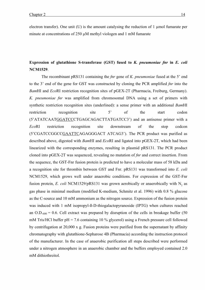
Chapter 2 16
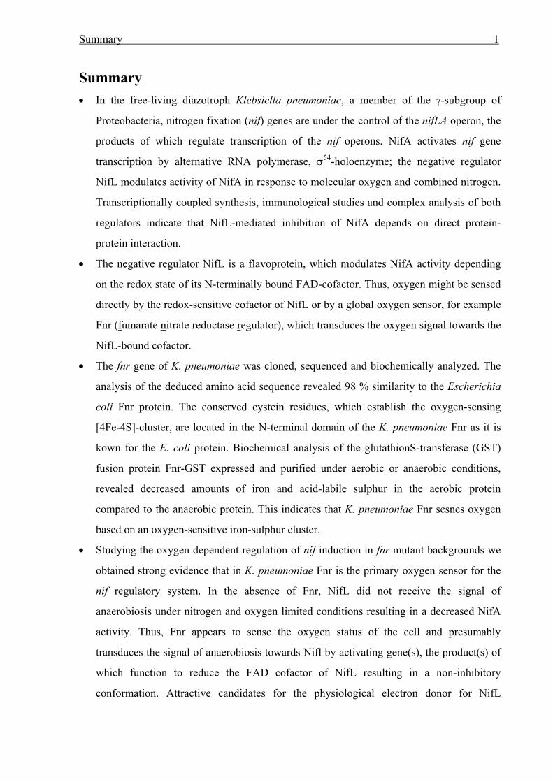
Figure 1: Organization of the cloned region of K. pneumoniae (A) and the sequence for the promoter and N-
terminal region of fnr (B). The deduced amino acid sequence is given in capital letters; the amino acid symbols
(one letter code) are written below the first nucleotide of the corresponding codon. Three of the four cystein
residues near the N-terminus, which are apparently required for the [4Fe-4S)-cluster ligation, are marled in grey.
A potential ribosome binding site (SD) is underlined and a putative s70-dependent promoter sequence is boxed.
The sequence of the cloned region has been submitted to GenBank under accession number AF220669.
K. pneumoniae fnr was identified on a 2.1 kbp EcoRI/BamHI fragment. This fragment was
subcloned into pSK+ Bluescript and the resulting plasmid designated pRS127. The insert of
pRS127 was entirely sequenced in both directions. Analysis of the sequence revealed two open
reading frames, orfA and orfB, and part of a third putative open reading frame (orfC') as shown
in Fig. 1. orfB showed high similarities to fnr from E. coli and was therefore designated as fnr.
The open reading frame upstream of fnr was identified as ogt by homology to the equivalent E.
coli gene and orfC' downstream of fnr shows homology to ydaA' of E. coli. The fnr gene of K.
pneumoniae is preceded by a weak ribosomal binding site, appropriately spaced from the start
codon; in addition, a sequence for a putative σ70-dependent promoter is located upstream of fnr
in position -61 to -32 (Fig. 1). The fnr gene (753 bp) codes for a polypeptide of 250 amino
acids with a predicted molecular mass of 27939 Da, which shows 98 % amino acid identity to
Fnr of E. coli (Shaw & Guest 1982). In addition Fnr of K. pneumoniae (FnrKp) contained all
four essential cysteine residues (Cys20, Cys22, Cys29 and Cys122) which are presumed to
comprise the oxygen-sensing [4Fe4S]2+-center in E. coli Fnr (FnrEc) (Spiro & Guest 1988).
. -35 . . -10 . . . ...GTTCGAGACTTACCTGCTCACCAAAAAGATGTTAAAATTGACCAATATCAATTAAAGCCT . . . . . . GAGCAGACTTATGATCCCGGAAAAGCGAATTATACGACGCATTCAGTCTGGCGGTTGTGC
SD M I P E K R I I R R I Q S G G C A
. fnr . . . . . AATCCATTGCCAGGATTGCAGCATTAGCCAGCTTTGCATCCCTTTTACTCTGAACGAGCA... I H C Q D C S I S Q L C I P F T L N E H
B)
A)
EcoRI
ogt fnr ydaA`
pRS127
1 640 1241 2022
BamHIHindIIIKpnI
Chapter 2 17
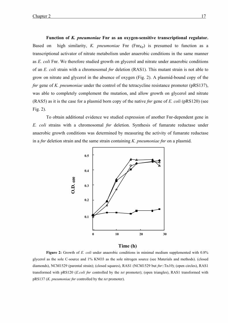
Function of K. pneumoniae Fnr as an oxygen-sensitive transcriptional regulator.
Based on high similarity, K. pneumoniae Fnr (FnrKp) is presumed to function as a
transcriptional activator of nitrate metabolism under anaerobic conditions in the same manner
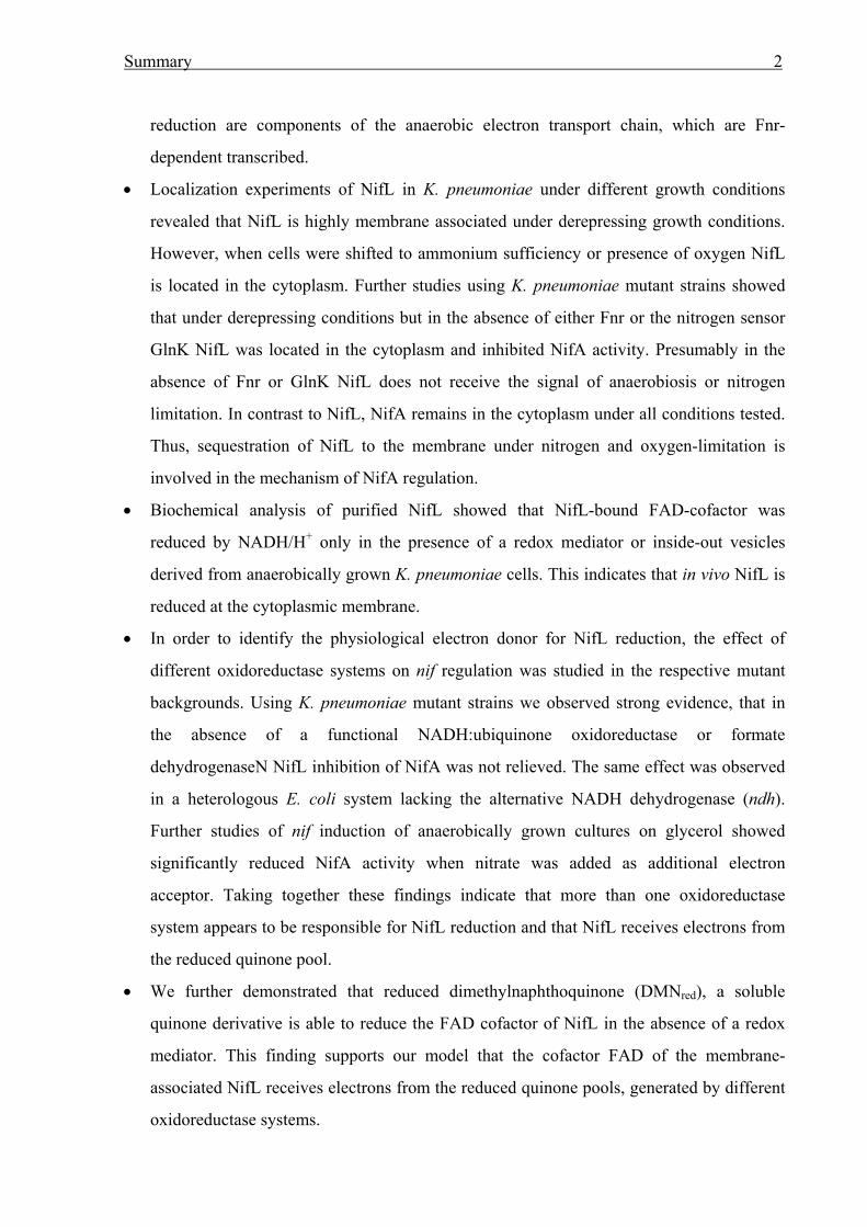
as E. coli Fnr. We therefore studied growth on glycerol and nitrate under anaerobic conditions
of an E. coli strain with a chromosomal fnr deletion (RAS1). This mutant strain is not able to
grow on nitrate and glycerol in the absence of oxygen (Fig. 2). A plasmid-bound copy of the
fnr gene of K. pneumoniae under the control of the tetracycline resistance promoter (pRS137),
was able to completely complement the mutation, and allow growth on glycerol and nitrate
(RAS5) as it is the case for a plasmid born copy of the native fnr gene of E. coli (pRS120) (see
Fig. 2).
To obtain additional evidence we studied expression of another Fnr-dependent gene in
E. coli strains with a chromosomal fnr deletion. Synthesis of fumarate reductase under
anaerobic growth conditions was determined by measuring the activity of fumarate reductase
in a fnr deletion strain and the same strain containing K. pneumoniae fnr on a plasmid.
Figure 2: Growth of E. coli under anaerobic conditions in minimal medium supplemented with 0.8%
glycerol as the sole C-source and 1% KNO3 as the sole nitrogen source (see Materials and methods). (closed
diamonds), NCM1529 (parental strain); (closed squares), RAS1 (NCM1529 but fnr::Tn10); (open circles), RAS1
transformed with pRS120 (E.coli fnr controlled by the tet promoter); (open triangles), RAS1 transformed with
pRS137 (K. pneumoniae fnr controlled by the tet promoter).
O.D
. 600
Time (h)
0.1
0.2
0.3
0.4
0.5
0 10 20 30
Chapter 2 18
Fumarate reductase activity in the fnr deletion strain (RAS1) was determined to be 23 mU /
mg cell extract protein, which was equivalent to 10 % of the activity in the parental strain
NCM1529 (231 mU / mg cell extract protein). When the fnr gene of K. pneumoniae was
expressed in trans under the control of the tet promoter (RAS5) it was able to complement the
fnr mutation in E. coli and allow significant higher fumarate reductase activity under anaerobic
conditions (174.5 mU / mg cell extract protein). These results indicate that FnrKp is functional
in E. coli and apparently acts as an oxygen-sensing transcriptional regulator.
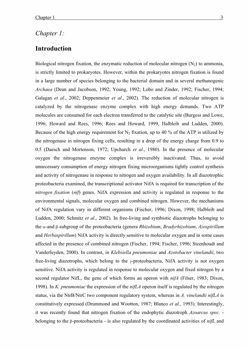
Purification and characterization of heterologous expressed FnrKp. In order to
characterize the iron-sulfur clusters we fused FnrKp to the glutathione S-transferase (GST) by
cloning the fnr gene into pGEX-2T (see Materials and Methods). The resulting plasmid, which
contains the gst-gene C-terminally fused to K. pneumoniae fnr under the control of the tac
promoter, was designated pRS131. After transforming pRS131 into E. coli NCM1529, which
grows well under anaerobic conditions (He et al. 1997), the fusion protein was synthesized in
minimal medium under aerobic and anaerobic growth conditions as described in Materials and
Methods with ammonium as nitrogen source. Under both growth conditions, induction of the
fusion protein at an optical density of O.D.600 = 0.6 resulted in a retarded growth. The
overexpressed fusion protein fractions were purified in the presence and absence of molecular
oxygen, respectively, by Glutathione-Sepharose 4B affinity chromatography and cleaved with
the site-specific protease thrombin. In both cases, homogeneous FnrKp preparations were
obtained, as revealed by sodium dodecylsulfate/polyacrylamide gel electrophoresis. The
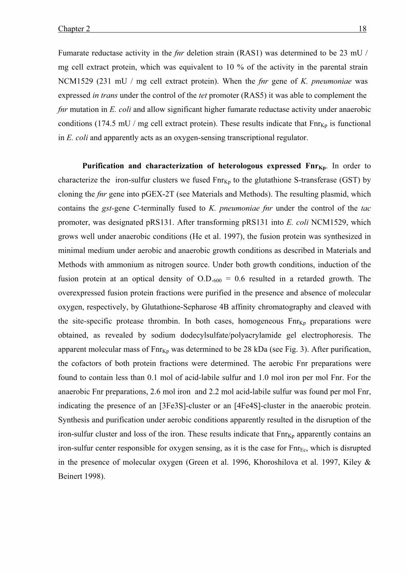
apparent molecular mass of FnrKp was determined to be 28 kDa (see Fig. 3). After purification,
the cofactors of both protein fractions were determined. The aerobic Fnr preparations were
found to contain less than 0.1 mol of acid-labile sulfur and 1.0 mol iron per mol Fnr. For the
anaerobic Fnr preparations, 2.6 mol iron and 2.2 mol acid-labile sulfur was found per mol Fnr,
indicating the presence of an [3Fe3S]-cluster or an [4Fe4S]-cluster in the anaerobic protein.
Synthesis and purification under aerobic conditions apparently resulted in the disruption of the
iron-sulfur cluster and loss of the iron. These results indicate that FnrKp apparently contains an
iron-sulfur center responsible for oxygen sensing, as it is the case for FnrEc, which is disrupted
in the presence of molecular oxygen (Green et al. 1996, Khoroshilova et al. 1997, Kiley &
Beinert 1998).
Chapter 2 19
Figure 3: Purification of K. pneumoniae Fnr fused to glutathion S-transferase (GST) and synthesized
under anaerobic conditions. Various stages in the purification are seperated by SDS-Page (12.8%). Lanes: 1 and 2,
whole cell extract before and after IPTG induction, respectively; 3, low-speed supernatant from cell extract; 4 and
10. low molecular mass marker (Pharmacia); 5, 6 and 7, wash fractions of GST-Fnr bound to Glutathione
Sepharose 4B; 8 and 9, flow through fractions following thrombin digest of GST-Fnr bound to Glutathione
Sepharose 4B; 11 and 12, GST fraction eluted with glutathion supplemented buffer. The gel was stained with
coomassie Brillant Blue R250.
In order to further analyse the iron-sulfur center of FnrKp we studied the spectroscopic
properties of the anaerobic FnrKp protein fraction. (i) UV-visible spectroscopy of the anaerobic
FnrKp protein showed no detectable absorption in the range of 400 to 420 nm. (ii) Using Low
Temperature EPR analyses, we revealed no signal typical for an iron sulfur cluster for the
anaerobic Fnr fraction (data not shown). This might be due to the low protein concentrations
we observed from the anaerobic protein purification (approximately 0.5 mg/ml) or due to
disruption of the iron-sulfur clusters during the purification procedure even when performed
under anaerobic conditions.
In summary. In order to characterize the oxygen-sensing system in K. pneumoniae we
have cloned and characterized the fnr gene of K. pneumoniae. Analyses of the K. pneumoniae
fnr gene showed high similarities to the E. coli fnr gene (98 % amino acid identity, Shaw &
Guest 1982). The ability of fnrKp to functionally complement fnrEc was shown in vivo by
restoration of growth on glycerol plus nitrate, and expression of Fnr-dependent genes
(frdABCD) in an E. coli fnr deletion strain transformed with a plasmid-bound copy of FnrKp.

Chapter 2 20
These results indicate that FnrKp activates transcription of genes in a similar way like E. coli
Fnr. They further suggest a similarity in the oxygen-sensing mechanism of the two organisms.
In addition, characterization of purified protein indicated the presence of an oxygen sensitive
[4Fe4S]2+-center in FnrKp: (i) The deduced amino acid sequence of K. pneumoniae fnr
contained all four essential cysteine residues near the N-terminus, which are required for the
oxygen-sensing function (Spiro & Guest 1988, Khoroshilova et al. 1997). (ii) Determination of
iron and acid-labile sulfur in aerobic- and anaerobic-purified protein fractions suggested the
presence of an iron-sulfur cluster, which is apparently disrupted upon the influence of oxygen.
Chapter 3 21
Chapter 3:
Fnr is required for NifL-dependent oxygen control of nif gene
expression in Klebsiella pneumoniae
Abstract
In Klebsiella pneumoniae, NifA dependent transcription of nitrogen fixation (nif) genes is
inhibited by NifL in response to molecular oxygen and combined nitrogen. We recently
showed that K. pneumoniae NifL is a flavoprotein, which apparently senses oxygen through a
redox-sensitive, conformational change. We have now studied the oxygen regulation of NifL
activity in Escherichia coli and K. pneumoniae strains by monitoring its inhibition of NifA-
mediated expression of K. pneumoniae ø(nifH’-’lacZ) fusions in different genetic
backgrounds. Strains of both organisms carrying fnr null mutations failed to release NifL
inhibition of NifA transcriptional activity under oxygen limitation: nif induction was similar
to the induction under aerobic conditions. When the transcriptional regulator Fnr was
synthesized from a plasmid, it was able to complement, i.e., to relieve NifL inhibition in the
fnr--backgrounds. Hence, Fnr appears to be involved, directly or indirectly, in NifL-dependent
oxygen regulation of nif gene expression in K. pneumoniae. The data indicate that in the
absence of Fnr NifL apparently does not receive the signal for anaerobiosis. We therefore
hypothesize that in the absence of oxygen, Fnr, as the primary oxygen sensor, activates
transcription of a gene(s) whose product(s) function to relieve NifL inhibition by reducing the
FAD cofactor under oxygen-limiting conditions.
Chapter 3 22
Introduction
In diazotrophic proteobacteria, transcription of the nitrogen fixation (nif) genes is mediated by
the nif-specific activator protein NifA, a member of a family of activators that functions with σ54
(Dixon, 1998, Fischer, 1994). Both the expression and the activity of NifA can be regulated in
response to the oxygen and / or combined nitrogen status of the cells; the mechanisms of the
regulation differ with the organism. In Klebsiella pneumoniae and Azotobacter vinelandii, NifA
transcriptional activity is regulated by a second regulatory protein, NifL. This negative
regulator of the nif genes inhibits the transcriptional activation by NifA in response to combined
nitrogen and or external molecular oxygen. The translationally-coupled synthesis of the two
regulatory proteins, immunological studies, complex analyses and studies using the two-hybrid
system in Saccharomyces cerivisiae imply that the inhibition of NifA activity by NifL
apparently occurs via direct protein-protein interaction (Govantes et al., 1998, Henderson et al.,
1989; Lei et al., 1999; Money et al., 1999). The mechanism by which nitrogen is sensed in
K. pneumoniae and A. vinelandii is currently the subject of extensive studies. Very recently, He
et al. (He et al., 1998), and Jack et al. (1999) provided evidence that in K. pneumoniae, the
second PII protein, GlnK, is required for relief of NifL inhibition under nitrogen-limiting
conditions. This indicates that GlnK regulates NifL inhibition of NifA in response to the
nitrogen status of the cells by interacting with NifL or NifA.
In both organisms, K. pneumoniae and A. vinelandii, the negative regulator NifL is a
flavoprotein with an N-terminally bound flavin adenine dinucleotide as a prosthetic group
(Hill et al., 1996; Klopprogge and Schmitz, 1999: Schmitz, 1997). In vitro, the oxidized form
of NifL inhibits NifA activity, whereas reduction of the FAD cofactor relieves NifL inhibition
(Hill et al., 1996; Macheroux et al., 1999). This indicates that NifL apparently acts as a redox
switch in response to the environmental oxygen status and allows NifA activity, only under
oxygen-limiting conditions. We recently showed that in vivo, the presence of iron is required
to relieve inhibitory effects of NifL on transcriptional activation by NifA and, additionally,
that iron is not present in NifL (Schmitz, 1997; Schmitz et al., 1996). Therefore, we have
postulated that an unidentified iron-containing protein may be the physiological reductant for
Chapter 3 23
NifL. This putative iron-containing protein is apparently not nif specific since NifL function is
regulated normally in response to cellular nitrogen and oxygen availability in Escherichia coli
in the absence of nif proteins other than NifA (He et al., 1998).
The key question concerning the oxygen signal transduction in K. pneumoniae is, whether
NifL senses oxygen directly via a redox-induced conformational change, or whether oxygen is
detected by a more general oxygen-sensing system, which then regulates NifL by inducing the
oxidation or reduction of the flavin cofactor. One candidate for a general oxygen sensor is the
transcriptional fumarate nitrate regulator (Fnr) (Spiro, 1994; Spiro and Guest, 1990), which in
the case of E. coli Fnr, senses oxygen via an oxygen-labile iron-sulfur ([4Fe-4S]+2)-cluster and
is involved in signal transduction of the cellular redox state (Green et al., 1996; Khoroshilova
et al., 1997; Melville and Gunsalus, 1990; Unden and Schirawski, 1997). Recently we cloned
and sequenced the fnr gene of K. pneumoniae and characterized the protein (Grabbe et al.,
2000). As the K. pneumoniae Fnr amino acid sequence is 98 % identical to the E. coli Fnr and
contains an iron-sulfur cluster, we have now tested the hypothesis that Fnr transduces the
oxygen signal to NifL. We present evidence that in the absence of Fnr, NifL inhibits NifA
activity under oxygen-limitation, suggesting that Fnr is required for relief of NifL inhibition
in K. pneumoniae under anaerobic conditions.
Materials and Methods
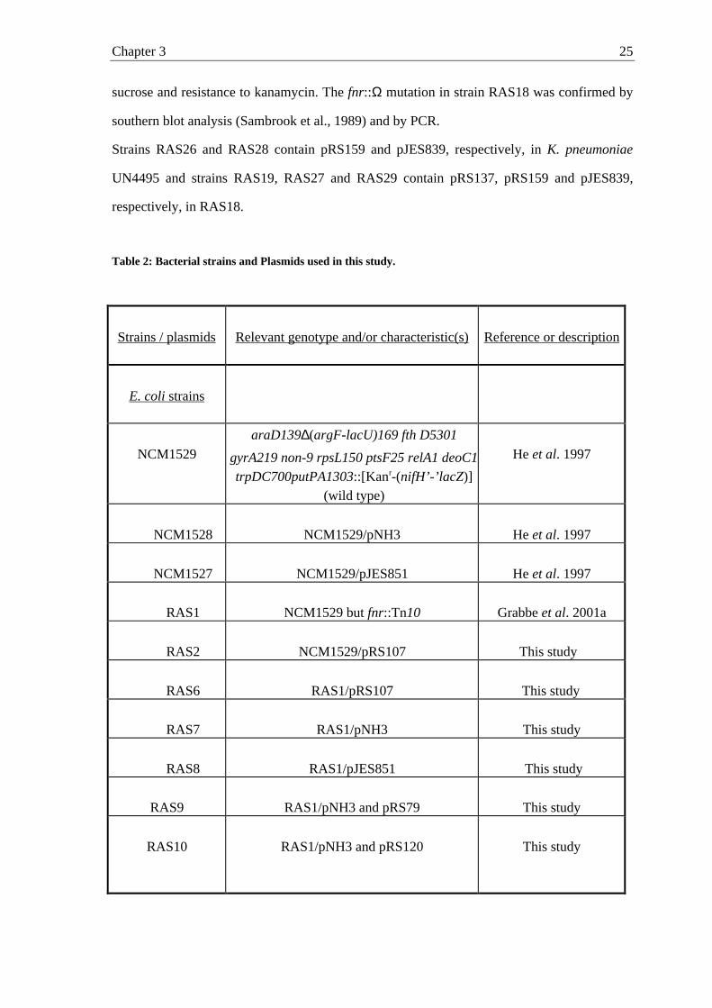
Bacterial strains and plasmids. The bacterial strains and plasmids used in this work are
listed in Table 2. Plasmid DNA was transformed into E. coli cells according to the method of
Inoue et al. (1990) and into K. pneumoniae cells by electroporation. Transduction by phage
P1 was performed as described previously (Silhavy et al., 1984).
E. coli strains. E. coli NCM1529, which contains a ø(nifH’-’lacZ) fusion (He et al. 1997),
and derivatives of NCM1529 were chosen to study NifA/NifL regulation in E. coli. The
fnr::Tn10 allele was transferred from the fnr::Tn10 derivative of M182 (Jayaraman et al.,
1988) into NCM1529 by P1-mediated transduction with selection for tetracycline resistance,
Chapter 3 24
resulting in RAS1 (Grabbe et al., 2001a). Strains RAS6, RAS7, RAS8, RAS9, RAS10,
RAS11 and RAS12 contain plasmids pRS107, pNH3, pJES851, pNH3 plus pRS79, pNH3
plus pRS120, pNH3 plus pMCL210, and pNH3 plus pACYC184, respectively, in RAS1. To
construct an independent second fnr null mutant, the [Kanr-(nifH’-’lacZ)] allele was
transferred from strain NCM1529 by P1-mediated transduction into the independent fnr
mutant strain RM101 (Sawers and Suppman, 1992) and into the parental strain MC4100 with
selection for kanamycin resistance, resulting in RAS13 and RAS21, respectively. Strains
RAS25, RAS14, RAS15, RAS16 and RAS17 contain plasmids pRS107, pNH3, pJES851,
pNH3 plus pRS120 and pNH3 plus pACYC184, respectively in RAS13.
Klebsiella strains. K. pneumoniae strains M5al (wild type) and UN4495 (ø(nifK-lacZ)5935
lac-4001 his D4226 Galr) (McNeil et al., 1981) were provided by Gary Roberts.
Construction of a fnr::Ω mutation: Strain RAS18 was obtained by insertion of a kanamycin
resistance cassette (Prentki et al., 1984) into the fnr gene of K. pneumoniae UN4495 as
achieved in the following steps. (i) The 2.1 kbp EcoRI/BamHI fragment, which carries the
ogt-fnr-ydaA'- region of K. pneumoniae, was subcloned into pBluescript SK+ to produce
pRS127. (ii) A 2.1 kb HindIII cassette containing an Ω interposon fragment with a kanamycin
resistance gene derived from plasmid pHP45Ω (Prentki et al., 1984) was cloned into the
HindIII site of fnr in pRS127 to yield plasmid pRS142. (iii) A 2.9 kb PCR fragment carrying
fnr::Ω was generated using pRS142 as template and a set of primers which were homologue
to the fnr flanking 5´- and 3´-regions with additional BamHI synthetic restriction recognition
sites (underlined) (5’ATATCAATGGATCCCTGAGCAGACTTA TGATCC3’, sense primer;
5'CTTATATGGATCCAATGAAACAGGGGAGGA3', antisense primer). The 2.9 kb PCR
product was cloned into the BamHI site of the sacB-containing vector pKNG101 (18),
creating plasmid pRS144. The correct insertion was analyzed by sequencing. (iv) pRS144 was
transformed into K. pneumoniae UN4495 and recombinant strains (generated by means of a
double cross over) were identified by the ability to grow on LB supplemented with 5%
Chapter 3 25
sucrose and resistance to kanamycin. The fnr::Ω mutation in strain RAS18 was confirmed by
southern blot analysis (Sambrook et al., 1989) and by PCR.
Strains RAS26 and RAS28 contain pRS159 and pJES839, respectively, in K. pneumoniae
UN4495 and strains RAS19, RAS27 and RAS29 contain pRS137, pRS159 and pJES839,
respectively, in RAS18.
Table 2: Bacterial strains and Plasmids used in this study.
Strains / plasmids Relevant genotype and/or characteristic(s) Reference or description
E. coli strains
NCM1529
araD139∆(argF-lacU)169 fth D5301
gyrA219 non-9 rpsL150 ptsF25 relA1 deoC1trpDC700putPA1303::[Kanr-(nifH’-’lacZ)]
(wild type)
He et al. 1997
NCM1528 NCM1529/pNH3 He et al. 1997
NCM1527 NCM1529/pJES851 He et al. 1997
RAS1 NCM1529 but fnr::Tn10 Grabbe et al. 2001a
RAS2 NCM1529/pRS107 This study
RAS6 RAS1/pRS107 This study
RAS7 RAS1/pNH3 This study
RAS8 RAS1/pJES851 This study
RAS9 RAS1/pNH3 and pRS79 This study
RAS10 RAS1/pNH3 and pRS120 This study
Chapter 3 26
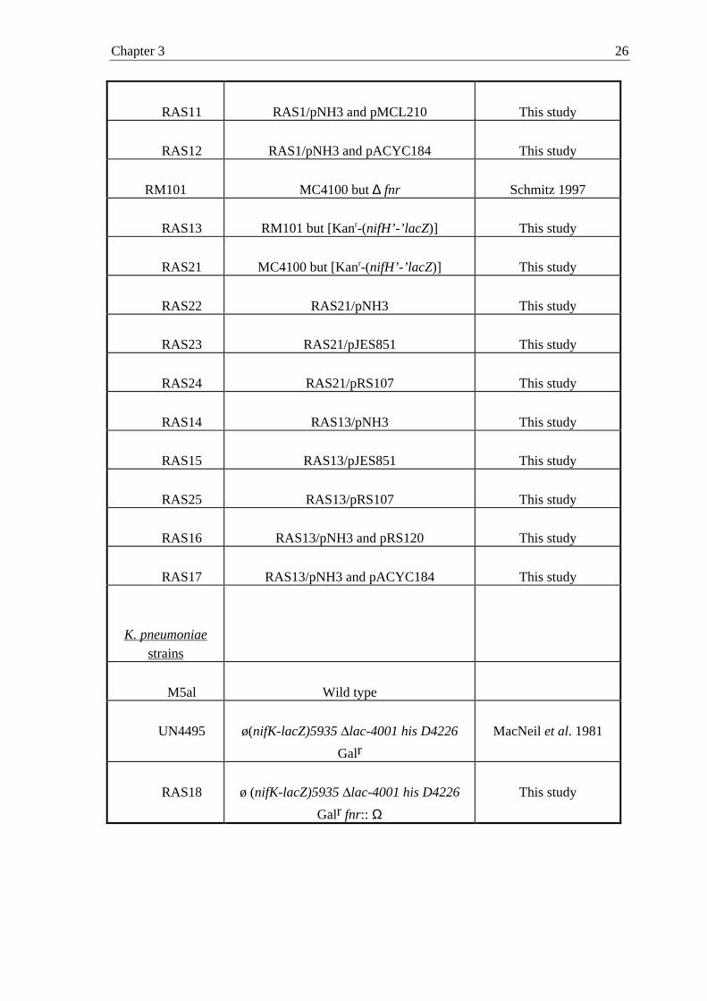
RAS11 RAS1/pNH3 and pMCL210 This study
RAS12 RAS1/pNH3 and pACYC184 This study
RM101 MC4100 but ∆ fnr Schmitz 1997
RAS13 RM101 but [Kanr-(nifH’-’lacZ)] This study
RAS21 MC4100 but [Kanr-(nifH’-’lacZ)] This study
RAS22 RAS21/pNH3 This study
RAS23 RAS21/pJES851 This study
RAS24 RAS21/pRS107 This study
RAS14 RAS13/pNH3 This study
RAS15 RAS13/pJES851 This study
RAS25 RAS13/pRS107 This study
RAS16 RAS13/pNH3 and pRS120 This study
RAS17 RAS13/pNH3 and pACYC184 This study
K. pneumoniae
strains
M5al Wild type
UN4495 ø(nifK-lacZ)5935 lac-4001 his D4226
GalrMacNeil et al. 1981
RAS18 ø (nifK-lacZ)5935 lac-4001 his D4226
Galr fnr:: ΩThis study
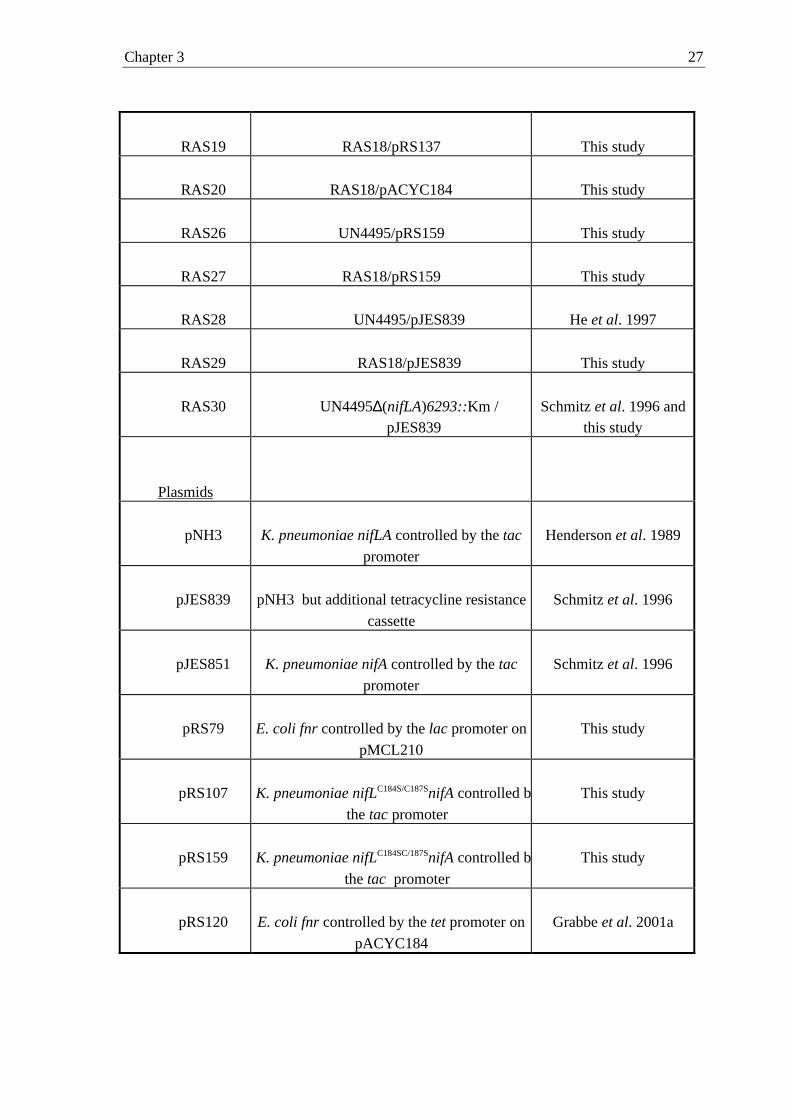
Chapter 3 27
RAS19 RAS18/pRS137 This study
RAS20 RAS18/pACYC184 This study
RAS26 UN4495/pRS159 This study
RAS27 RAS18/pRS159 This study
RAS28 UN4495/pJES839 He et al. 1997
RAS29 RAS18/pJES839 This study
RAS30 UN4495∆(nifLA)6293::Km /pJES839
Schmitz et al. 1996 andthis study
Plasmids
pNH3 K. pneumoniae nifLA controlled by the tac
promoter
Henderson et al. 1989
pJES839 pNH3 but additional tetracycline resistance
cassette
Schmitz et al. 1996
pJES851 K. pneumoniae nifA controlled by the tac
promoter
Schmitz et al. 1996
pRS79 E. coli fnr controlled by the lac promoter on
pMCL210
This study
pRS107 K. pneumoniae nifLC184S/C187SnifA controlled by
the tac promoter
This study
pRS159 K. pneumoniae nifLC184SC/187SnifA controlled by
the tac promoter
This study
pRS120 E. coli fnr controlled by the tet promoter on
pACYC184
Grabbe et al. 2001a
Chapter 3 28
pRS127 2.1 kbp fragment in pBluescript SK+
containing K. pneumoniae fnr
Grabbe et al. 2001a
pRS137 K. pneumoniae fnr controlled by thetet
promoter on pACYC184Grabbe et al. 2001a
pACYC184 Low copy vector New England Biolabs,UK
pMCL210 Low copy vector Nakano et al. 1995
pBluescript
SK+
Cloning vector Stratagene, La Jolla, US
Construction of plasmids. Plasmid pRS107 contains the K. pneumoniae nifLC184S/C187SnifA-
operon under the control of the tac promoter, in which the Cys184 and Cys187 of nifL are
changed to serine (Ser184-Ala-Asp-Ser187). It was constructed from pNH3 (Henderson et al.,
1989) by introducing the double mutation into nifL by site directed mutagenesis. Site directed
mutagenesis was performed using the GeneEditor System (Promega) according to the
protocol of the manufacturer. The double mutation was confirmed by sequencing. Plasmid
pRS159 was constructed by inserting a tetracycline-resistance cassette (Schmitz et al., 1996)
into the ScaI site of plasmid pRS107. Plasmid pRS79 contains the E. coli fnr gene inserted
into the BamHI and PstI site of pMCL210 (Nakano et al., 1995) under the control of the lac
promoter. pRS120 and pRS137 contain E. coli fnr gene and K. pneumoniae fnr gene,
respectively, inserted into the SalI and BamHI site of pACYC184 and thereby expressed from
the tet promoter (Grabbe et al., 2001a).
Growth. K. pneumoniae and E. coli strains were grown under anaerobic conditions with N2 as
gas phase at 30° C in minimal medium (Schmitz et al., 1996) supplemented with 4 mM
glutamine, 10 mM Na2CO
3, 0.3 mM sulfide and 0.002 % resazurine to monitor anaerobiosis.
The medium was further supplemented with 0.004% histidine and with 0.4% sucrose as sole
Chapter 3 29
carbon source for K. pneumoniae strains. For E. coli strains, the medium was supplemented
with 0.1 mM tryptophane and 0.8 % glucose as the carbon source. Precultures were grown
overnight in closed bottles with N2 as gas phase, in medium lacking sulfide and resazurine but
supplemented with 4 mM ammonium acetate in addition to glutamine; both ammonium and
glutamine were completely utilized during growth of precultures. The cultures (25 ml) were
grown in closed bottles with N2 as gas phase at 30° C under strictly anaerobic conditions
without shaking. Samples for monitoring growth at 600 nm and determining ß-galactosidase
activity were taken anaerobically. In E. coli strains carrying a plasmid encoding NifL and
NifA (pNH3 (12)), NifLC184S/C187S and NifA (pRS107) or a plasmid encoding NifA alone
(pJES851 (Schmitz et al., 1996)) expression of nifLA, nifLC184SC/187SnifA or nifA was
induced from the tac promoter with 10 µM IPTG (isopropyl-ß-D-thiogalactopyranoside).
Fnr phenotypes of RAS1, RAS13, RAS18 and the respective complemented strains RAS9,
RAS10, RAS16 and RAS19 were tested anaerobically using glycerol and nitrate (0.5%) as
sole carbon and nitrogen source in minimal medium.
ß-Galactosidase assay. NifA-mediated activation of transcription from the nifHDK promoter
in K. pneumoniae UN4495 and E. coli strains was monitored by measuring the differential
rate of ß-galactosidase synthesis during exponential growth (units per milliliter per OD600)
(Schmitz et al., 1996). Inhibitory effects of NifL on NifA activity were assessed by virtue of a
decrease in nifH expression.
Western blot analysis. Cells were grown anaerobically in minimal medium with glutamine
as nitrogen source, when the culture reached a turbidity of 0.4 to 0.7 at 660 nm, 1 ml samples
of the exponentially growing cultures were harvested and concentrated 20-fold into sodium
dodecyl sulfate (SDS) gel-loading buffer (Laemmli, 1970). Samples were separated by
SDS/polyacrylamide (12%) gel electrophoresis and transferred to nitrocellulose membranes as
described previously (Sambrook et al., 1989). Membranes were exposed to polyclonal rabbit
antisera directed against the NifL or NifA proteins of K. pneumoniae, protein bands were
Chapter 3 30
detected with secondary antibodies directed against rabbit immunoglobulin G and coupled to
horseradish peroxidase (BioRad Laboratories). Purified NifA and NifL from K. pneumoniae
and prestained protein markers (New England Biolabs, UK) were used as standards.
Data deposition. K. pneumoniae fnr sequence has been submitted to GenBank under
accession number AF220669.
Results
We recently showed that in vivo iron is specifically required for nif-induction in
K. pneumoniae, and additionally, that iron is not present in NifL (Schmitz, 1997; Schmitz et
al., 1996). In order to examine whether oxygen is detected by a more general system rather
than by NifL directly we chose to examine the possible influence of Fnr on the nif-induction
in a heterologous E. coli system. We performed all experiments under nitrogen limiting-
growth conditions to exclude NifA inhibition by NifL in response of ammonium presence. If
Fnr is indeed the primary oxygen sensor, which transduces the oxygen signal to NifL, the iron
requirement for the nif-induction under oxygen-limiting conditions may be based on the iron
requirement for the assembly of iron sulfur clusters of Fnr.
Studying the effect of Fnr on the nif-induction in a heterologous E. coli system. In order
to study the effect of Fnr on nif regulation in response to oxygen we chose a heterologous
E. coli system. Strain NCM1529 carrying a chromosomal nifH’-‘lacZ fusion was used as
parental strain (He et al., 1997). NifL and NifA were induced independent of the Ntr system
from plasmids which carried the K. pneumoniae nifLA (pNH3) and nifA (pJES851) genes
under the control of the tac promoter. The two regulatory proteins were induced with 10 µM
IPTG to levels at which NifL function is regulated normally in response to oxygen and
combined nitrogen in E. coli in the absence of nif proteins other than NifA (He et al., 1997).
To study the effect of an fnr null mutation on the regulation of NifL activity in response to
oxygen, an fnr null allele (fnr::Tn10) was introduced by P1 transduction into the parental
Chapter 3 31
strain NCM1529 carrying the ø(nifH’-’lacZ) fusion as described in Materials and Methods,
resulting in strain RAS1. After introducing nifLA and nifA on plasmids, the resulting strains
were generally grown in mineral medium with glucose as sole carbon source and under
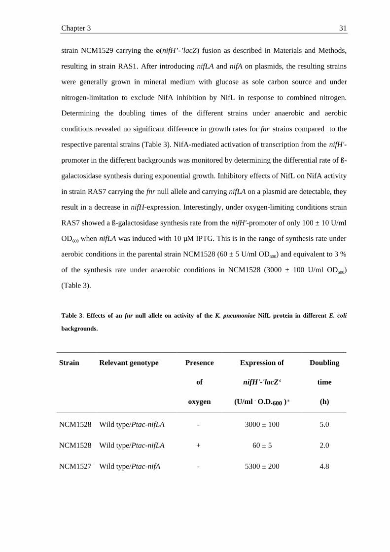
nitrogen-limitation to exclude NifA inhibition by NifL in response to combined nitrogen.
Determining the doubling times of the different strains under anaerobic and aerobic
conditions revealed no significant difference in growth rates for fnr- strains compared to the
respective parental strains (Table 3). NifA-mediated activation of transcription from the nifH'-
promoter in the different backgrounds was monitored by determining the differential rate of ß-
galactosidase synthesis during exponential growth. Inhibitory effects of NifL on NifA activity
in strain RAS7 carrying the fnr null allele and carrying nifLA on a plasmid are detectable, they
result in a decrease in nifH-expression. Interestingly, under oxygen-limiting conditions strain
RAS7 showed a ß-galactosidase synthesis rate from the nifH'-promoter of only 100 ± 10 U/ml
OD600 when nifLA was induced with 10 µM IPTG. This is in the range of synthesis rate under
aerobic conditions in the parental strain NCM1528 (60 ± 5 U/ml OD600) and equivalent to 3 %
of the synthesis rate under anaerobic conditions in NCM1528 (3000 ± 100 U/ml OD600)
(Table 3).
Table 3: Effects of an fnr null allele on activity of the K. pneumoniae NifL protein in different E. coli
backgrounds.
Strain Relevant genotype Presence
of
oxygen
Expression of
nifH'-'lacZ‘
(U/ml . O.D.600 ) a
Doubling
time
(h)
NCM1528 Wild type/Ptac-nifLA - 3000 ± 100 5.0
NCM1528 Wild type/Ptac-nifLA + 60 ± 5 2.0
NCM1527 Wild type/Ptac-nifA - 5300 ± 200 4.8
Chapter 3 32
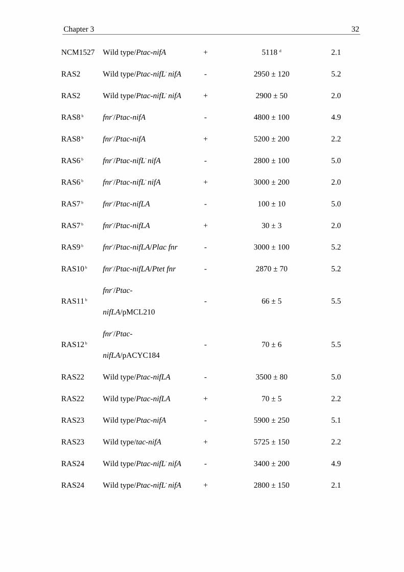
NCM1527 Wild type/Ptac-nifA + 5118 d 2.1
RAS2 Wild type/Ptac-nifL- nifA - 2950 ± 120 5.2
RAS2 Wild type/Ptac-nifL- nifA + 2900 ± 50 2.0
RAS8 b fnr-/Ptac-nifA - 4800 ± 100 4.9
RAS8 b fnr-/Ptac-nifA + 5200 ± 200 2.2
RAS6 b fnr-/Ptac-nifL- nifA - 2800 ± 100 5.0
RAS6 b fnr-/Ptac-nifL- nifA + 3000 ± 200 2.0
RAS7 b fnr-/Ptac-nifLA - 100 ± 10 5.0
RAS7 b fnr-/Ptac-nifLA + 30 ± 3 2.0
RAS9 b fnr-/Ptac-nifLA/Plac fnr - 3000 ± 100 5.2
RAS10 b fnr-/Ptac-nifLA/Ptet fnr - 2870 ± 70 5.2
RAS11 b
fnr-/Ptac-
nifLA/pMCL210
- 66 ± 5 5.5
RAS12 b
fnr-/Ptac-
nifLA/pACYC184- 70 ± 6 5.5
RAS22 Wild type/Ptac-nifLA - 3500 ± 80 5.0
RAS22 Wild type/Ptac-nifLA + 70 ± 5 2.2
RAS23 Wild type/Ptac-nifA - 5900 ± 250 5.1
RAS23 Wild type/tac-nifA + 5725 ± 150 2.2
RAS24 Wild type/Ptac-nifL- nifA - 3400 ± 200 4.9
RAS24 Wild type/Ptac-nifL- nifA + 2800 ± 150 2.1
Chapter 3 33
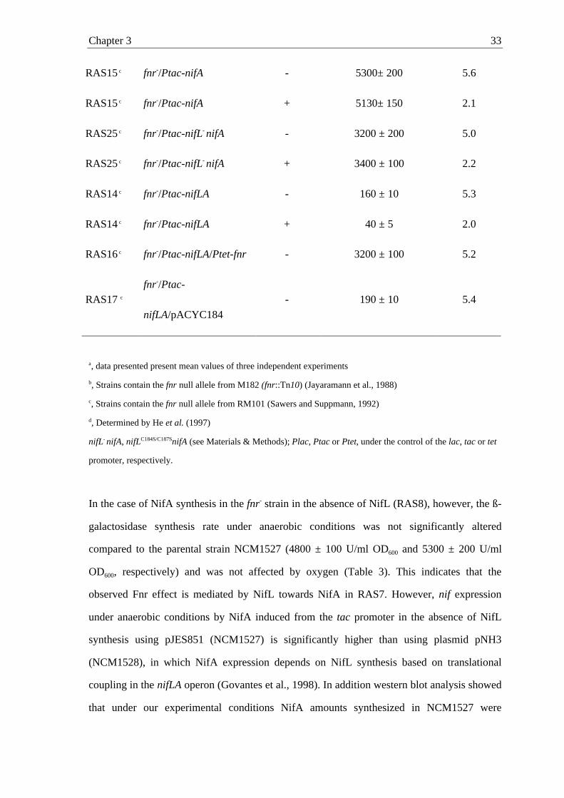
RAS15 c fnr-/Ptac-nifA - 5300± 200 5.6
RAS15 c fnr-/Ptac-nifA + 5130± 150 2.1
RAS25 c fnr-/Ptac-nifL- nifA - 3200 ± 200 5.0
RAS25 c fnr-/Ptac-nifL- nifA + 3400 ± 100 2.2
RAS14 c fnr-/Ptac-nifLA - 160 ± 10 5.3
RAS14 c fnr-/Ptac-nifLA + 40 ± 5 2.0
RAS16 c fnr-/Ptac-nifLA/Ptet-fnr - 3200 ± 100 5.2
RAS17 c
fnr-/Ptac-
nifLA/pACYC184
- 190 ± 10 5.4
a, data presented present mean values of three independent experiments
b, Strains contain the fnr null allele from M182 (fnr::Tn10) (Jayaramann et al., 1988)
c, Strains contain the fnr null allele from RM101 (Sawers and Suppmann, 1992)
d, Determined by He et al. (1997)
nifL- nifA, nifLC184S/C187SnifA (see Materials & Methods); Plac, Ptac or Ptet, under the control of the lac, tac or tet
promoter, respectively.
In the case of NifA synthesis in the fnr- strain in the absence of NifL (RAS8), however, the ß-
galactosidase synthesis rate under anaerobic conditions was not significantly altered
compared to the parental strain NCM1527 (4800 ± 100 U/ml OD600 and 5300 ± 200 U/ml
OD600, respectively) and was not affected by oxygen (Table 3). This indicates that the
observed Fnr effect is mediated by NifL towards NifA in RAS7. However, nif expression
under anaerobic conditions by NifA induced from the tac promoter in the absence of NifL
synthesis using pJES851 (NCM1527) is significantly higher than using plasmid pNH3
(NCM1528), in which NifA expression depends on NifL synthesis based on translational
coupling in the nifLA operon (Govantes et al., 1998). In addition western blot analysis showed
that under our experimental conditions NifA amounts synthesized in NCM1527 were
Chapter 3 34
approximately 30 - 40 % higher compared to NifA amounts synthesized in NCM1528 (data
not shown). To rule out that nif expression in the fnr mutant using pJES851 (RAS8) is not due
to this increase in NifA expression we additionally constructed pRS107 containing
nifLC184S/C187SnifA translationally coupled under the control of the tac promoter (see Materials
and Methods). IPTG induction in NCM1529 containing pRS107 (RAS2) resulted in NifA
expression comparable to NCM1528 (data not shown) and expression of NifLC184S/C187S, which
completely lost its nitrogen and oxygen regulatory function (Klopprogge and Schmitz,
unpublished). Determination of ß-galactosidase synthesis rates showed, that nif-induction by
NifA expressed from pRS107 in the absence of a functional NifL protein was again not
affected by the fnr mutation (compare RAS2 with RAS6) and was in the range of nif
induction in NCM1528 under anaerobic conditions (Table 3). These findings indicate that the
fnr null allele is not affecting NifA activity directly in the absence of functional NifL. In the
presence of both regulatory proteins, however, NifL inhibits NifA activity under oxygen-
limiting conditions when Fnr is absent, suggesting that the Fnr effect is mediated through
NifL to NifA.
The finding that in the absence of Fnr NifL inhibits NifA activity under oxygen-limiting
conditions to the same amount as under aerobic growth conditions indicates that NifL
apparently does not receive the signal of anaerobiosis, when Fnr is absent. To confirm this
observation, we analyzed the nif-induction under anaerobic conditions in a different fnr
mutant strain (RAS13). After introduction of nifLA, nifA and nifLC184S/C187SnifA and on
plasmids, the respective strains RAS14, RAS15 and RAS25 were grown under oxygen-
limitation. By determining the ß-galactosidase synthesis rates from the nifH'-promoter in
RAS14, we observed that in this independent fnr mutant strain the nif-induction was 160 ± 10
U/ml OD600, when nifLA was expressed under anaerobic conditions. This nif-induction is
again significantly lower than in the parental strain RAS22 (3500 ± 80 U/ml OD600) and is in
the range of aerobic nif-induction in the parental strain (70 ± 5 U/ml OD600) (Table 3). Similar
to RAS8 and RAS6 the ß-galactosidase synthesis rate in the case of NifA synthesis in the
absence of a functional NifL was not affected by the fnr- mutation (RAS15 compared to
RAS23 and RAS25 compared to RAS24).
Chapter 3 35
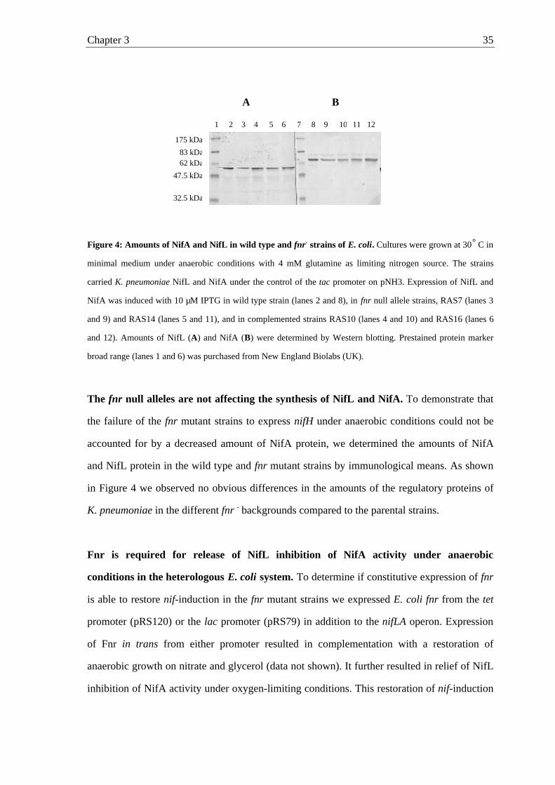
Figure 4: Amounts of NifA and NifL in wild type and fnr- strains of E. coli. Cultures were grown at 30° C in
minimal medium under anaerobic conditions with 4 mM glutamine as limiting nitrogen source. The strains
carried K. pneumoniae NifL and NifA under the control of the tac promoter on pNH3. Expression of NifL and
NifA was induced with 10 µM IPTG in wild type strain (lanes 2 and 8), in fnr null allele strains, RAS7 (lanes 3
and 9) and RAS14 (lanes 5 and 11), and in complemented strains RAS10 (lanes 4 and 10) and RAS16 (lanes 6
and 12). Amounts of NifL (A) and NifA (B) were determined by Western blotting. Prestained protein marker
broad range (lanes 1 and 6) was purchased from New England Biolabs (UK).
The fnr null alleles are not affecting the synthesis of NifL and NifA. To demonstrate that
the failure of the fnr mutant strains to express nifH under anaerobic conditions could not be
accounted for by a decreased amount of NifA protein, we determined the amounts of NifA
and NifL protein in the wild type and fnr mutant strains by immunological means. As shown
in Figure 4 we observed no obvious differences in the amounts of the regulatory proteins of
K. pneumoniae in the different fnr - backgrounds compared to the parental strains.
Fnr is required for release of NifL inhibition of NifA activity under anaerobic
conditions in the heterologous E. coli system. To determine if constitutive expression of fnr
is able to restore nif-induction in the fnr mutant strains we expressed E. coli fnr from the tet
promoter (pRS120) or the lac promoter (pRS79) in addition to the nifLA operon. Expression
of Fnr in trans from either promoter resulted in complementation with a restoration of
anaerobic growth on nitrate and glycerol (data not shown). It further resulted in relief of NifL
inhibition of NifA activity under oxygen-limiting conditions. This restoration of nif-induction
1 3 6 7542 10 1198 12
175 kDa
83 kDa62 kDa
47.5 kDa
32.5 kDa
A B
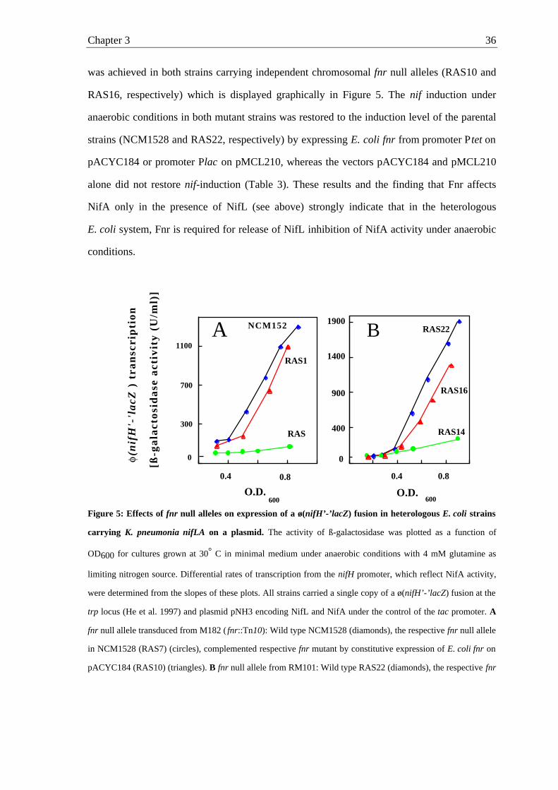
Chapter 3 36
was achieved in both strains carrying independent chromosomal fnr null alleles (RAS10 and
RAS16, respectively) which is displayed graphically in Figure 5. The nif induction under
anaerobic conditions in both mutant strains was restored to the induction level of the parental
strains (NCM1528 and RAS22, respectively) by expressing E. coli fnr from promoter Ptet on
pACYC184 or promoter Plac on pMCL210, whereas the vectors pACYC184 and pMCL210
alone did not restore nif-induction (Table 3). These results and the finding that Fnr affects
NifA only in the presence of NifL (see above) strongly indicate that in the heterologous
E. coli system, Fnr is required for release of NifL inhibition of NifA activity under anaerobic
conditions.
Figure 5: Effects of fnr null alleles on expression of a ø(nifH’-’lacZ) fusion in heterologous E. coli strains
carrying K. pneumonia nifLA on a plasmid. The activity of ß-galactosidase was plotted as a function of
OD600 for cultures grown at 30° C in minimal medium under anaerobic conditions with 4 mM glutamine as
limiting nitrogen source. Differential rates of transcription from the nifH promoter, which reflect NifA activity,
were determined from the slopes of these plots. All strains carried a single copy of a ø(nifH’-’lacZ) fusion at the
trp locus (He et al. 1997) and plasmid pNH3 encoding NifL and NifA under the control of the tac promoter. A
fnr null allele transduced from M182 ( fnr::Tn10): Wild type NCM1528 (diamonds), the respective fnr null allele
in NCM1528 (RAS7) (circles), complemented respective fnr mutant by constitutive expression of E. coli fnr on
pACYC184 (RAS10) (triangles). B fnr null allele from RM101: Wild type RAS22 (diamonds), the respective fnr
O.D.600
300
700
1100
0
0.4 0.8
RAS1
NCM152
RAS
A
O.D.600
1400
1900
B0, 0, 1
400
900
0
0.80.4
RAS22
RAS16
RAS14
B
[ß-g
ala
cto
sid
ase
act
ivit
y (
U/m
l)]
(nif
H'-
'la
cZ )
tra
nsc
rip
tio
n
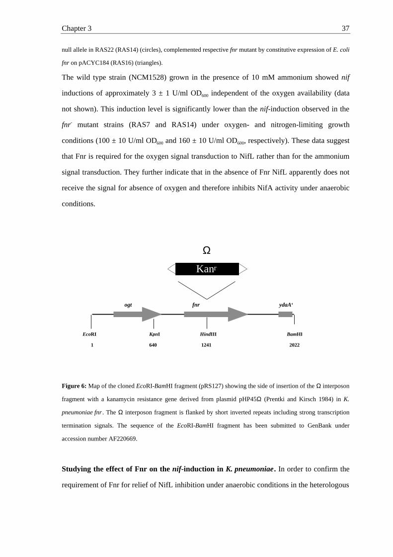
Chapter 3 37
null allele in RAS22 (RAS14) (circles), complemented respective fnr mutant by constitutive expression of E. coli
fnr on pACYC184 (RAS16) (triangles).
The wild type strain (NCM1528) grown in the presence of 10 mM ammonium showed nif
inductions of approximately 3 ± 1 U/ml OD600 independent of the oxygen availability (data
not shown). This induction level is significantly lower than the nif-induction observed in the
fnr- mutant strains (RAS7 and RAS14) under oxygen- and nitrogen-limiting growth
conditions (100 ± 10 U/ml OD600 and 160 ± 10 U/ml OD600, respectively). These data suggest
that Fnr is required for the oxygen signal transduction to NifL rather than for the ammonium
signal transduction. They further indicate that in the absence of Fnr NifL apparently does not
receive the signal for absence of oxygen and therefore inhibits NifA activity under anaerobic
conditions.
Figure 6: Map of the cloned EcoRI-BamHI fragment (pRS127) showing the side of insertion of the Ω interposon
fragment with a kanamycin resistance gene derived from plasmid pHP45Ω (Prentki and Kirsch 1984) in K.
pneumoniae fnr . The Ω interposon fragment is flanked by short inverted repeats including strong transcription
termination signals. The sequence of the EcoRI-BamHI fragment has been submitted to GenBank under
accession number AF220669.
Studying the effect of Fnr on the nif-induction in K. pneumoniae. In order to confirm the
requirement of Fnr for relief of NifL inhibition under anaerobic conditions in the heterologous
BamHIEcoRI KpnI HindIII
ogt ydaA‘fnr
1 640 1241 2022
Kanr
Ω
Chapter 3 38
E. coli system, we constructed a chromosomal fnr null allele in K. pneumoniae. We used
K. pneumoniae strain UN4495 carrying nifLA and a nifK-lacZ fusion on the chromosome,
which allows monitoring of NifA-mediated transcription from the nifHDK-promoter by
measuring the differential rate of ß-galactosidase synthesis (Schmitz et al., 1996). The fnr
deletion was constructed on a plasmid by inserting an Ω interposon fragment with a
kanamycin resistance gene into K. pneumoniae fnr (Figure 6), which was than introduced into
the chromosome by marker exchange using the sac system (see Materials and Methods). The
disruption of the fnr gene was confirmed by PCR and southern blot analysis (data not shown).
Klebsiella strains with the exception of RAS26 and RAS27 were generally grown in minimal
medium under nitrogen limitation to exclude NifA inhibition by NifL in response to
ammonium. The fnr::Ω mutation in K. pneumoniae UN4495 did not result in a significant
growth-rate reduction, but did reduce the nif-induction under oxygen-limiting conditions to 10
% of the nif-induction in the parental strain. The observed induction level of the
K. pneumoniae fnr mutant strain (RAS18) under anaerobic conditions (400 ± 20 U / ml OD600)
again is in the same range as the nif-induction in the presence of oxygen in the parental K.
pneumoniae strain (220 ± 20 U / ml OD600) (Table 4). Determination of NifA and NifL
proteins in the fnr- mutant strain revealed no differences in the amount of the regulatory
proteins compared to the parental strain (data not shown), indicating that the failure to express
nifH could not be accounted for by a decrease of NifA expression. Normal NifL/NifA-
dependent regulation was restored by introduction of the K. pneumoniae fnr gene expressed
from the tet promoter on pRS137 into the fnr mutant (Figure 7). nif induction in the
complemented mutant (RAS19) was determined to be 3800 ± 50 U / ml OD600, whereas the
low copy vector pACYC184 alone did not result in complementation (RAS20). These
findings in the native background again suggest that Fnr is required for nif expression in
K. pneumoniae under anaerobic conditions.
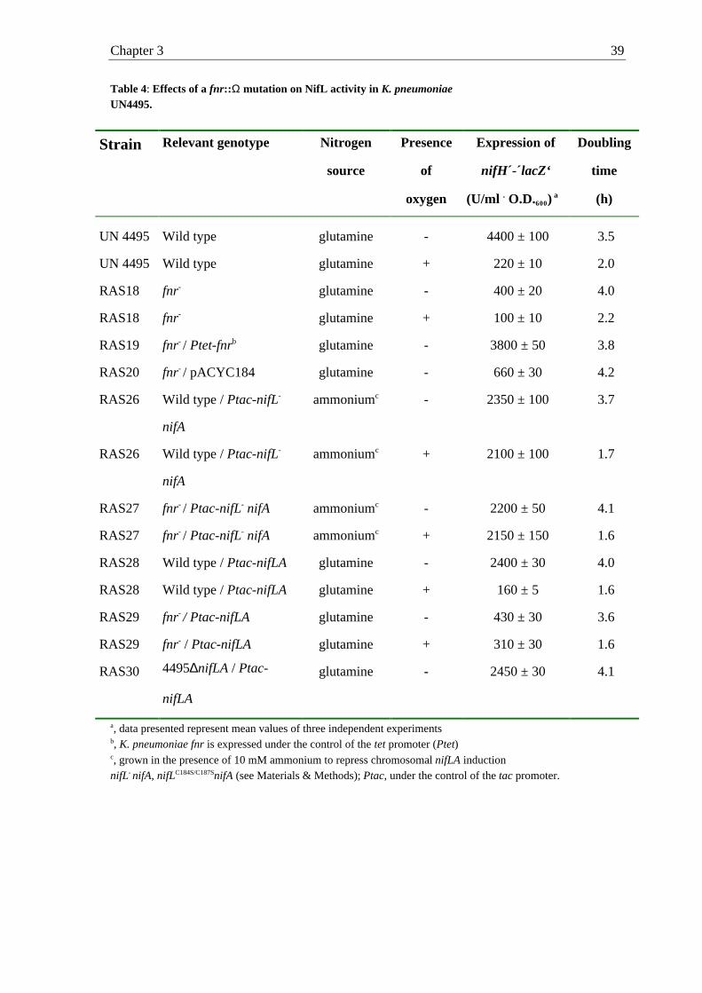
Chapter 3 39
Table 4: Effects of a fnr::Ω mutation on NifL activity in K. pneumoniaeUN4495.
Strain Relevant genotype Nitrogen
source
Presence
of
oxygen
Expression of
nifH´-´lacZ‘
(U/ml . O.D.600) a
Doubling
time
(h)
UN 4495 Wild type glutamine - 4400 ± 100 3.5
UN 4495 Wild type glutamine + 220 ± 10 2.0
RAS18 fnr- glutamine - 400 ± 20 4.0
RAS18 fnr- glutamine + 100 ± 10 2.2
RAS19 fnr- / Ptet-fnrb glutamine - 3800 ± 50 3.8
RAS20 fnr- / pACYC184 glutamine - 660 ± 30 4.2
RAS26 Wild type / Ptac-nifL-
nifA
ammoniumc - 2350 ± 100 3.7
RAS26 Wild type / Ptac-nifL-
nifA
ammoniumc + 2100 ± 100 1.7
RAS27 fnr- / Ptac-nifL- nifA ammoniumc - 2200 ± 50 4.1
RAS27 fnr- / Ptac-nifL- nifA ammoniumc + 2150 ± 150 1.6
RAS28 Wild type / Ptac-nifLA glutamine - 2400 ± 30 4.0
RAS28 Wild type / Ptac-nifLA glutamine + 160 ± 5 1.6
RAS29 fnr- / Ptac-nifLA glutamine - 430 ± 30 3.6
RAS29 fnr- / Ptac-nifLA glutamine + 310 ± 30 1.6
RAS30 4495∆nifLA / Ptac-
nifLA
glutamine - 2450 ± 30 4.1
a, data presented represent mean values of three independent experimentsb, K. pneumoniae fnr is expressed under the control of the tet promoter (Ptet)c, grown in the presence of 10 mM ammonium to repress chromosomal nifLA inductionnifL- nifA, nifLC184S/C187SnifA (see Materials & Methods); Ptac, under the control of the tac promoter.
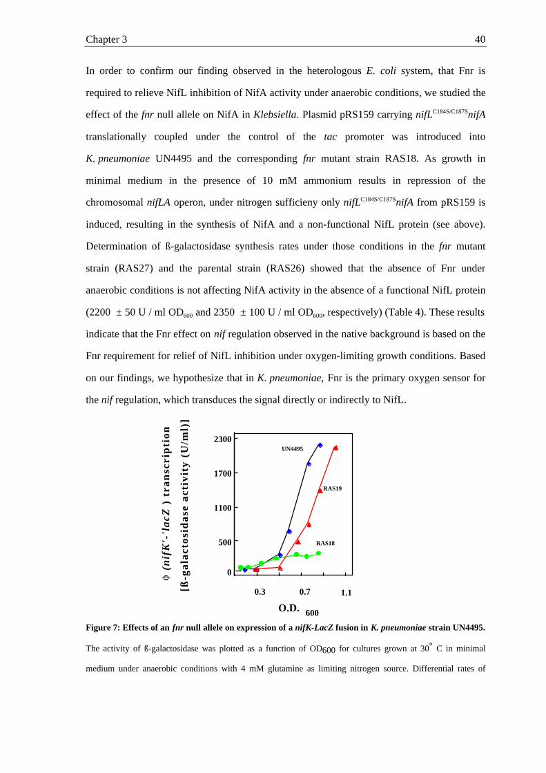
Chapter 3 40
In order to confirm our finding observed in the heterologous E. coli system, that Fnr is
required to relieve NifL inhibition of NifA activity under anaerobic conditions, we studied the
effect of the fnr null allele on NifA in Klebsiella. Plasmid pRS159 carrying nifLC184S/C187SnifA
translationally coupled under the control of the tac promoter was introduced into
K. pneumoniae UN4495 and the corresponding fnr mutant strain RAS18. As growth in
minimal medium in the presence of 10 mM ammonium results in repression of the
chromosomal nifLA operon, under nitrogen sufficieny only nifLC184S/C187SnifA from pRS159 is
induced, resulting in the synthesis of NifA and a non-functional NifL protein (see above).
Determination of ß-galactosidase synthesis rates under those conditions in the fnr mutant
strain (RAS27) and the parental strain (RAS26) showed that the absence of Fnr under
anaerobic conditions is not affecting NifA activity in the absence of a functional NifL protein
(2200 ± 50 U / ml OD600 and 2350 ± 100 U / ml OD600, respectively) (Table 4). These results
indicate that the Fnr effect on nif regulation observed in the native background is based on the
Fnr requirement for relief of NifL inhibition under oxygen-limiting growth conditions. Based
on our findings, we hypothesize that in K. pneumoniae, Fnr is the primary oxygen sensor for
the nif regulation, which transduces the signal directly or indirectly to NifL.
Figure 7: Effects of an fnr null allele on expression of a nifK-LacZ fusion in K. pneumoniae strain UN4495.
The activity of ß-galactosidase was plotted as a function of OD600 for cultures grown at 30° C in minimal
medium under anaerobic conditions with 4 mM glutamine as limiting nitrogen source. Differential rates of
500
1100
1700
2300
O.D. 600
0
0.3 0.7 1.1
UN4495
RAS19
RAS18
[ß-g
ala
cto
sid
ase
act
ivit
y (
U/m
l)]
(nif
K'-
'la
cZ )
tra
nsc
rip
tio
n
Chapter 3 41
transcription from the nifHDK promoter were determined from the slopes of these plots. Wild type UN4495
(diamonds), the fnr mutant strain of UN4495 (RAS18) (circles), complemented respective fnr mutant by
constitutive expression of K. pneumoniae fnr on pACYC184 (RAS19) (triangles).
Discussion
Our goal is to determine how K. pneumoniae NifL perceives the oxygen status of the cells in
order to regulate NifA activity in response to environmental oxygen. The main question
concerning the oxygen signal transduction is whether NifL senses oxygen directly via a
redox-induced conformational change, or whether oxygen is detected by a more general
system. After receiving the oxygen signal, directly or indirectly, the redox state of the
flavoprotein NifL is thought to influence the ability of NifL to modulate the NifA activity in
response to environmental oxygen, and to allow NifA activity only in the absence of oxygen
(Hill et al., 1996; Macheroux et al., 1998; Schmitz 1996). We recently showed that iron is
specifically required for nif- induction, but is not present in NifL (Schmitz, 1997; Schmitz et
al., 1996). To determine whether this iron requirement for nif induction could be accounted
for by the role of Fnr in transducing the oxygen signal to NifL, we determined the effect of an
fnr null allele on nif regulation. Using different genetic backgrounds and independent fnr null
alleles, we were able to show that the absence of Fnr effects the nif regulation dramatically.
The nif-induction in the absence of Fnr was low, similar to the nif-induction under aerobic
conditions, even though cells were growing under oxygen limitation. Normal nif regulation
was achieved in the mutant strains by introduction of a low-copy vector expressing fnr
constitutively (Figures 6 and 8). These data indicate that Fnr is required to relieve NifL
inhibition of NifA activity under anaerobic conditions and this appears to account for the iron
requirement of nif induction (Schmitz et al.,1996). Therefore, in addition to the rhizobial
homologous Fnr proteins, FnrN and FixK, which are known to be involved in regulation of
nitrogen fixation in the symbiotic bacteria (Fischer, 1994 and therein cited papers, Guiterrez
et al., 1997), in K. pneumoniae the transcriptional activator Fnr is apparently also involved in
regulation of nitrogen fixation. These results are in contrast to the report of Hill (1985), that
Chapter 3 42
redox regulation of nif expression in a heterologous E. coli strain is independent of the E. coli
fnr gene product. This discrepancy may be due to experimental differences. We determined
NifA-mediated transcriptional activation by measuring differential rates of β-galactosidase
expression from a chromosomal nifK-lacZ fusion in order to monitor nif induction. In
contrast, Hill determined acetylene reduction by nitrogenase after growing heterologous
E. coli fnr- strains carrying the Nif+ plasmid pRD1 under derepressing conditions. Also, as
plasmid pRD1 contains in addition to the nif genes non-identified K. pneumoniae genes
(Dixon et al., 1976) we cannot completely rule out that K. pneumoniae fnr is encoded on the
plasmid. Apart from these experimental differences concerning the heterologous E. coli
systems we confirmed the Fnr requirement for the nif regulation in the native genetic
background K. pneumoniae.
We further showed that the general oxygen sensor Fnr is required for relief of NifL inhibition
under anaerobic growth conditions and that the presence of ammonium results in significantly
lower nif-inductions in the wilde type strain than observed in fnr mutant strains under
nitrogen- and oxygen-limitation. Both these findings suggest, that the oxygen signal is not
detected by NifL directly but by Fnr, which transduces the signal - directly or indirectly - to
NifL. However, at this state of experimental data we cannot completely rule out that the Fnr
requirement might be due to some Fnr-dependent metabolic signals not directly related to the
lack of oxygen. If Fnr is indeed the primary oxygen sensor for the nif regulation in
K. pneumoniae, it still remains to be explained how the oxygen signal is transmitted to NifL.
Fnr is either transducing the oxygen signal by directly interacting with NifL in the absence of
oxygen or under anaerobic conditions Fnr is activating the transcription of gene(s) whose
product(s) mediate the signal to NifL. As Fnr is a transcriptional activator and can be
excluded as the physiological electron donor for NifL reduction it is more reasonably that
under anaerobic conditions Fnr transduces the signal by transcriptional activation.
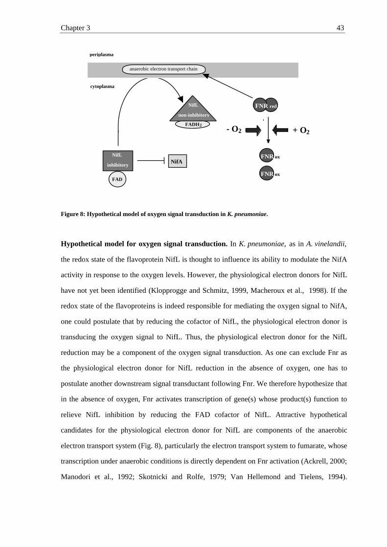
Chapter 3 43
Figure 8: Hypothetical model of oxygen signal transduction in K. pneumoniae.
Hypothetical model for oxygen signal transduction. In K. pneumoniae, as in A. vinelandii,
the redox state of the flavoprotein NifL is thought to influence its ability to modulate the NifA
activity in response to the oxygen levels. However, the physiological electron donors for NifL
have not yet been identified (Klopprogge and Schmitz, 1999, Macheroux et al., 1998). If the
redox state of the flavoproteins is indeed responsible for mediating the oxygen signal to NifA,
one could postulate that by reducing the cofactor of NifL, the physiological electron donor is
transducing the oxygen signal to NifL. Thus, the physiological electron donor for the NifL
reduction may be a component of the oxygen signal transduction. As one can exclude Fnr as
the physiological electron donor for NifL reduction in the absence of oxygen, one has to
postulate another downstream signal transductant following Fnr. We therefore hypothesize that
in the absence of oxygen, Fnr activates transcription of gene(s) whose product(s) function to
relieve NifL inhibition by reducing the FAD cofactor of NifL. Attractive hypothetical
candidates for the physiological electron donor for NifL are components of the anaerobic
electron transport system (Fig. 8), particularly the electron transport system to fumarate, whose
transcription under anaerobic conditions is directly dependent on Fnr activation (Ackrell, 2000;
Manodori et al., 1992; Skotnicki and Rolfe, 1979; Van Hellemond and Tielens, 1994).
periplasma
cytoplasma
anaerobic electron transport chain
NifL
inhibitory
FAD
FADH2
NifL
non-inhibitory
NifA
FNR red
FNR ox
FNR ox
- O2 + O2

Chapter 3 44
Preliminary data, which indicate that K. pneumoniae NifL under anaerobic conditions is
membrane-associated, whereas in the presence of oxygen NifL is in the cytosolic fraction,
support this model (Klopprogge and Schmitz, unpublished). Studies of the anaerobic electron
transport system components as potential physiological electron donors for NifL are in
process.
Chapter 4 45
Chapter 4:
Membrane association of Klebsiella pneumoniae NifL is affected
by molecular oxygen and combined nitrogen
KAI KLOPPROGGE , ROMAN GRABBE , MICHEAL HOPPERT, AND RUTH A. SCHMITZ, Abstract In the diazotroph Klebsiella pneumoniae, NifL and NifA regulate transcription of the nitrogen
fixation genes in response to molecular oxygen and combined nitrogen. We recently showed
that Fnr is the primary oxygen sensor, which transduces the oxygen signal towards the
negative regulator NifL by activating genes whose products reduce the FAD moiety of NifL
under anaerobic conditions. Potentially, these Fnr-dependent gene products could be
membrane-bound components of the anaerobic electron transport chain; consequently, in this
study we now examine the localization of NifL within the cell under various growth
conditions. In K. pneumoniae grown under oxygen- and nitrogen-limited conditions,
approximately 55 % of the total NifL protein were found in the membrane fraction. However,
when the cells were grown aerobically or shifted to nitrogen sufficiency, less than 10 % of
total NifL was membrane-associated. In contrast to NifL, NifA was located in the cytoplasm
under all growth conditions tested. Further studies using K. pneumoniae mutant strains
showed that under derepressing conditions but in the absence of either the primary oxygen
sensor Fnr or the primary nitrogen sensor GlnK and the ammonium transporter AmtB, NifL
was located in the cytoplasm and inhibited NifA activity. These findings suggest that under
nitrogen- and oxygen-limitation, a significant higher membrane affinity of NifL may create a
spatial gap between NifL and its cytoplasmic target protein NifA thereby impairing inhibition
of NifA by NifL. Localization of GlnK further showed that under nitrogen-limited conditions,
but independent of oxygen presence, 15 - 20 % of the total GlnK is membrane associated. Introduction The free-living diazotroph Klebsiella pneumoniae is able to fix molecular nitrogen under
anaerobic and nitrogen-limited growth conditions. To avoid unnecessary consumption of
energy synthesis of nitrogenase is tightly controlled by the regulatory nitrogen fixation operon
nifLA (for review see Dixon, 1998; Schmitz et al., 2001). Products of the nifLA operon
regulate transcription of the other nif genes in response to environmental signals. NifA is the
Chapter 4 46
transcriptional activator of all of the nif operons except the nifLA operon, which is under
control of the global nitrogen regulatory system, ntr (Drummond and Wootton, 1987; Blanco
et al., 1993); NifL antagonizes the transcriptional activity of NifA in response to combined
nitrogen and molecular oxygen by direct protein-protein interaction with NifA (Merrick et al.,
1982; Hill et al., 1981; Money et al., 1999; Lei et al., 1999; Money et al., 2001; Little et al.,
2000, Barrett et al., 2001).
External nitrogen availability is apparently perceived by K. pneumoniae through changes in
the internal glutamine pool (Schmitz, 2000), which are subsequently mediated towards the nif
regulatory system. Recent evidence strongly suggests that the nitrogen status of the cells is
transduced towards the NifL/NifA regulatory system in K. pneumoniae and Azotobacter
vinelandii by the GlnK protein, a paralogue PII-protein (He et al., 1998; Jack et al., 1999;
Arcondéguy et al., 1999 and 2000; Little et al., 2000; Reyes-Ramirez et al., 2001). The effect
of GlnK, which apparently interacts with NifL or affects the NifL/NifA-complex via direct
protein-protein interaction, appears to be contradictory in K. pneumoniae and A. vinelandii
(He et al., 1998; Jack et al., 1999; Little et al., 2000; Reyes-Ramirez et al., 2001). The oxygen
signal is received by NifL, which contains an N-terminally bound flavin adenine dinucleotide
(FAD) as a prosthetic group. Recent work has shown that the flavoprotein NifL acts as a
redox-sensitive regulatory protein, which modulates NifA activity in response to the redox
state of its FAD cofactor and allows NifA activity only in the absence of oxygen (Hill et al.,
1996; Schmitz, 1997; Macheroux et al., 1998; Little et al., 2000). Thus, under anaerobic
conditions in the absence of combined nitrogen, reduction of the flavin moiety of NifL results
in a non-inhibitory conformation of the NifL protein. We have recently shown that in K.
pneumoniae, the global transcriptional regulator Fnr is required to mediate the signal of
anaerobiosis to NifL (Grabbe et al., 2001b). Thus, we proposed that in the absence of oxygen
the primary oxygen sensor Fnr activates transcription of a gene or genes whose product or
products reduce the FAD cofactor of NifL, resulting in a non-inhibitory conformation of the
protein, assuming the absence of a sufficient nitrogen source. Candidates for the physiological
electron donor for NifL reduction include those components of the anaerobic electron
transport system with Fnr-dependent synthesis (Grabbe et al., 2001b). This model implies that
under anaerobic conditions, NifL will contact the cytoplasmic membrane during the reduction
of its flavin cofactor. If NifL reduction indeed occurs by a membrane-associated electron
donor, this provides a potential mechanism for the signal transduction of anaerobiosis that is
similar to the signal transduction of oxygen presence proposed for the Escherichia coli FAD-
containing aerotaxis protein Aer (Bibikov et al. 1997; Rebbapragada et al.,
Chapter 4 47
1997).
Our goal is to analyze the reduction process of NifL-bound FAD and the subsequent
conformational change of the protein. We therefore localized of NifL protein in K.
pneumoniae and K. pneumoniae mutant strains grown under various conditions. We report
here that under derepressing conditions, NifL shows a significantly higher association with
the cytoplasmic membrane than in the presence of either molecular oxygen or combined
nitrogen. As the presence of molecular oxygen or combined nitrogen results in nif gene
repression, these findings imply that a spatial separation of cytoplasmic NifA and its
antagonist NifL may be responsible for nif gene induction under oxygen- and nitrogen-limited
conditions.
Materials and methods Bacterial strains and plasmids
K. pneumoniae strains M5al (wild-type), M5al containing plasmid pNH3, and K. pneumoniae
UN4495 (ø(nifK-lacZ)5935 ∆lac-4001 hisD4226 Galr) (MacNeil et al., 1981) and mutant
derivatives (K. pneumoniae UN4495 fnr::Ω (RAS18, Grabbe et al., 2001b), K. pneumoniae
UN4495 glnK::KIXX) were used in this study. The glnK::KIXX allele was transferred from
K. pneumoniae UNF3433 (Jack et al., 1999) into K. pneumoniae UN4495 by P1-mediated
transduction with selection for kanamycin resistance, resulting in RAS36. Plasmid pNH3
carries the K. pneumoniae nifLA operon under the control of the tac promoter (Henderson et
al., 1989).
Growth conditions
K. pneumoniae strains were grown anaerobically with molecular nitrogen (N2) as gas phase at
30°C in minimal medium supplemented with either 2 mM glutamine (nitrogen limitation) or
10 mM ammonium (nitrogen sufficiency) as the sole nitrogen source, 0.004 % histidine, 10
mM Na2CO3, 0.3 mM sulfide and 1 % sucrose as the sole carbon source (Schmitz et al.,
1996). To monitor anaerobiosis, the medium was further supplemented with 0.002 %
resazurin. Precultures were grown overnight in closed bottles with N2 as gas phase in the
same medium but lacking sulfide and resazurin. The 1-l main cultures were inoculated from
precultures and incubated in closed bottles with molecular nitrogen as gas phase under strictly
anaerobic conditions without shaking. Samples were taken anaerobically for monitoring the
optical density at 600 nm and determining ß-galactosidase activity. Aerobic 1-l cultures were
incubated in 2-l flasks with vigorous shaking (130 rpm) using the same medium and culture
Chapter 4 48
supplements as described for the anaerobic growth but lacking Na2CO3, sulfide and resazurin.
For ammonium shift experiments, 1-l cultures growing under nitrogen limitation in the
presence of 2 mM glutamine were shifted to nitrogen sufficiency by the addition of 10 mM
NH4Cl during mid-exponential growth; the shifted cultures were further incubated for 2 h
before the cells were harvested.
β-Galactosidase assay
NifA-mediated activation of transcription from the nifHDK promoter in K. pneumoniae
UN4495 and mutant derivatives (UN4495 fnr::Ω and UN4495 glnK::KIXX) was monitored
by measuring the differential rate of β-galactosidase synthesis during exponential growth (U
ml-1 (OD600) -1) as described by Schmitz et al. (1996). Inhibitory effects of NifL on NifA
activity were assessed by a decrease in nifH expression.
Electron microscopy
For electron microscopy, K. pneumoniae strain M5al carrying a plasmid-borne nifLA-operon
under the control of the tac promoter was used (pNH3, Henderson et al., 1989) in addition to
the chromosomal nifLA operon. Cultures (50 ml) were incubated anaerobically in minimal
medium supplemented with either 2 mM glutamine or 10 mM NH4Cl as the sole nitrogen
source, as described above. During growth additional NifL and NifA synthesis from the
plasmid was induced by the addition of 10 µM IPTG. When cultures reached an OD600 of 0.8,
cells were harvested by centrifugation at 10.000 x g under anaerobic conditions. The resulting
cell pellet was resuspended in 50 mM potassium phosphate buffer, pH 7.3, and cells were
chemically fixed in 0.2 % (w/v) formaldehyde and 0.3 % (w/v) glutardialdehyde solution for
90 min at 0 °C in a closed reaction cup under anaerobic conditions. Finally, the cells were
dehydrated in a graded methanol series and embedded in Lowicryl K4M resin under air (Roth
et al., 1981; Hoppert and Holzenburg, 1998). Resin sections of 80 - 100 nm in thickness were
cut with glass knives. NifL was localized in resin sections using specific polyclonal antisera
directed against NifL and goat-anti-rabbit-IgG linked to colloidal gold (10 nm in diameter,
BBI, Cardiff, UK), essentially as described by Roth et al. (1978) with some modifications
(Hoppert and Holzenburg, 1998). Electron micrographs were taken, at calibrated
magnifications, with a Philips EM 301 (Philips, Eindhoven, The Netherlands).
Membrane preparation
Chapter 4 49
To localize of NifL synthesized from the single chromosomal copy of the nifL gene,
cytoplasmic and membrane fractions of K. pneumoniae UN4495 and mutant derivatives were
separated by several centrifugation steps. Membranes from anaerobically grown cells were
prepared under strictly anaerobic conditions in the presence of 2 mM dithiothreitol under a
nitrogen atmosphere; aerobic membranes were prepared under aerobic conditions in the
absence of dithiothreitol. To separate the membrane and cytoplasmic fractions, exponentially
growing 1-l cultures were harvested by centrifugation, resuspended in 30 ml B buffer (2 mM
Epps (N-[2-hydroxyethyl]piperazine-N'-3-propanesulfonic acid), 25 mM potassium glutamate,
5 % glycerol, pH 8.0) and disrupted using a French pressure cell. Cell debris were sedimented
by centrifugation twice at 20,000 x g for 30 min each time. The resulting cell-free cell extract
was centrifuged twice at 120,000 x g for 2 h to sediment the membrane fraction. The
membrane fraction was subsequently washed two times with 10 ml B-buffer followed by
centrifugation at 120,000 x g for 2 h. The resulting supernatants of all ultracentrifugation
steps were combined (a totat volume 50 ml), designated the cytoplasmic fraction and stored at
4 °C for further studies. The resulting hydrophobic pellets were resuspended in 10 ml B-
buffer containing 3 mM Triton X-100. The membrane-bound and membrane-associated
proteins were solubilized out of the membrane fraction by incubating the resuspended
membrane pellet for 30 min at 4 °C under vigorous shaking. After this solubilization step, the
phospholipids were subsequently separated from the solubilized protein by centrifugation at
120,000 x g for 2 h. The supernatant of a total volume of 10 ml containing the solubilized
proteins was designated the membrane fraction and stored at 4 °C for further studies. Protein
concentration of the membrane and cytoplasmic fraction was determined via the method of
Bradford (1976) with the BioRad protein assay using bovine serum albumin as standard.
The quality of the membrane preparations was evaluated by determination of the malate
dehydrogenase activity in both the membrane and the cytoplasmic fraction, according to
Bergmayer (1983). The oxidation of NADH was measured at room temperature in 1-ml test
assays containing 100 mM HEPES pH 7.4, 0.44 mM NADH, and 100 µl of the respective
samples. The reactions were started by the addition of 1.8 mM oxaloacetate. The oxidation of
the NADH was monitored at 365 nm using a Jasco V550 UV/Vis-spectrophotometer. In
addition, quinoproteins were specifically detected by a redox-cycle stain assay to detect
leakage of membrane proteins into the cytoplasmic fraction. Aliquots (5 µl) of membrane and
cytoplasmic fractions were spotted on a nitrocellulose membrane and stained using 0.24 mM
nitroblue tetrazolium in 2 M potassium glycinate (pH 10) as described by Flückiger et al.
(1995). The nitrocellulose membrane was immersed in the nitroblue tetrazolium/glycinate
Chapter 4 50
solution in the dark for 45 min, resulting in a blue-purple stain of quinoproteins. Subsequently
protein was stained red with Ponceau S (0.1 % in 5 % acetic acid); the already-stained
quinoproteins remained blue-purple.
Western blot analysis and quantification of NifL, NifA and GlnK in membrane and
cytoplasmic fractions
Samples of the membrane and cytoplasmic fractions were diluted 1:1 with gel-loading buffer
containing or lacking SDS and subsequently separated by SDS-polyacrylamide (12 %) gel
electrophoresis (Laemmli, 1970) or native polyacrylamide (12.5 %) gel electrophoresis
(Atkinson et al., 1994), respectively. Prestained protein markers (New England Biolabs, UK)
were used as molecular mass standards. After separation, proteins were transferred to
nitrocellulose membranes as described previously (Sambrook et al., 1989). Membranes were
exposed to specific polyclonal rabbit antisera directed against the NifL, NifA or GlnK
proteins of K. pneumoniae. Polyclonal antibodies directed against NifL, NifA and GlnK from
K. pneumoniae were specific for the K. pneumoniae proteins NifL, NifA and GlnK,
respectively. Polyclonal GlnK antibody was used in a very high dilution range, conditions
under which cross-reaction with GlnB was approximately negligible as confirmed by
separating purified GlnB and GlnK by isoeletric focusing and western blot analysis.
Protein bands were detected with secondary antibodies directed against rabbit
immunoglobulin G and coupled to horseradish peroxidase (BioRad Laboratories). The bands
were visualized using the ECLplus system (Amersham Pharmacia) with a fluoroimager
(Storm, Molecular Dynamics). The protein bands were quantified for each growth condition
in three independent membrane preparations using the ImageQuant v1.2 software (Molecular
Dynamics) and known amounts of the respective purified proteins. The calibration with
purified K. pneumoniae proteins showed that quantification of NifL and NifA was linear
within absolute amounts of 0.5 to 10 µg per lane and GlnK within 0.5 to 5 µg; all
quantifications of those proteins in K. pneumoniae cell fractions have been performed within
this linear range of the detection system. For calculation of the protein amounts in the
fractions the quantifications were normalized to the actual volume for both membrane and
cytoplasmic fractions. This was done either by initially applying 20 µl of the cytoplasmic and
4 µl of the membrane fraction onto the gels or applying equal amounts of the fractions onto
the gel and considering the higher total volume of the cytoplasmic fraction in the calculation.
Relative amounts of protein in the respective fraction to total amount were calculated by
setting the absolute amounts in both the cytoplasmic and membrane fraction of a membrane
preparation as 100 %.
Chapter 4 51
Analysis of GlnK uridylylation by native gel electrophoresis
For the analysis of GlnK modification, the different mobilities of the uridylylated and
unmodified protein in non-denaturating polyacrylamide gels was investigated (Forchhammer
and Hedler, 1997). Portein samples were separated by native gel electrohoresis using 12.5 %
polyacrylamide gels (29:1, acrylamide:bisacrylamide) with 5% stacking gels. The buffer for
the running gels was 187.5 mM Tris/HCl, pH 8.9, the buffer for the stacking gels was 62.5
mM Tris/HCl, pH 7.5, and the running buffer was 82.6 mM Tris/HCl, pH 9.4, containing 33
mM glycine. After gelelectrophoresis using a BioRad Miniprotein I electrophoresis apparatus
and proteins were subsequently transferred on nitrocellulose membranes for western blot
analysis. In general, uridylylated forms of GlnK proteins show higher mobilities in non-
denaturing polyacrylamide gels resulting in a protein band with an apparent lower molecular
mass than the respective non-modified protein.
Results In our current working model for the oxygen signal transduction in K. pneumoniae, we
hypothesize that under anaerobic conditions, the FAD moiety of NifL is reduced by a
component of the anaerobic electron transport chain, which is transcriptionally controlled by
Fnr. If the reduction of NifL indeed occurs by a membrane-bound electron donor, then NifL
must contact the cell membrane. We therefore localized NifL in K. pneumoniae and mutant
strains growing under various conditions.
Localization of NifL in K. pneumoniae cells by electron microscopy. We localized NifL in
K. pneumoniae strain M5a1 grown anaerobically under nitrogen limitation or nitrogen-
sufficient conditions in the presence of 2 mM glutamine or 10 mM ammonium, respectively.
The detection of NifL synthezised from the chromosomal nifL gene could not be analyzed
statistically by electron microscopy as the level of expression was too low (data not shown).
We therefore induced additional NifL expression from the plasmid pNH3 in K. pneumoniae
M5a1 with 10 µM IPTG to levels at which NifL function is regulated normally in response to
oxygen and combined nitrogen in K. pneumoniae (Schmitz et al., 1996). Cells in mid
exponential phase grown anaerobically were harvested in the absence of oxygen, and prepared
for electron microscopy under a nitrogen atmosphere in a glove box, as described in Materials
and Methods. Immunogold detection by electron microscopy analysis of the overexpressed
protein in 50 independent cells showed that approximately 76.4 % of total NifL were found in
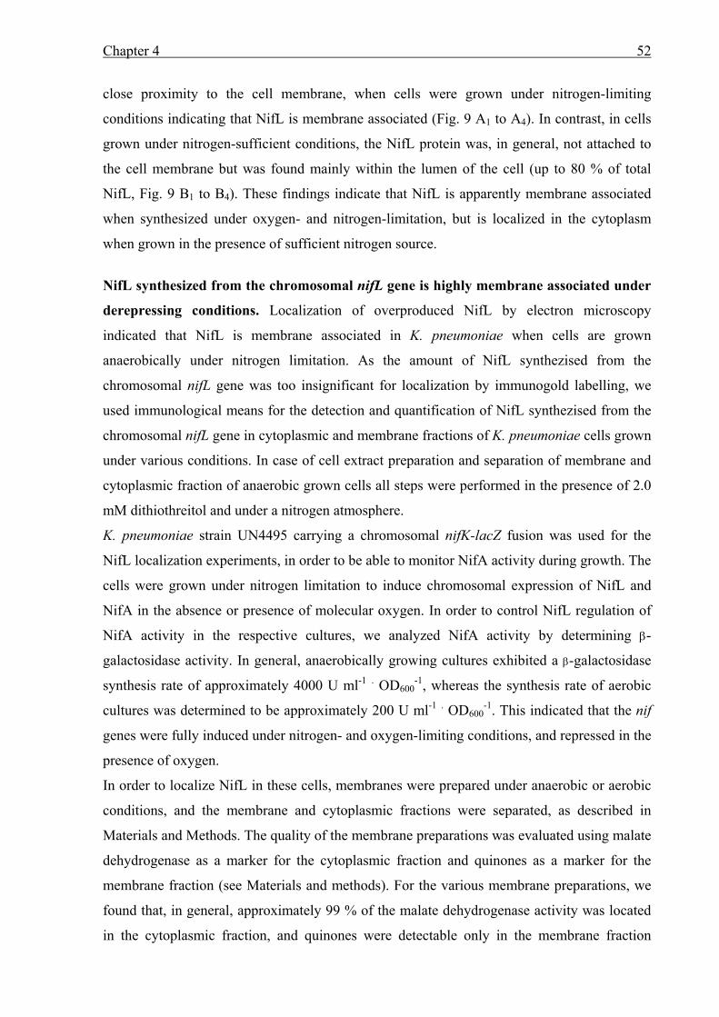
Chapter 4 52
close proximity to the cell membrane, when cells were grown under nitrogen-limiting
conditions indicating that NifL is membrane associated (Fig. 9 A1 to A4). In contrast, in cells
grown under nitrogen-sufficient conditions, the NifL protein was, in general, not attached to
the cell membrane but was found mainly within the lumen of the cell (up to 80 % of total
NifL, Fig. 9 B1 to B4). These findings indicate that NifL is apparently membrane associated
when synthesized under oxygen- and nitrogen-limitation, but is localized in the cytoplasm
when grown in the presence of sufficient nitrogen source.
NifL synthesized from the chromosomal nifL gene is highly membrane associated under
derepressing conditions. Localization of overproduced NifL by electron microscopy
indicated that NifL is membrane associated in K. pneumoniae when cells are grown
anaerobically under nitrogen limitation. As the amount of NifL synthezised from the
chromosomal nifL gene was too insignificant for localization by immunogold labelling, we
used immunological means for the detection and quantification of NifL synthezised from the
chromosomal nifL gene in cytoplasmic and membrane fractions of K. pneumoniae cells grown
under various conditions. In case of cell extract preparation and separation of membrane and
cytoplasmic fraction of anaerobic grown cells all steps were performed in the presence of 2.0
mM dithiothreitol and under a nitrogen atmosphere.
K. pneumoniae strain UN4495 carrying a chromosomal nifK-lacZ fusion was used for the
NifL localization experiments, in order to be able to monitor NifA activity during growth. The
cells were grown under nitrogen limitation to induce chromosomal expression of NifL and
NifA in the absence or presence of molecular oxygen. In order to control NifL regulation of
NifA activity in the respective cultures, we analyzed NifA activity by determining β-
galactosidase activity. In general, anaerobically growing cultures exhibited a β-galactosidase
synthesis rate of approximately 4000 U ml-1 . OD600-1, whereas the synthesis rate of aerobic
cultures was determined to be approximately 200 U ml-1 . OD600-1. This indicated that the nif
genes were fully induced under nitrogen- and oxygen-limiting conditions, and repressed in the
presence of oxygen.
In order to localize NifL in these cells, membranes were prepared under anaerobic or aerobic
conditions, and the membrane and cytoplasmic fractions were separated, as described in
Materials and Methods. The quality of the membrane preparations was evaluated using malate
dehydrogenase as a marker for the cytoplasmic fraction and quinones as a marker for the
membrane fraction (see Materials and methods). For the various membrane preparations, we
found that, in general, approximately 99 % of the malate dehydrogenase activity was located
in the cytoplasmic fraction, and quinones were detectable only in the membrane fraction
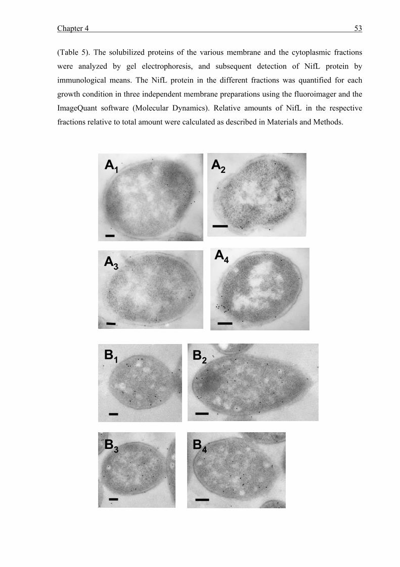
Chapter 4 53
(Table 5). The solubilized proteins of the various membrane and the cytoplasmic fractions
were analyzed by gel electrophoresis, and subsequent detection of NifL protein by
immunological means. The NifL protein in the different fractions was quantified for each
growth condition in three independent membrane preparations using the fluoroimager and the
ImageQuant software (Molecular Dynamics). Relative amounts of NifL in the respective
fractions relative to total amount were calculated as described in Materials and Methods.
A1A1 A2A2
A3A3A4A4
B1B1 B2B2
B3B3 B4B4
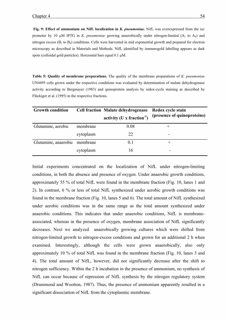
Chapter 4 54
Fig. 9: Effect of ammonium on NifL localization in K. pneumoniae. NifL was overexpressed from the tac
promoter by 10 µM IPTG in K. pneumoniae growing anaerobically under nitrogen-limited (A1 to A4) and
nitrogen excess (B1 to B4) conditions. Cells were harvested in mid exponential growth and prepared for electron
microscopy as described in Materials and Methods. NifL identified by immunogold labelling appears as dark
spots (colloidal gold particles). Horizontal bars equal 0.1 µM.
Table 5: Quality of membrane preparations. The quality of the membrane preparations of K. pneumoniae
UN4495 cells grown under the respective conditions was evaluated by determination of malate dehydrogenase
activity according to Bergmayer (1983) and quinoprotein analysis by redox-cycle staining as described by
Flückiger et al. (1995) in the respective fractions.
Growth condition Cell fraction Malate dehydrogenase
activity (U x fraction-1)
Redox cycle stain (presence of quinoproteins)
Glutamine, aerobic membrane
cytoplasm
0.08
22
+
-
Glutamine, anaerobic membrane
cytoplasm
0.1
16
+
-
Initial experiments concentrated on the localization of NifL under nitrogen-limiting
conditions, in both the absence and presence of oxygen. Under anaerobic growth conditions,
approximately 55 % of total NifL were found in the membrane fraction (Fig. 10, lanes 1 and
2). In contrast, 6 % or less of total NifL synthesized under aerobic growth conditions was
found in the membrane fraction (Fig. 10, lanes 5 and 6). The total amount of NifL synthesized
under aerobic conditions was in the same range as the total amount synthesized under
anaerobic conditions. This indicates that under anaerobic conditions, NifL is membrane-
associated, whereas in the presence of oxygen, membrane association of NifL significantly
decreases. Next we analyzed anaerobically growing cultures which were shifted from
nitrogen-limited growth to nitrogen-excess conditions and grown for an additional 2 h when
examined. Interestingly, although the cells were grown anaerobically, also only
approximately 10 % of total NifL was found in the membrane fraction (Fig. 10, lanes 3 and
4). The total amount of NifL, however, did not significantly decrease after the shift to
nitrogen sufficiency. Within the 2 h incubation in the presence of ammonium, no synthesis of
NifL can occur because of repression of NifL synthesis by the nitrogen regulatory system
(Drummond and Wootton, 1987). Thus, the presence of ammonium apparently resulted in a
significant dissociation of NifL from the cytoplasmic membrane.
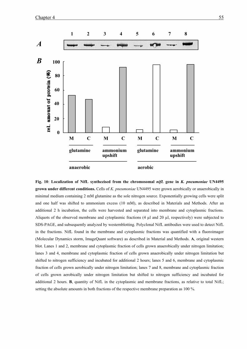
Chapter 4 55
Fig. 10: Localization of NifL synthezised from the chromosomal nifL gene in K. pneumoniae UN4495
grown under different conditions. Cells of K. pneumoniae UN4495 were grown aerobically or anaerobically in
minimal medium containing 2 mM glutamine as the sole nitrogen source. Exponentially growing cells were split
and one half was shifted to ammonium excess (10 mM), as described in Materials and Methods. After an
additional 2 h incubation, the cells were harvested and separated into membrane and cytoplasmic fractions.
Aliquots of the observed membrane and cytoplasmic fractions (4 µl and 20 µl, respectively) were subjected to
SDS-PAGE, and subsequently analyzed by westernblotting. Polyclonal NifL antibodies were used to detect NifL
in the fractions. NifL found in the membrane and cytoplasmic fractions was quantified with a fluoroimager
(Molecular Dynamics storm, ImageQuant software) as described in Material and Methods. A, original western
blot. Lanes 1 and 2, membrane and cytoplasmic fraction of cells grown anaerobically under nitrogen limitation;
lanes 3 and 4, membrane and cytoplasmic fraction of cells grown anaerobically under nitrogen limitation but
shifted to nitrogen sufficiency and incubated for additional 2 hours; lanes 5 and 6, membrane and cytoplasmic
fraction of cells grown aerobically under nitrogen limitation; lanes 7 and 8, membrane and cytoplasmic fraction
of cells grown aerobically under nitrogen limitation but shifted to nitrogen sufficiency and incubated for
additional 2 hours. B, quantity of NifL in the cytoplasmic and membrane fractions, as relative to total NifL;
setting the absolute amounts in both fractions of the respective membrane preparation as 100 %.
A
B
0.0
0.2
0.4
0.6
0.8
1.0
Membraneanaerob
glutamine
Cytoplasmanaerob
glutamine
Membraneanaerob
ammonium
Cytoplasmanaerob
ammonium
Membraneaerob gln
Cytoplasmaerob
glutamine
Membraneaerob
ammonium
Cytoplasmaerob
ammoniumM C M C M C M C
glutamine ammoniumupshift
glutamine ammoniumupshift
anaerobic aerobic
100
0
80
60
40
20
1 2 3 4 5 6 7 8
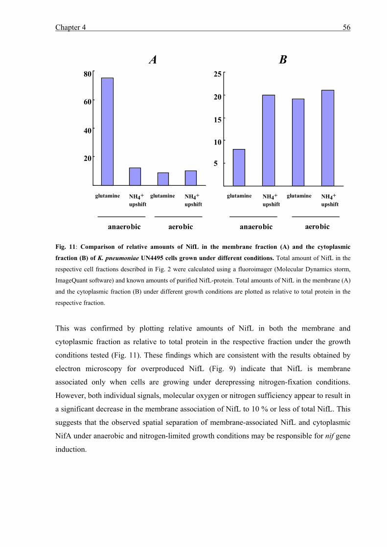
Chapter 4 56
Fig. 11: Comparison of relative amounts of NifL in the membrane fraction (A) and the cytoplasmic
fraction (B) of K. pneumoniae UN4495 cells grown under different conditions. Total amount of NifL in the
respective cell fractions described in Fig. 2 were calculated using a fluoroimager (Molecular Dynamics storm,
ImageQuant software) and known amounts of purified NifL-protein. Total amounts of NifL in the membrane (A)
and the cytoplasmic fraction (B) under different growth conditions are plotted as relative to total protein in the
respective fraction.
This was confirmed by plotting relative amounts of NifL in both the membrane and
cytoplasmic fraction as relative to total protein in the respective fraction under the growth
conditions tested (Fig. 11). These findings which are consistent with the results obtained by
electron microscopy for overproduced NifL (Fig. 9) indicate that NifL is membrane
associated only when cells are growing under derepressing nitrogen-fixation conditions.
However, both individual signals, molecular oxygen or nitrogen sufficiency appear to result in
a significant decrease in the membrane association of NifL to 10 % or less of total NifL. This
suggests that the observed spatial separation of membrane-associated NifL and cytoplasmic
NifA under anaerobic and nitrogen-limited growth conditions may be responsible for nif gene
induction.
80
60
40
20
25
20
15
10
5
A B
glutamine glutamine glutamine glutamine
anaerobic aerobic anaerobic aerobic
NH4+upshift
NH4+upshift
NH4+upshift
NH4+upshift
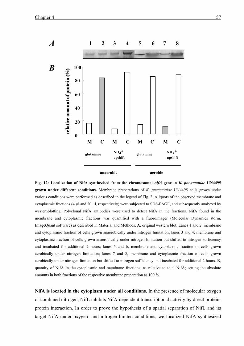
Chapter 4 57
Fig. 12: Localization of NifA synthezised from the chromosomal nifA gene in K. pneumoniae UN4495
grown under different conditions. Membrane preparations of K. pneumoniae UN4495 cells grown under
various conditions were performed as described in the legend of Fig. 2. Aliquots of the observed membrane and
cytoplasmic fractions (4 µl and 20 µl, respectively) were subjected to SDS-PAGE, and subsequently analyzed by
westernblotting. Polyclonal NifA antibodies were used to detect NifA in the fractions. NifA found in the
membrane and cytoplasmic fractions was quantified with a fluoroimager (Molecular Dynamics storm,
ImageQuant software) as described in Material and Methods. A, original western blot. Lanes 1 and 2, membrane
and cytoplasmic fraction of cells grown anaerobically under nitrogen limitation; lanes 3 and 4, membrane and
cytoplasmic fraction of cells grown anaerobically under nitrogen limitation but shifted to nitrogen sufficiency
and incubated for additional 2 hours; lanes 5 and 6, membrane and cytoplasmic fraction of cells grown
aerobically under nitrogen limitation; lanes 7 and 8, membrane and cytoplasmic fraction of cells grown
aerobically under nitrogen limitation but shifted to nitrogen sufficiency and incubated for additional 2 hours. B,
quantity of NifA in the cytoplasmic and membrane fractions, as relative to total NifA; setting the absolute
amounts in both fractions of the respective membrane preparation as 100 %.
NifA is located in the cytoplasm under all conditions. In the presence of molecular oxygen
or combined nitrogen, NifL inhibits NifA-dependent transcriptional activity by direct protein-
protein interaction. In order to prove the hypothesis of a spatial separation of NifL and its
target NifA under oxygen- and nitrogen-limited conditions, we localized NifA synthesized
glutamine glutamine
anaerobic aerobic
NH4+
upshiftNH4+
upshift
MC MCM CM C
80
60
40
20
100
1 2 3 4 5 6 7 8A
B
0
Chapter 4 58
from the chromosomal nifLA operon using the same membrane and cytoplasmic fractions in
which we had localized NifL. We found that approximately 12 ± 3 % of total NifA are located
in the membrane fraction under all growth conditions tested (Fig. 12). As shown for NifL, no
difference in total NifA protein was detected under the various growth conditions. Taking into
account that (i) NifA has to be localized in the cytoplasm to activate nif transcription, and (ii)
the NifA membrane-associated fraction is under all conditions in the same range as the
membrane association of NifL in the presence of oxygen or ammonium, a membrane
association in the range of 10 % may be based on non-specific binding of hydrophobic
regions of the two proteins to the membrane.
Under derepressing conditions but in the absence of either Fnr or GlnK ant AmtB, NifL
is located in the cytoplasm. In order to observe additional evidence that both individual
signals, molecular oxygen and nitrogen sufficiency, result in a significant decrease in the
membrane association of NifL, we localized NifL synthesized from the chromosomal nifL
gene under derepressing conditions in the absence of either the oxygen sensory protein Fnr or
the nitrogen sensory protein GlnK and AmtB. K. pneumonaie UN4495 carrying an fnr null-
allele (K. pneumoniae UN4495 fnr::Ω, RAS18) and K. pneumoniae UN4495 carrying an
glnKamtB null-allele (K. pneumoniae UN4495 glnK::KIXX, RAS36) were grown under
nitrogen- and oxygen-limited conditions. During growth NifA activity was analyzed by
determining β-galactosidase synthesis rates.
As expected from previous studies (Grabbe et al., 2001b; He et al., 1998; Jack et al., 1999), in
the absence of the primary oxygen sensor Fnr (RAS18) or in the absence the primary nitrogen
sensor GlnK (RAS36), NifL inhibited NifA activity (Table 6). No obvious differences in the
total amounts of NifA or NifL in the mutant backgrounds compared to the parental strain
were detected, indicating that the failure of the mutant strains to express nifH under
derepressed conditions could not be accounted for by a decreased amount of NifA protein.
Localization of NifL in the two mutant strains under nitrogen and oxygen limitation in three
independent membrane praparations showed that in both, the fnr mutant and the glnKamtB
mutant, approximately 90 % of NifL is localized in the cytoplasmic fraction (Table 6).
Additional shifts to nitrogen excess did not change the NifL location significantly. Thus, in
the absence of Fnr or GlnK plus the ammonium transporter AmtB, NifL does not receive the
signal for oxygen- or nitrogen limitation, respectively, resulting in a NifL protein which is
located in the cytoplasm and inhibits NifA activity. These findings strongly support our model
that under derepressing conditions NifL is membrane associated, however either signal,
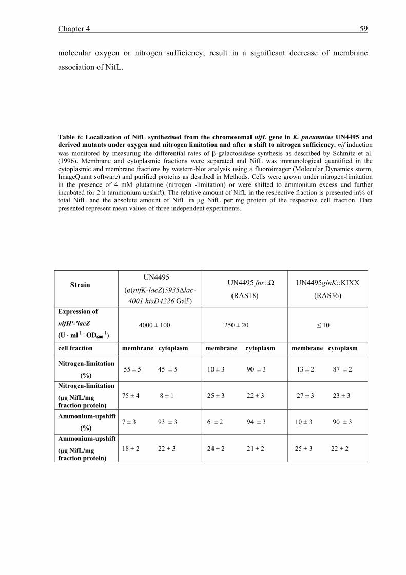
Chapter 4 59
molecular oxygen or nitrogen sufficiency, result in a significant decrease of membrane
association of NifL.
Table 6: Localization of NifL synthezised from the chromosomal nifL gene in K. pneumniae UN4495 and derived mutants under oxygen and nitrogen limitation and after a shift to nitrogen sufficiency. nif induction was monitored by measuring the differential rates of β-galactosidase synthesis as described by Schmitz et al. (1996). Membrane and cytoplasmic fractions were separated and NifL was immunological quantified in the cytoplasmic and membrane fractions by western-blot analysis using a fluoroimager (Molecular Dynamics storm, ImageQuant software) and purified proteins as desribed in Methods. Cells were grown under nitrogen-limitation in the presence of 4 mM glutamine (nitrogen -limitation) or were shifted to ammonium excess und further incubated for 2 h (ammonium upshift). The relative amount of NifL in the respective fraction is presented in% of total NifL and the absolute amount of NifL in µg NifL per mg protein of the respective cell fraction. Data presented represent mean values of three independent experiments.
Strain
UN4495
(ø(nifK-lacZ)5935∆lac-4001 hisD4226 Galr)
UN4495 fnr::Ω
(RAS18)
UN4495glnK::KIXX
(RAS36)
Expression of
nifH'-'lacZ
(U · ml-1 . OD600-1)
4000 ± 100 250 ± 20 ≤ 10
cell fraction membrane cytoplasm membrane cytoplasm membrane cytoplasm
Nitrogen-limitation
(%) 55 ± 5 45 ± 5 10 ± 3 90 ± 3 13 ± 2 87 ± 2
Nitrogen-limitation
(µg NifL/mg fraction protein)
75 ± 4 8 ± 1 25 ± 3 22 ± 3 27 ± 3 23 ± 3
Ammonium-upshift
(%) 7 ± 3 93 ± 3 6 ± 2 94 ± 3 10 ± 3 90 ± 3
Ammonium-upshift
(µg NifL/mg fraction protein)
18 ± 2 22 ± 3 24 ± 2 21 ± 2 25 ± 3 22 ± 2
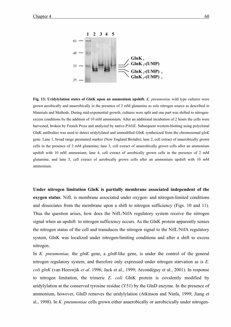
Chapter 4 60
Fig. 13: Uridylylation states of GlnK upon an ammonium upshift. K. pneumoniae wild type cultures were
grown aerobically and anaerobically in the presence of 2 mM glutamine as sole nitrogen source as described in
Materials and Methods. During mid-exponential growth, cultures were split and one part was shifted to nitrogen-
excess conditions by the addition of 10 mM ammonium. After an additional incubation of 2 hours the cells were
harvested, broken by French Press and analyzed by native-PAGE. Subsequent western-blotting using polyclonal
GlnK antibodies was used to detect uridylylated and unmodified GlnK synthesized from the chromosomal glnK
gene. Lane 1, broad range prestained marker (New England Biolabs); lane 2, cell extract of anaerobically grown
cells in the presence of 2 mM glutamine; lane 3, cell extract of anaerobically grown cells after an ammonium
upshift with 10 mM ammonium; lane 4, cell extract of aerobically grown cells in the presence of 2 mM
glutamine, and lane 5, cell extract of aerobically grown cells after an ammonium upshift with 10 mM
ammonium.
Under nitrogen limitation GlnK is partially membrane associated independent of the
oxygen status. NifL is membrane associated under oxygen- and nitrogen-limited conditions
and dissociates from the membrane upon a shift to nitrogen sufficiency (Figs. 10 and 11).
Thus the question arises, how does the NifL/NifA regulatory system receive the nitrogen
signal when an upshift to nitrogen sufficiency occurs. As the GlnK protein apparently senses
the nitrogen status of the cell and transduces the nitrogen signal to the NifL/NifA regulatory
system, GlnK was localized under nitrogen-limiting conditions and after a shift to excess
nitrogen.
In K. pneumoniae, the glnK gene, a glnB-like gene, is under the control of the general
nitrogen regulatory system, and therefore only expressed under nitrogen starvation as is E.
coli glnK (van Heeswjik et al. 1996; Jack et al., 1999; Arcondéguy et al., 2001). In response
to nitrogen limitation, the trimeric E. coli GlnK protein is covalently modified by
uridylylation at the conserved tyrosine residue (Y51) by the GlnD enzyme. In the presence of
ammonium, however, GlnD removes the uridylylation (Atkinson and Ninfa, 1999; Jiang et
al., 1998). In K. pneumoniae cells grown either anaerobically or aerobicically under nitrogen-
GlnK 3
GlnK 3-(UMP)
GlnK 3-(UMP) 2
GlnK 3-(UMP) 3
1 2 3 4 5
33
48
83
25
Chapter 4 61
limiting conditions, GlnK trimers were up to 80 % uridylylated (GlnK3-(UMP)3) as detected
by native gel electrophoresis and subsequent western blot analysis (Fig. 13, lanes 2 and 4); the
uridylylation apparently changes the overall charge of the trimers resulting in a faster
migration of the uridylylated forms compared to the non-modified trimers. Two hours after an
ammonium upshift, the same cultures showed fully deuridylylated GlnK trimers (Fig. 13,
compare lanes 2 and 4 with lanes 3 and 5). These findings show that the uridylylation state of
K. pneumoniae GlnK, like that of E. coli GlnK, is dependent on the nitrogen status of the cell.
In order to analyze and localize GlnK trimers after a shift to nitrogen sufficiency, we
performed ammonium upshift experiments on K. pneumoniae cells grown under nitrogen-
limited conditions in the presence or absence of oxygen. Exponentially growing cultures were
split and one part was shifted to nitrogen sufficiency by the addition of 10 mM ammonium
and further incubated for 2 h. The glnKamtB operon is subject to nitrogen control at the
transcriptional level mediated by NtrC (Jack et al., 1999), thus within the 2 h incubation in the
presence of ammonium, no expression of glnK can occur. The membrane and cytoplasmic
fractions before and after the ammonium upshift were subjected to native PAGE and
subsequent western blot analysis to separate and quantify the GlnK trimers in the different
fractions. In the cell-free extracts under nitrogen-limiting conditions approximately 80 %
GlnK was found in its completely uridylylated form (GlnK3-(UMP)3). 15 to 20 % of the total
GlnK protein was found in the membrane fraction, in both the anaerobic and the aerobic
preparation (Fig. 14B, lanes 1 and 2, lanes 5 and 6). The membrane-bound GlnK and
cytoplasmic GlnK, however, showed no difference in the uridylylation pattern, indicating that
membrane association is not dependent on a defined uridylylation state of GlnK (Fig. 14A,
lanes 1 and 2, lanes 5 and 6). This observed membrane association of GlnK under nitrogen
limitation is of special interest, since the GlnK protein shows little if any hydrophobicity, and
is a highly soluble protein.
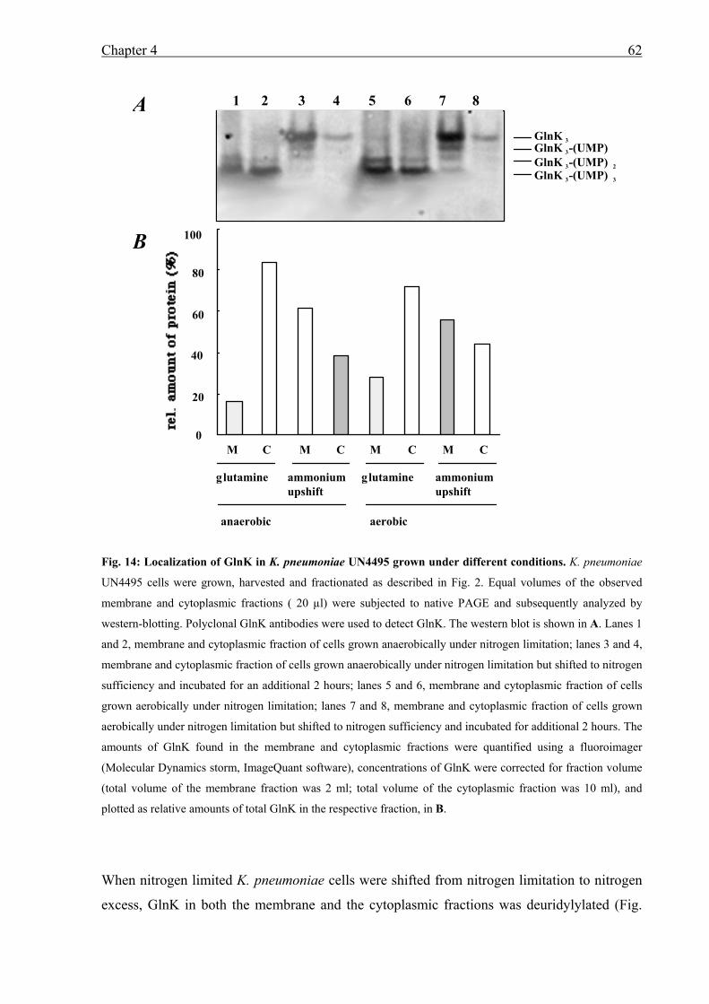
Chapter 4 62
Fig. 14: Localization of GlnK in K. pneumoniae UN4495 grown under different conditions. K. pneumoniae
UN4495 cells were grown, harvested and fractionated as described in Fig. 2. Equal volumes of the observed
membrane and cytoplasmic fractions ( 20 µl) were subjected to native PAGE and subsequently analyzed by
western-blotting. Polyclonal GlnK antibodies were used to detect GlnK. The western blot is shown in A. Lanes 1
and 2, membrane and cytoplasmic fraction of cells grown anaerobically under nitrogen limitation; lanes 3 and 4,
membrane and cytoplasmic fraction of cells grown anaerobically under nitrogen limitation but shifted to nitrogen
sufficiency and incubated for an additional 2 hours; lanes 5 and 6, membrane and cytoplasmic fraction of cells
grown aerobically under nitrogen limitation; lanes 7 and 8, membrane and cytoplasmic fraction of cells grown
aerobically under nitrogen limitation but shifted to nitrogen sufficiency and incubated for additional 2 hours. The
amounts of GlnK found in the membrane and cytoplasmic fractions were quantified using a fluoroimager
(Molecular Dynamics storm, ImageQuant software), concentrations of GlnK were corrected for fraction volume
(total volume of the membrane fraction was 2 ml; total volume of the cytoplasmic fraction was 10 ml), and
plotted as relative amounts of total GlnK in the respective fraction, in B.
When nitrogen limited K. pneumoniae cells were shifted from nitrogen limitation to nitrogen
excess, GlnK in both the membrane and the cytoplasmic fractions was deuridylylated (Fig.
100
0
60
80
40
20
glutamine ammoniumupshift
glutamine ammoniumupshift
anaerobic aerobic
M C M C M C M C
A
B
1 2 3 4 5 6 7 8
GlnK 3GlnK 3-(UMP)GlnK 3-(UMP) 2
GlnK 3-(UMP) 3
100
Chapter 4 63
14A, lanes 3 and 4, lanes 7 and 8). However, it appeared that after a shift to nitrogen-excess
independent of oxygen availability the cytoplasmic GlnK fraction decreased significantly. In
contrast, the amount of membrane-associated GlnK apparently did not change (Fig. 14A,
compare lanes 2 and 4, and lanes 6 and 8), thus shifting the ratio of membrane-associated
GlnK to cytoplasmic GlnK from 15 % / 85 % under nitrogen limitation, to approximately 60
% / 40 % in the presence of excess nitrogen (Fig. 14B). As no new GlnK synthesis occurred
during the ammonium upshift the ratio between membrane-bound and cytoplasmic GlnK
should not change unless increased degradation of one fraction occurs. However, one cannot
rule out that unspecific proteolysis of the cytoplasmic GlnK fraction occurred during the time
of separating the two fractions, as the buffers used for cell breakage and membrane
preparation were not supplemented with protease inhibitors. Thus, further analysis of the
apparent faster degradation of cytoplasmic GlnK after a shift to nitrogen sufficiency is
required.
Discussion
Regulatory proteins that are membrane bound and transmit an environmental signal via a
cytoplasmic transmitter domain are a common principle in bacterial signal transduction. In a
variety of such regulatory proteins or transducers of both prokaryotic and eukaryotic origin,
conserved sequence motifs, so called PAS domains, have been identified (for review see
Taylor and Zhulin, 1999). Most bacterial sensory proteins containing a PAS domain are
histidine kinase sensor proteins of two-component regulatory systems, and usually contain
one or more transmembrane domains (Zhulin et al., 1997; Taylor and Zhulin, 1999). The
regulatory protein NifL contains a C-terminal histidine-kinase-like transmitter domain
(Drummond and Wootton, 1987; Parkinson and Kofoid, 1992; Woodley and Drummond,
1994) and its N-terminal domain contains the conserved motifs of the PAS domain (Zhulin et
al., 1997). NifL differs, however, in that no membrane-spanning domain can be predicted
from amino acid sequence data of the protein (Drummond and Wootton, 1987). Thus, NifL is
considered to be a solely cytoplasmic protein that receives and transduces the oxygen and
nitrogen signal to the transcriptional activator NifA in the cytoplasm (Dixon, 1998). We have
recently shown that Fnr of K. pneumoniae is the primary oxygen sensor for nitrogen fixation,
which apparently transduces the oxygen signal to NifL by activating transcription of genes,
whose products reduce the NifL-bound FAD under anaerobic conditions (Grabbe et al.,
2001b). In addition, preliminary studies indicated that K. pneumoniae NifL is membrane-
Chapter 4 64
associated during anaerobic growth. Thus we proposed that the physiological electron donor
for the reduction of NifL during anaerobic growth is a component of the anaerobic electron
transport chain. In order to characterize a potential membrane association of NifL as a part of
the regulatory process further, we localized NifL in cells grown anaerobically and aerobically,
both in the absence and presence of combined nitrogen.
Spatial separation as the potential regulatory principle in nif regulation by NifA and
NifL. We present three lines of evidence that both conditions, nitrogen limitation and the
absence of oxygen, are required for significant membrane association of K. pneumoniae NifL.
Either signal alone is not sufficient for NifL association with the membrane. (i) Electron
microscopy analysis of NifL overproduced in K. pneumoniae indicated that under oxygen-
and nitrogen-limiting conditions, NifL is significantly membrane associated, whereas under
nitrogen sufficiency NifL is located in the cell lumen (Fig. 9). (ii) Immunological
quantifications of NifL synthesized from the chromosomal nifL gene confirmed that under
oxygen and nitrogen limitation, approximately 55 % of the total NifL protein is located in the
membrane fraction. A shift to nitrogen sufficiency or the presence of molecular oxygen,
however, resulted in a significant decrease in membrane association of NifL, to approximately
10 % (Fig. 10). (iii) In the absence of eiher the primary oxygen sensor Fnr or the primary
nitrogen sensor GlnK plus the ammonium transporter AmtB, NifL is located in the cytoplasm
(Table 6). Thus, in addition to nitrogen limitation, the reduced conformation of NifL appears
to be critically important for the membrane affinity of the protein. With oxidation, the
membrane affinity of NifL significantly decreases, and NifL is again located in the cytoplasm.
Determination of malate dehydrogenase activity and detection of quinoproteins in the
different membrane and cytoplasmic fractions ruled out that the analyzed membrane fractions
were contaminated with cytoplasmic proteins (Table 5). Thus, the basal amount of a
maximum of 10 % membrane-bound NifL, detected under all conditions except under
oxygen- and nitrogen-limitation, appears to be based on non-specific binding of the
hydrophobic regions of the NifL protein to the cell membrane. This is consistent with the
amounts we observed for the NifA protein in the same membrane and cytoplasmic fractions;
approximately 10 % of total NifA were membrane associated under all conditions tested (Fig.
12), although NifA is a transcriptional activator and is therefore expected to be a soluble
protein located in the cytoplasm (Austin et al., 1990; Lee et al., 1993). This suggests that the
observed fractions of NifA and NifL, which appear to be membrane associated under all
conditions, are fractions of both regulatory proteins, which bind, independent from each other,
non-specifically to the membrane and are not functionally involved in the regulatory process.
Chapter 4 65
The observed decrease in cytoplasmic NifL under anaerobic and nitrogen-limited conditions,
conditions under which no change of NifA location is detectable, suggests that membrane
association of NifL plays a critical role in the regulation of NifA activity. The spatial
separation of membrane-bound NifL and cytoplasmic NifA under nitrogen and oxygen
limitation may be responsible for the release of the NifL inhibition of NifA resulting in nif
gene induction. We therefore propose a NifL conformation that integrates the oxygen and
nitrogen signal in such a way that the overall conformation of the protein under anaerobic and
nitrogen-limited conditions is able to bind to the cytoplasmic membrane, creating a spatial
gap between NifL and its target NifA. A comparable regulatory mechanism is discussed for
the transcriptional regulator PutA, which is involved in proline catabolism in Salmonella
typhimurium and Escherichia coli (Maloy, 1987). PutA associates with the membrane and
catalyzes the two-step oxidation of proline to glutamate when the intracellular proline
concentration is high (Muro-Pastor et al., 1997; Wood, 1987); when the intracellular proline
concentration decreases, PutA dissociates from the membrane and represses transcription of
the proline utilization (put) operon by binding to an operator (Ostrovsky et al., 1991; Brown
and Wood, 1993). In contrast to the observed membrane affinity of NifL in K. pneumoniae
under oxygen and nitrogen limitation, no membrane association for A. vinelandii NifL has
been reported to date (Dixon, 1998).
Hypothetical function for GlnK in nif regulation
Concerning the nif regulation by combined nitrogen, we observed evidence that a shift from
nitrogen limitation to nitrogen sufficiency results in a decrease in membrane association of
NifL (Figs. 9 and 10). Thus the question arises, how does the presence of sufficient nitrogen
change the membrane affinity of NifL. We therefore localized GlnK, a highly soluble protein,
which is responsible for the detection of the internal nitrogen status and for the transduction
of the nitrogen signal to the nif regulatory system (Xu et al., 1998, He, et al., 1998; Jack et al.,
1999; Arcondéguy et al., 1999 and 2001). Unexpected, we observed a significant membrane
association of GlnK under nitrogen-limiting conditions (approximately 15 - 20 %, Fig. 14),
which may result from its interaction with the ammonium transporter AmtB and is not
dependent on a defined uridylylation state of GlnK. At the actual experimental status
however, one cannot decide if the observed membrane-association of GlnK is directly linked
to the NifL location or whether GlnK is regulating the NifL location indirectly as a
consequence of its role in controlling the interaction of NifL with NifA. Under nitrogen
excess in the absence of GlnK, cytoplasmic NifL inhibits NifA by complex formation. If

Chapter 4 66
GlnK transduces the signal of nitrogen limitation either by interacting with NifL or NifA
resulting in the dissociation of the NifL/NifA complex, NifL would be able to interact with
the putative membrane bound electron donor and stays membrane-associated. In the presence
of oxygen however, the oxidized form of NifL would be preferentially located in the
cytoplasm and this conformation may interact with NifA even when GlnK is present.
Chapter 5 67
Chapter 5:
Oxygen Control of nif Gene Expression in Klebsiella pneumoniae
is dependent on NifL reduction at the cytoplasmic membrane by
electrons derived from the reduced quinone pool
ROMAN GRABBE AND RUTH A. SCHMITZ*
Abstract
In Klebsiella pneumoniae, NifA mediated transcriptional activation of the nitrogen fixation
(nif) genes is inhibited in the presence of molecular oxygen by the negative regulator NifL.
The primary oxygen sensor Fnr transduces the signal of anaerobiosis to the negative regulator
resulting in the non-inhibitory, reduced conformation of the flavoprotein NifL. We have
recently demonstrated that membrane sequestration of NifL under anaerobic and nitrogen-
limited conditions impairs inhibition of cytoplasmic NifA by NifL and thus seems to be
involved in the regulatory mechanism for oxygen dependent nif-regulation in K. pneumoniae.
We have now investigated the influence of different membrane-bound oxidoreductases of the
anaerobic electron transport chain on nif-regulation in K. pneumoniae by biochemical analysis
of purified NifL and by monitoring NifA-mediated expression of nifH’-’lacZ reporter fusions
in different genetic backgrounds. In vitro analysis showed that NifL-bound FAD-cofactor was
reduced by NADH/H+ only in the presence of either a redox mediator or anaerobic inside-out
vesicles derived from anaerobically grown K. pneumoniae cells, indicating that in vivo NifL is
reduced by a membrane-bound component of the anaerobic electron transport chain. This
mechanism is further supported by three lines of evidence: First, Klebsiella strains carrying
null mutations of fdnG or nuoCD showed significantly reduced nif induction under
derepressing conditions, indicating that NifL inhibition of NifA was not relieved in the
absence of formate dehydrogenaseN or NADH:ubiquinone oxidoreductase. The same effect
was observed in a heterologous E. coli system carrying a ndh null allele (coding for NADH
dehydrogenase II). Second, studying nif induction in K. pneumoniae under different growth
conditions revealed that the presence of nitrate during anaerobic growth on glycerol under
nitrogen limitation resulted in a significant decrease of nif induction. However, when growing
Chapter 5 68
on sucrose or glucose, nitrate did not effect nif regulation. The final line of evidence is that a
reduced quinone derivative, dimethylnaphthoquinonered (DMNred) is able to transfer electrons
to the FAD-moiety of purified NifL resulting in the reduced conformation of NifL. On the
basis of these data, we postulate that under anaerobic and nitrogen-limiting conditions NifL
inhibition on NifA activity is relieved by reduction of the FAD-cofactor at the cytoplasmic
membrane through the reduced quinone pool of the anaerobic electron transport chain.
INTRODUCTION
In the free-living diazotroph Klebsiella pneumoniae, a member of the γ-subgroup of
proteobacteria, nitrogen (N2) fixation is tightly controlled to avoid unnecessary consumption
of energy. The transcriptional activator NifA and the inhibitor NifL, both under the control of
the NtrB/C-system, regulate the transcription of the nitrogen fixation (nif) operons according
to the environmental signals molecular oxygen and combined nitrogen (for review see Dixon
1998, Schmitz et al. 2002). Under oxygen and nitrogen limitation the inhibitor NifL stays in
the non-inhibitory conformation and nif-gene expression is activated by NifA. In the presence
of oxygen or combined nitrogen, NifL antagonizes the activity of NifA resulting in a decrease
of nif-gene expression. The translationally coupled synthesis of nifL and nifA in addition to
evidence from immunological studies of complex formation, imply that the inhibition of NifA
activity by NifL occurs via a direct protein-protein interaction (Govantes et al. 1998;
Henderson et al. 1989). Recently, in the diazotroph Azotobacter vinelandii formation of
NifL/NifA complexes has been demonstrated by in vitro co-chromatography in the presence
of adenine nucleotides and using the yeast-two-hybrid system (Money et al. 1999 and 2001,
Lei et al. 1999).
Recent studies revealed that the nitrogen signal in K. pneumoniae and A. vinelandii is
transduced towards the regulatory proteins NifL and NifA by the GlnK protein, a paralogue
PII-protein. However, the mechanism appears to be opposite in K. pneumoniae and A.
vinelandii. In K. pneumoniae, relief of NifL inhibition under nitrogen limiting conditions
depends on the presence of GlnK, the uridylyation state of which appears not to be essential
for its nitrogen signaling function (He et al. 1998, Jack et al. 1999, Arcondeguy et al. 1999
and 2000). It is currently not known, whether GlnK interacts with NifL or NifA alone or
affects the NifL/NifA-complex in K. pneumoniae. In contrast to K. pneumoniae, non-
uridylylated GlnK protein appears to activate the inhibitory function of A. vinelandii NifL
under nitrogen excess, whereas under nitrogen limitation the inhibitory activity of NifL is

Chapter 5 69
apparently relieved by elevated levels of 2-oxoglutarate (Little et al. 2000, Reyes-Ramirez et
al. 2001). Very recently interactions between A. vinelandii GlnK and NifL was demonstrated
using the yeast-two-hybrid system and in vitro studies further indicated that the non-
uridylylated form of A. vinelandii GlnK directly interacts with NifL preventing nif-gene
expression (Little et al. 2002, Rudnick et al. 2002).
For the oxygen-signaling pathway it was shown that A. vinelandii NifL and K. pneumoniae
NifL act as redox-sensitive regulatory proteins. NifL modulates NifA activity in response to
the redox-state of its N-terminal bound FAD-cofactor and allows NifA activity only in the
absence of molecular oxygen, when the flavin cofactor is reduced (Hill et al. 1996, Schmitz
1997, Dixon 1998, Macheroux et al. 1998, Klopprogge and Schmitz 1999). Thus, under
anaerobic conditions in the absence of combined nitrogen, reduction of the flavin moiety of
NifL results in a non-inhibitory conformation of the NifL protein. Recently, we have
demonstrated that in K. pneumoniae the global regulator Fnr is required to mediate the signal
of anaerobiosis to NifL (Grabbe et al. 2001b). Thus, we proposed that in the absence of
oxygen the primary oxygen sensor Fnr activates transcription of gene(s) the product(s) of
which reduce the NifL-bound FAD-cofactor resulting in a non-inhibitory conformation of
NifL, which allows NifA activity. Further localization analyses of NifL under various growth
conditions showed that only under derepressing conditions NifL is highly membrane-
associated impairing the inhibition of cytoplasmic NifA. This indicates that sequestration of
NifL to the membrane under anaerobic and nitrogen-limited conditions is involved in the
regulation of NifA activity by NifL (Klopprogge et al. 2002). Based on these findings the
question arises, whether NifL reduction occurs at the cytoplasmic membrane by a component
of the anaerobic electron transport chain during membrane association of NifL. In order to
verify this hypothesis and to identify the electron donor - potentially localized in the
cytoplasmic membrane - we analyzed the effects of different membrane-bound
oxidoreductases of the anaerobic electron transport chain on nif-regulation in K. pneumoniae
and in a heterologous E. coli system. In addition in vitro reduction of purified NifL was
studied using artificial electron donors or NADH/H+ in the presence of inverted vesicles
derived from K. pneumoniae cells.
Chapter 5 70
MATERIAL AND METHODS
Bacterial strains and Plasmids
The bacterial strains and plasmids used in this study are listed in Table 7. Plasmid DNA was
transformed into E. coli cells according to the method of Inoue et al. (1990) and into K.
pneumoniae cells by electroporation. Transduction by phage P1 was performed as described
previously (Silhavy et al. 1984).
Table 1. Bacterial strains and plasmids used in this study Strain or plasmid Relevant genotype Source, reference Strains: Klebisella pneumoniae:
M5a1 Wild type MacNeil et al. 1981UN4495 φ (nifK-lacZ)5935 ∆lac-4001 his D4226 Galr MacNeil et al. 1981RAS 18 UN4495, but fnr::Ω Grabbe et al. 2001 RAS46 UN4495, but spontaneous streptomycine
resistance This study
RAS47 UN4495, but nuoCD::tet This study RAS48 UN4495, but fdnG::tet This study RAS49 UN4495, but frdA::tet This study E. coli:
NCM1529 araD139∆(argF-lacU)169 fthD5301 gyrA219 non-9 rspL150 ptsF25 relA1 deoC1 trpDC700putPA1303::[Kanr-(nifH-lacZ)] (Wild type)
He et al. 1998
NCM1528 NCM1529/pNH3 He et al. 1998 NCM1527 NCM1529/pJES851 He et al. 1998 RAS50 NCM1529, but ndh::tet This study RAS51 RAS50 + pNH3 This study RAS52 RAS50 + pJES851 This study RAS53 NCM1529, but frd::tet This study RAS54 RAS53/pNH3 This study RAS55 RAS53/pJES851 This study Plasmids:
pBSK+ cloning vector Stratagene pCR 2.1 Topo-TA cloning vector Invitrogen pKAS46 allelic exchange vector, oriR6K;
rpsL*(Streps), Ampr, Kanr Skorupsky K. & R.K. Taylor, 1996
pNH3 K. pneumoniae nifLA under the control of the tac promoter
Henderson et al. 1989
pJES851 K. pneumoniae nifA under the control of tac promoter
Schmitz et al. 1996
pJES794 K. pneumoniae malE-nifL under the control of the tac promoter
Narberhaus et al. 1995
Chapter 5 71
pRS167 EcoRI/HindIII fdnG fragment (K. pneumoniae
M5a1) in pBSK+ This study
pRS177 pRS167, but fdnG::tet This study pRS187 frdA fragment (K. pneumoniae M5a1) in
pCR2.1 This study
pRS191 EcoRI/HindIII nuoCD fragment (K. pneumoniae M5a1) in pBSK+
This study
pRS193 fdnG::tet fragment from pRS177 in pKAS46 This study pRS194 pRS191, but nuoCD::tet This study pRS197 nuoCD::tet fragment from pRS194 in pKAS46 This study pRS214 pRS187, but frdA::tet This study pRS215 frdA::tet fragment from pRS214 in pKAS46 This study
(i) E. coli strains:
E. coli NCM1529, containing a chromosomal nifH´-lacZ´ fusion (He et al. 1997) was chosen
to study NifA and NifL regulation in E. coli. The ndhII::tet allele was transferred from
ANN001 (T. Friedrich, unpublished) into NCM1529 by P1 mediated transduction with
selection for tetracycline resistance, resulting in RAS50. Strains RAS51 and RAS52 contain
plasmid pNH3 and plasmid pJES851, respectively. (ii) K. pneumoniae strains:
K. pneumoniae strain M5al (wild type, N2-fixing) and strain UN4495 [φ(nifK-lacZ) 5935
∆lac-4001 his D4226 Galr] (McNeill et al. 1981) were provided by Gary Roberts. The
spontaneous streptomycin resistant UN4495 strain, RAS46, carrying a rpsL mutation was
isolated by plating UN4495 on a Luria-Bertani (LB) agar plate containing 100 µg
streptomycin per ml. K. pneumoniae subsp. pneumoniae (DSM No. 4799, not N2-fixing) and
K. oxytoca (DSM No. 4798, not N2-fixing) were obtained from the Deutsche Sammlung von
Mikroorganismen und Zellkulturen GmbH (Braunschweig, Germany).
Mutant strains of UN4495 were in general constructed by cloning the respective genes by
PCR-techniques, inserting a tetracycline resistance cassette derived from the MiniTn5
(DeLorenzo et al. 1990), cloning the respective interrupted genes into the suicide vector
pKAS46 (Skorukpski and Taylor 1996) followed by transformation into the streptomycin
resistant K. pneumoniae UN4495 strain (RAS46). Recombinant strains (generated by means
of a double cross over) were identified by the ability to grow on LB supplemented with 400
µg streptomycin per ml and resistance to tetracycline (Skorukpski and Taylor 1996); the
respective chromosomal mutations were confirmed by PCR and Southern blot analysis
(Sambrock et al. 1989). For generating homologous primer for PCR amplification sequence
information for genes of K. pneumoniae MG478578 (subsp. pneumoniae, not N2-fixing) were
Chapter 5 72
obtained from the database of the Genome Sequencing Center, Washington University, St.
Louis (Genome Sequencing Center, personal communication) and using the database ERGO
(Integrated Genomics, Inc.) (http://www.integratedgenomics.com).
nuoCD mutant: RAS47 was constructed as follows, (i) a 1.6 kb fragment carrying the nuoCD
genes of K. pneumoniae M5a1 was amplified by PCR using primers with additional synthetic
restriction recognition sites (underlined) nuoC/D ERI (5'CAGCGCGAATTCTCGCCG-
GCA3') and primer nuoC/D HindIII (5'CTGCTGAAGCTTGCGCAGACTCTG') and cloned
into pBluescript SK+ producing pRS191, (ii) a 2.2 kb fragment containing the tetracycline
resistance cassette (DeLorenzo et al. 1990) was inserted into the EcoRV site of nuoCD gene
region in pRS191 yielding pRS194, (iii) the 3.8 kb EcoRI/KpnI fragment of pRS191 carrying
the interrupted nuoCD region was transferred into the allelic exchange vector pKAS46
(Skorukpski and Taylor 1996) creating plasmid pRS197; the correct insertion of the
tetracycline cassette was checked by sequencing, (iv) pRS197 was transformed into RAS46
and recombinant strains carrying the chromosomally inserted plasmid by means of single
homologous recombination were identified by their inability to grow on streptomycin agar
plates as a consequence of the plasmid encoded rpsL mutation. Overnight selection of single
colonies in liquid LB medium containing 400 µg streptomycin per ml resulted in the loss of
the integrated plasmid with an integration frequency of the interrupted nuoCD region in 50 %
of the integrands.
fdnG mutant: Primer fdnG 5‘ EcoRI (5'CCGACTGATGAATTCCGACCGCGA3') and
primer fdnG 3‘ HindIII (5'GCCGAGCAGAAGCTTGATCATCGC3') were used to clone a 1
kb fdnG fragment from K. pneumoniae M5a1 into pBSK+ vector creating pRS167, followed
by insertion of the tetracycline resistance cassette into the EcoRV site of fdnG fragment
resulting in pRS177. The 3.2 kb EcoRI/KpnI fragment of pRS177 including the fdnG::tet
region was cloned into pKAS46. The construction of the K. pneumoniae chromosomal mutant
was performed using the same strategy as described in detail above, yielding RAS48. Growth conditions. E. coli and K pneumoniae strains were grown anaerobically with
molecular nitrogen (N2) as gas phase at 30 °C in minimal medium supplemented with 4 mM
glutamine as the sole nitrogen source (nitrogen limitation), 10 mM Na2CO3, 0.3 mM sulfide
and 0.002% resazurin to monitor anaerobiosis (Schmitz et al. 1996). The medium was further
supplemented with. 0.5 % sucrose and 0.004 % histidine for K. pneumoniae strains and 1%
glucose and 0.002 % tryptophane for E. coli strains. Precultures were grown overnight in
closed bottles with N2 as gas phase in the same medium but lacking sulfide and resazurin. 25
ml main cultures were inoculated from precultures and incubated under a nitrogen atmosphere
Chapter 5 73
and strictly anoxic conditions without shaking. Samples were taken anaerobically for
monitoring the optical density at 600 nm and determining ß-galactosidase activity. In E. coli
strains carrying a plasmid encoding NifL and NifA (pNH3) or NifA alone (pJES851)
expression of nifLA or nifA from the tac promoter was induced by the addition of 10 µM
IPTG (isopropyl-ß-D-thiogalactopyranoside). ß-Galactosidase assay. NifA-mediated activation of transcription from the nifHDK promoter
in K. pneumoniae UN4495 and E. coli strains was monitored by measuring the differential
rate of ß-galactosidase synthesis during exponential growth (units per ml per optical density at
600 nm (OD600) (Schmitz et al. 1996)). Inhibitory effects of NifL on NifA activity were
assessed by virtue of a decrease in nifH expression. Purification of MBP-NifL. The fusion protein between maltose binding protein (MBP) and
NifL was synthesized in NCM1529 carrying plasmid pJES794 (Narberhaus et al. 1995)
growing aerobically at 30 °C in maximal induction medium (Mott et al. 1985) supplemented
with 0.5 mM riboflavin. Expression of the fusion protein was induced with 100 µM IPTG
when cultures reached an OD600 of 0.6. After harvesting and disruption in B buffer (20 mM
Epps (N-[2-hydroxyethyl]piperazine-N'-3-propanesulfonic acid), 125 mM potassium
glutamate, 5 % glycerol, 1.5 mM dithiothreitol, pH 8.0) using a French pressure cell, cells
debris were sedimented by centrifugation at 20,000 x g for 30 min and fusion proteins were
purified from the supernatant by amylose affinity chromatography. All purification steps were
performed at 4 °C in the dark preventing degradation of the FAD moiety. The purified
protein was dialyzed overnight into B buffer containing 25 mM potassium glutamate and
subsequently used for biochemical analysis. The amount of FAD cofactor of the NifL
fractions was calculated using a UV/Vis spectrum at 450 nm and the extinction coefficient
∈450 = 11.3 mM–1cm-1 (Whitby 1953). In general an FAD content of 0.4 to 0.6 mol FAD / mol
purified MBP-NifL was obtained. Spectral analysis of purified MBP-NifL. Purified MBP-NifL was reduced under a N2
atmosphere in the presence of NADH/H+ and methyl viologen. The standard 0.2 ml assay was
performed in B buffer (25 mM potassium glutamate, pH 8.0) under a nitrogen atmosphere
using 40 µM MBP-NifL. Reduction of fully oxidized MBP-NifL at room temperature was
followed using a spectrophotometer with an integrated diode array detector (J&M Analytische
Meß- und Regeltechnik, Aalen, Germany). As reductants 1.25 mM NADH/H+ (final
concentration) in the presence of 0.2 µM methyl viologen or inverted vesicles (10 mg/ml)
derived from K. pneumoniae cells and 0.12 mM (final concentration) non-physiological
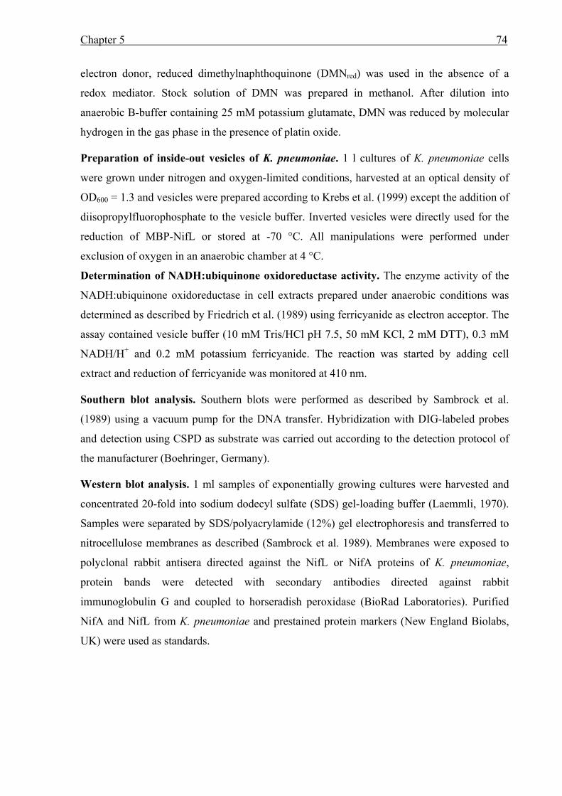
Chapter 5 74
electron donor, reduced dimethylnaphthoquinone (DMNred) was used in the absence of a
redox mediator. Stock solution of DMN was prepared in methanol. After dilution into
anaerobic B-buffer containing 25 mM potassium glutamate, DMN was reduced by molecular
hydrogen in the gas phase in the presence of platin oxide. Preparation of inside-out vesicles of K. pneumoniae. 1 l cultures of K. pneumoniae cells
were grown under nitrogen and oxygen-limited conditions, harvested at an optical density of
OD600 = 1.3 and vesicles were prepared according to Krebs et al. (1999) except the addition of
diisopropylfluorophosphate to the vesicle buffer. Inverted vesicles were directly used for the
reduction of MBP-NifL or stored at -70 °C. All manipulations were performed under
exclusion of oxygen in an anaerobic chamber at 4 °C.
Determination of NADH:ubiquinone oxidoreductase activity. The enzyme activity of the
NADH:ubiquinone oxidoreductase in cell extracts prepared under anaerobic conditions was
determined as described by Friedrich et al. (1989) using ferricyanide as electron acceptor. The
assay contained vesicle buffer (10 mM Tris/HCl pH 7.5, 50 mM KCl, 2 mM DTT), 0.3 mM
NADH/H+ and 0.2 mM potassium ferricyanide. The reaction was started by adding cell
extract and reduction of ferricyanide was monitored at 410 nm. Southern blot analysis. Southern blots were performed as described by Sambrock et al.
(1989) using a vacuum pump for the DNA transfer. Hybridization with DIG-labeled probes
and detection using CSPD as substrate was carried out according to the detection protocol of
the manufacturer (Boehringer, Germany). Western blot analysis. 1 ml samples of exponentially growing cultures were harvested and
concentrated 20-fold into sodium dodecyl sulfate (SDS) gel-loading buffer (Laemmli, 1970).
Samples were separated by SDS/polyacrylamide (12%) gel electrophoresis and transferred to
nitrocellulose membranes as described (Sambrock et al. 1989). Membranes were exposed to
polyclonal rabbit antisera directed against the NifL or NifA proteins of K. pneumoniae,
protein bands were detected with secondary antibodies directed against rabbit
immunoglobulin G and coupled to horseradish peroxidase (BioRad Laboratories). Purified
NifA and NifL from K. pneumoniae and prestained protein markers (New England Biolabs,
UK) were used as standards.
Chapter 5 75
RESULTS
Under oxygen and nitrogen limitation reduction of the flavin moiety of NifL results in a non-
inhibitory conformation of the NifL-protein. Localization analysis of K. pneumoniae NifL
revealed that under those derepressed conditions NifL is membrane-associated, indicating that
sequestration of NifL to the membrane is involved in the regulation of NifA activity by NifL.
In order to analyze whether the association of NifL to the cytoplasmic membrane is
accompanied with the reduction of NifL by a membrane-bound electron donor, we studied
reduction of purified MBP-NifL in vitro and analyzed the influence of different
oxidoreductases of the anaerobic electron transport chain on NifL reduction.
K. pneumoniae NifL is reduced by NADH/H+ in the presence of a redox-mediator or
anaerobic inside-out vesicles. In order to demonstrate whether NADH/H+ is a potential
electron donor in vivo, reduction of purified NifL was studied in vitro. In general, NifL was
synthesized in maximal induction medium under aerobic conditions fused to the maltose
binding protein (MBP) to keep NifL in a more soluble state. Subsequently MBP-NifL was
purified to apparent homogeneity by affinity chromatography. The FAD content of those
purified fractions was in the range of 0.4 - 0.6 FAD per MBP-NifL. Fully oxidized MBP-NifL
(40 µM) was incubated in an anaerobic cuvette under a nitrogen atmosphere in a total volume
of 200 µl B-buffer containing 25 mM glutamate. The absorption spectra were recorded online
using a diode array detector. In the absence of a redox mediator, the addition of 1.25 mM
NADH/H+ (final concentration) did not result in reduction of the NifL-bound FAD-cofactor
even after long incubation periods up to 25 min (data not shown). However, in the presence of
0.2 µM methyl viologen, significant reduction of the flavin-moiety of NifL by NADH/H+ was
observed. After the addition of NADH/H+ the flavin-specific absorbance at 450 nm decreased
constantly within 50 min indicating that the flavin cofactor of NifL was reduced by electrons
derived from NADH/H+ (Fig. 15). This was further supported by the difference spectrum of
oxidized MBP-NifL before the addition of NADH/H+ corrected versus the spectrum 50 min
after NADH/H+ addition, which clearly showed the flavin-specific absorption maximum at
450 nm (inset of Fig. 15) and the 420 nm absorbance which is generally found in NifL
preparations synthesized under nitrogen sufficiency (Klopprogge and Schmitz, 1999). These
findings strongly indicate that NADH/H+ is a potential electron donor for NifL reduction in
vivo, however it appears that the reducing equivalents derived from NADH/H+ have to be
transferred to NifL through an additional oxidoreductase system.
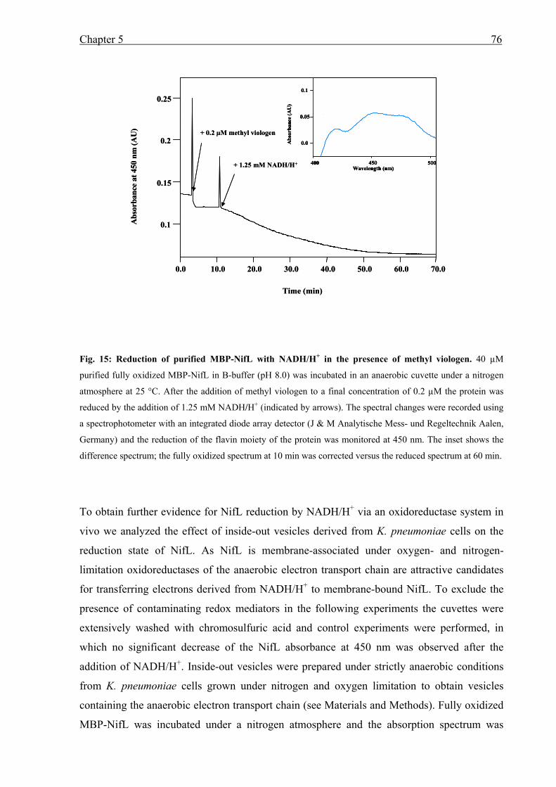
Chapter 5 76
Fig. 15: Reduction of purified MBP-NifL with NADH/H+ in the presence of methyl viologen. 40 µM
purified fully oxidized MBP-NifL in B-buffer (pH 8.0) was incubated in an anaerobic cuvette under a nitrogen
atmosphere at 25 °C. After the addition of methyl viologen to a final concentration of 0.2 µM the protein was
reduced by the addition of 1.25 mM NADH/H+ (indicated by arrows). The spectral changes were recorded using
a spectrophotometer with an integrated diode array detector (J & M Analytische Mess- und Regeltechnik Aalen,
Germany) and the reduction of the flavin moiety of the protein was monitored at 450 nm. The inset shows the
difference spectrum; the fully oxidized spectrum at 10 min was corrected versus the reduced spectrum at 60 min.
To obtain further evidence for NifL reduction by NADH/H+ via an oxidoreductase system in
vivo we analyzed the effect of inside-out vesicles derived from K. pneumoniae cells on the
reduction state of NifL. As NifL is membrane-associated under oxygen- and nitrogen-
limitation oxidoreductases of the anaerobic electron transport chain are attractive candidates
for transferring electrons derived from NADH/H+ to membrane-bound NifL. To exclude the
presence of contaminating redox mediators in the following experiments the cuvettes were
extensively washed with chromosulfuric acid and control experiments were performed, in
which no significant decrease of the NifL absorbance at 450 nm was observed after the
addition of NADH/H+. Inside-out vesicles were prepared under strictly anaerobic conditions
from K. pneumoniae cells grown under nitrogen and oxygen limitation to obtain vesicles
containing the anaerobic electron transport chain (see Materials and Methods). Fully oxidized
MBP-NifL was incubated under a nitrogen atmosphere and the absorption spectrum was
0.1
0.15
0.2
0.25
Abs
orba
nce
at 4
50 n
m (A
U)
0.0 10.0 20.0 30.0 40.0 50.0 60.0 70.0
Time (min)
+ 0.2 µM methyl viologen
+ 1.25 mM NADH/H+
0.0
0.05
0.1
400 450 500
Abs
orba
nce
(AU
)
Wavelength (nm)
0.1
0.15
0.2
0.25
Abs
orba
nce
at 4
50 n
m (A
U)
0.0 10.0 20.0 30.0 40.0 50.0 60.0 70.0
Time (min)
+ 0.2 µM methyl viologen
+ 1.25 mM NADH/H+
0.0
0.05
0.1
400 450 500
Abs
orba
nce
(AU
)
Wavelength (nm)
0.0
0.05
0.1
400 450 500
Abs
orba
nce
(AU
)
Wavelength (nm)
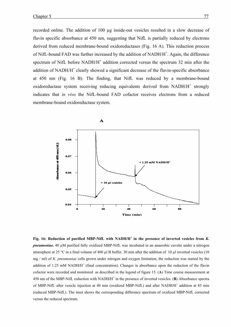
Chapter 5 77
recorded online. The addition of 100 µg inside-out vesicles resulted in a slow decrease of
flavin specific absorbance at 450 nm, suggesting that NifL is partially reduced by electrons
derived from reduced membrane-bound oxidoreductases (Fig. 16 A). This reduction process
of NifL-bound FAD was further increased by the addition of NADH/H+. Again, the difference
spectrum of NifL before NADH/H+ addition corrected versus the spectrum 32 min after the
addition of NADH/H+ clearly showed a significant decrease of the flavin-specific absorbance
at 450 nm (Fig. 16 B). The finding, that NifL was reduced by a membrane-bound
oxidoreductase system receiving reducing equivalents derived from NADH/H+ strongly
indicates that in vivo the NifL-bound FAD cofactor receives electrons from a reduced
membrane-bound oxidoreductase system.
Fig. 16: Reduction of purified MBP-NifL with NADH/H+ in the presence of inverted vesicles from K.
pneumoniae. 40 µM purified fully oxidized MBP-NifL was incubated in an anaerobic cuvette under a nitrogen
atmosphere at 25 °C in a final volume of 400 µl B buffer. 30 min after the addition of 10 µl inverted vesicles (10
mg / ml) of K. pneumoniae cells grown under nitrogen and oxygen limitation, the reduction was started by the
addition of 1.25 mM NADH/H+ (final concentration). Changes in absorbance upon the reduction of the flavin
cofactor were recorded and monitored as described in the legend of figure 15. (A) Time course measurement at
450 nm of the MBP-NifL reduction with NADH/H+ in the presence of inverted vesicles. (B) Absorbance spectra
of MBP-NifL after vesicle injection at 40 min (oxidized MBP-NifL) and after NADH/H+ addition at 85 min
(reduced MBP-NifL). The inset shows the corresponding difference spectrum of oxidized MBP-NifL corrected
versus the reduced spectrum.
0.05
0.06
0.07
0.08
0 20 40 60 80
Abs
orba
nce
at 4
50 n
m (A
U)
Time (min)
+ 10 µl vesicles
+ 1.25 mM NADH/H+
A
0.04
0.05
0.06
0.07
0.08
0 20 40 60 80
Abs
orba
nce
at 4
50 n
m (A
U)
Time (min)
+ 10 µl vesicles
+ 1.25 mM NADH/H+
A
0.04
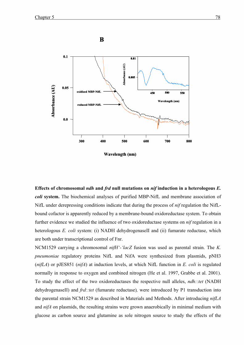
Chapter 5 78
Effects of chromosomal ndh and frd null mutations on nif induction in a heterologous E.
coli system. The biochemical analyses of purified MBP-NifL and membrane association of
NifL under derepressing conditions indicate that during the process of nif regulation the NifL-
bound cofactor is apparently reduced by a membrane-bound oxidoreductase system. To obtain
further evidence we studied the influence of two oxidoreductase systems on nif regulation in a
heterologous E. coli system: (i) NADH dehydrogenaseII and (ii) fumarate reductase, which
are both under transcriptional control of Fnr.
NCM1529 carrying a chromosomal nifH‘-‘lacZ fusion was used as parental strain. The K.
pneumoniae regulatory proteins NifL and NifA were synthesized from plasmids, pNH3
(nifLA) or pJES851 (nifA) at induction levels, at which NifL function in E. coli is regulated
normally in response to oxygen and combined nitrogen (He et al. 1997, Grabbe et al. 2001).
To study the effect of the two oxidoreductases the respective null alleles, ndh::tet (NADH
dehydrogenaseII) and frd::tet (fumarate reductase), were introduced by P1 transduction into
the parental strain NCM1529 as described in Materials and Methods. After introducing nifLA
and nifA on plasmids, the resulting strains were grown anaerobically in minimal medium with
glucose as carbon source and glutamine as sole nitrogen source to study the effects of the
0.0
0.05
0.1
300 400 500 600 700 800
Abs
orba
nce
( AU
)
Wavelength (nm)
B
Abs
orba
nce
(AU
)
Wavelength (nm)
0.01
450 500 550
0.005
reduced MBP-NifL
oxidized MBP-NifL
0.0
0.05
0.1
300 400 500 600 700 800
Abs
orba
nce
( AU
)
Wavelength (nm)
B
Abs
orba
nce
(AU
)
Wavelength (nm)
0.01
450 500 550
0.005
0.01
450 500 550
0.005
reduced MBP-NifL
oxidized MBP-NifL
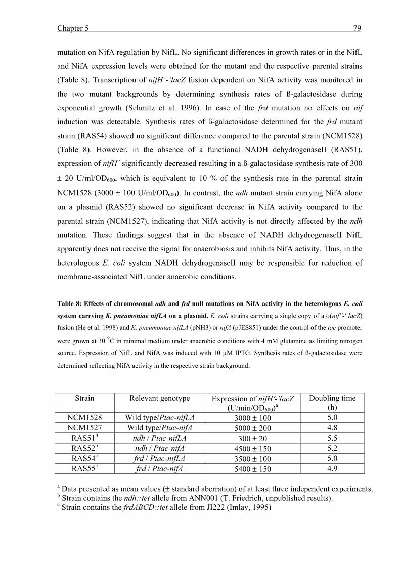
Chapter 5 79
mutation on NifA regulation by NifL. No significant differences in growth rates or in the NifL
and NifA expression levels were obtained for the mutant and the respective parental strains
(Table 8). Transcription of nifH‘-‘lacZ fusion dependent on NifA activity was monitored in
the two mutant backgrounds by determining synthesis rates of ß-galactosidase during
exponential growth (Schmitz et al. 1996). In case of the frd mutation no effects on nif
induction was detectable. Synthesis rates of ß-galactosidase determined for the frd mutant
strain (RAS54) showed no significant difference compared to the parental strain (NCM1528)
(Table 8). However, in the absence of a functional NADH dehydrogenaseII (RAS51),
expression of nifH´ significantly decreased resulting in a ß-galactosidase synthesis rate of 300
± 20 U/ml/OD600, which is equivalent to 10 % of the synthesis rate in the parental strain
NCM1528 (3000 ± 100 U/ml/OD600). In contrast, the ndh mutant strain carrying NifA alone
on a plasmid (RAS52) showed no significant decrease in NifA activity compared to the
parental strain (NCM1527), indicating that NifA activity is not directly affected by the ndh
mutation. These findings suggest that in the absence of NADH dehydrogenaseII NifL
apparently does not receive the signal for anaerobiosis and inhibits NifA activity. Thus, in the
heterologous E. coli system NADH dehydrogenaseII may be responsible for reduction of
membrane-associated NifL under anaerobic conditions.
Table 8: Effects of chromosomal ndh and frd null mutations on NifA activity in the heterologous E. coli
system carrying K. pneumoniae nifLA on a plasmid. E. coli strains carrying a single copy of a φ(nif'′-' lacZ)
fusion (He et al. 1998) and K. pneumoniae nifLA (pNH3) or nifA (pJES851) under the control of the tac promoter
were grown at 30 °C in minimal medium under anaerobic conditions with 4 mM glutamine as limiting nitrogen
source. Expression of NifL and NifA was induced with 10 µM IPTG. Synthesis rates of ß-galactosidase were
determined reflecting NifA activity in the respective strain background.
Strain Relevant genotype Expression of nifH′-′lacZ (U/min/OD600)a
Doubling time (h)
NCM1528 Wild type/Ptac-nifLA 3000 ± 100 5.0 NCM1527 Wild type/Ptac-nifA 5000 ± 200 4.8 RAS51b ndh / Ptac-nifLA 300 ± 20 5.5 RAS52b ndh / Ptac-nifA 4500 ± 150 5.2 RAS54c frd / Ptac-nifLA 3500 ± 100 5.0 RAS55c frd / Ptac-nifA 5400 ± 150 4.9
a Data presented as mean values (± standard aberration) of at least three independent experiments. b Strain contains the ndh::tet allele from ANN001 (T. Friedrich, unpublished results). c Strain contains the frdABCD::tet allele from JI222 (Imlay, 1995)
Chapter 5 80
NADH:ubiquinone oxidoreductase and formate dehydrogenaseN are effecting nif
regulation in K. pneumoniae. As the findings using the heterologous E. coli system indicated
that NADH dehydrogenaseII might be the physiological electron donor for NifL reduction,
we intended to construct a chromosomal null allele of the respective homologous gene in K.
pneumoniae. In order to clone the ndh gene by PCR techniques we generated primers based
on the 5' and 3' end sequences of the ndh gene of K. pneumoniae MGH78578 (subsp.
pneumoniae); sequence information was obtained from the database from the Genome
Sequencing Center, Washington University St. Louis (Genome Sequencing Center, personal
communication). Using chromosomal DNA from K. pneumoniae subsp. pneumoniae (DSM
No. 4799) as control, the ndh gene was amplified. However, no corresponding PCR product
was obtained using chromosomal DNA from K. pneumoniae M5a1, although different pairs
of homologous and degenerated primers were tested under various PCR-assay conditions. To
confirm this finding we performed Southern blot analysis of SmaI digested chromosomal
DNA derived from K. pneumoniae M5a1, K. oxytoca (DSM No. 4798) and K. pneumoniae
subsp. pneumoniae using a DIG-labeled ndh probe derived from K. pneumoniae subsp.
pneumoniae. Hybridization resulted in a single hybridization signal (4 kbp) for K.
pneumoniae subsp. pneumoniae DNA (Fig. 17), which is in accordance with the expected
fragment size based on the knowledge of the genomic sequence. However, no signal was
obtained for DNA derived from K. pneumoniae M5a1 and K. oxytoca even under conditions
of low stringency. These findings strongly indicate that the nitrogen fixing strain K.
pneumoniae M5a1 and K. oxytoca do not exhibit an NADH-dehydrogenaseII. Thus, we
decided to examine the influence of two other membrane-bound oxidoreductases of the
anaerobic respiration on nif regulation in K. pneumoniae.
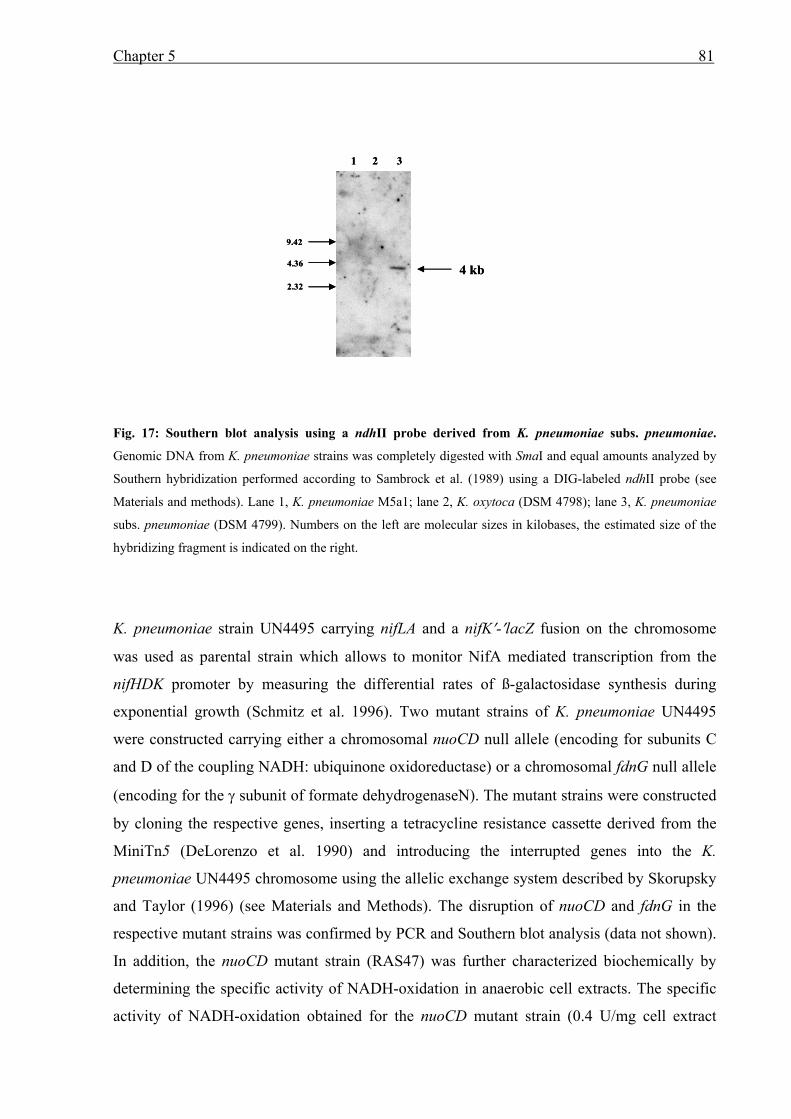
Chapter 5 81
Fig. 17: Southern blot analysis using a ndhII probe derived from K. pneumoniae subs. pneumoniae.
Genomic DNA from K. pneumoniae strains was completely digested with SmaI and equal amounts analyzed by
Southern hybridization performed according to Sambrock et al. (1989) using a DIG-labeled ndhII probe (see
Materials and methods). Lane 1, K. pneumoniae M5a1; lane 2, K. oxytoca (DSM 4798); lane 3, K. pneumoniae
subs. pneumoniae (DSM 4799). Numbers on the left are molecular sizes in kilobases, the estimated size of the
hybridizing fragment is indicated on the right.
K. pneumoniae strain UN4495 carrying nifLA and a nifK′-′lacZ fusion on the chromosome
was used as parental strain which allows to monitor NifA mediated transcription from the
nifHDK promoter by measuring the differential rates of ß-galactosidase synthesis during
exponential growth (Schmitz et al. 1996). Two mutant strains of K. pneumoniae UN4495
were constructed carrying either a chromosomal nuoCD null allele (encoding for subunits C
and D of the coupling NADH: ubiquinone oxidoreductase) or a chromosomal fdnG null allele
(encoding for the γ subunit of formate dehydrogenaseN). The mutant strains were constructed
by cloning the respective genes, inserting a tetracycline resistance cassette derived from the
MiniTn5 (DeLorenzo et al. 1990) and introducing the interrupted genes into the K.
pneumoniae UN4495 chromosome using the allelic exchange system described by Skorupsky
and Taylor (1996) (see Materials and Methods). The disruption of nuoCD and fdnG in the
respective mutant strains was confirmed by PCR and Southern blot analysis (data not shown).
In addition, the nuoCD mutant strain (RAS47) was further characterized biochemically by
determining the specific activity of NADH-oxidation in anaerobic cell extracts. The specific
activity of NADH-oxidation obtained for the nuoCD mutant strain (0.4 U/mg cell extract
9.42
4.36
2.32
1 2 3
4 kb
9.42
4.36
2.32
1 2 3
4 kb
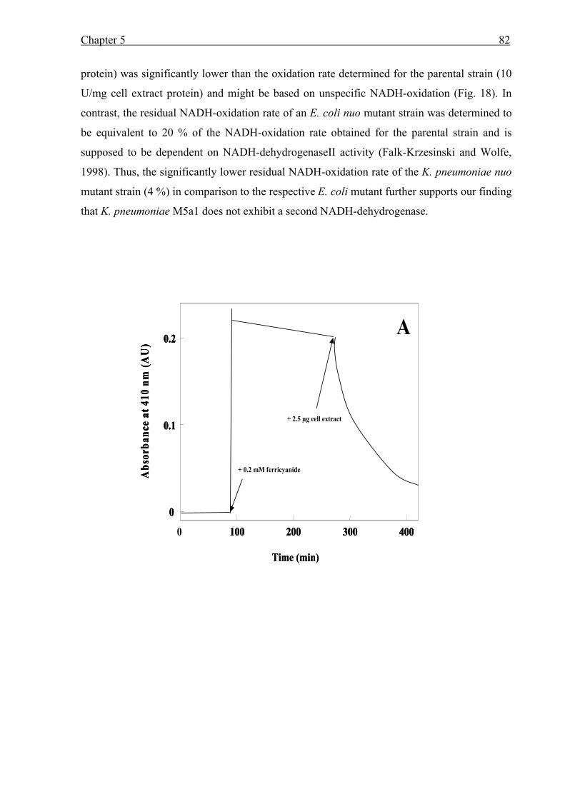
Chapter 5 82
protein) was significantly lower than the oxidation rate determined for the parental strain (10
U/mg cell extract protein) and might be based on unspecific NADH-oxidation (Fig. 18). In
contrast, the residual NADH-oxidation rate of an E. coli nuo mutant strain was determined to
be equivalent to 20 % of the NADH-oxidation rate obtained for the parental strain and is
supposed to be dependent on NADH-dehydrogenaseII activity (Falk-Krzesinski and Wolfe,
1998). Thus, the significantly lower residual NADH-oxidation rate of the K. pneumoniae nuo
mutant strain (4 %) in comparison to the respective E. coli mutant further supports our finding
that K. pneumoniae M5a1 does not exhibit a second NADH-dehydrogenase.
0.1
0.2
0 100 200 300 400
Abs
orba
nce
at 4
10 n
m (A
U)
0
A
+ 0.2 mM ferricyanide
+ 2.5 µg cell extract
Time (min)
0.1
0.2
0 100 200 300 400
Abs
orba
nce
at 4
10 n
m (A
U)
0
A
+ 0.2 mM ferricyanide
+ 2.5 µg cell extract0.1
0.2
0 100 200 300 400
Abs
orba
nce
at 4
10 n
m (A
U)
0
A
+ 0.2 mM ferricyanide
+ 2.5 µg cell extract
Time (min)
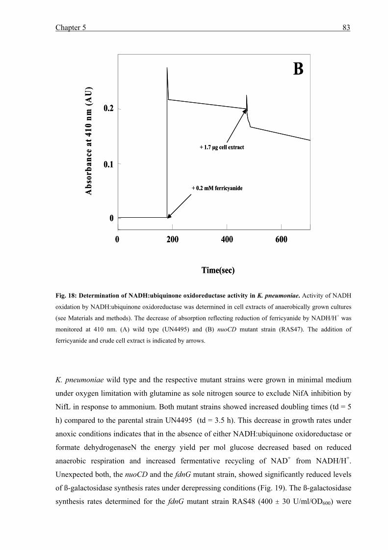
Chapter 5 83
Fig. 18: Determination of NADH:ubiquinone oxidoreductase activity in K. pneumoniae. Activity of NADH
oxidation by NADH:ubiquinone oxidoreductase was determined in cell extracts of anaerobically grown cultures
(see Materials and methods). The decrease of absorption reflecting reduction of ferricyanide by NADH/H+ was
monitored at 410 nm. (A) wild type (UN4495) and (B) nuoCD mutant strain (RAS47). The addition of
ferricyanide and crude cell extract is indicated by arrows.
K. pneumoniae wild type and the respective mutant strains were grown in minimal medium
under oxygen limitation with glutamine as sole nitrogen source to exclude NifA inhibition by
NifL in response to ammonium. Both mutant strains showed increased doubling times (td = 5
h) compared to the parental strain UN4495 (td = 3.5 h). This decrease in growth rates under
anoxic conditions indicates that in the absence of either NADH:ubiquinone oxidoreductase or
formate dehydrogenaseN the energy yield per mol glucose decreased based on reduced
anaerobic respiration and increased fermentative recycling of NAD+ from NADH/H+.
Unexpected both, the nuoCD and the fdnG mutant strain, showed significantly reduced levels
of ß-galactosidase synthesis rates under derepressing conditions (Fig. 19). The ß-galactosidase
synthesis rates determined for the fdnG mutant strain RAS48 (400 ± 30 U/ml/OD600) were
Abs
orba
nce
at 4
10 n
m (A
U)
Time(sec)
0
0.1
0.2
0 200 400 600
B
+ 0.2 mM ferricyanide
+ 1.7 µg cell extract
Abs
orba
nce
at 4
10 n
m (A
U)
Time(sec)
0
0.1
0.2
0 200 400 600
B
+ 0.2 mM ferricyanide
+ 1.7 µg cell extract
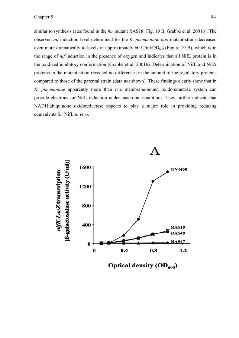
Chapter 5 84
similar to synthesis rates found in the fnr mutant RAS18 (Fig. 19 B, Grabbe et al. 2001b). The
observed nif induction level determined for the K. pneumoniae nuo mutant strain decreased
even more dramatically to levels of approximately 60 U/ml/OD600 (Figure 19 B), which is in
the range of nif induction in the presence of oxygen and indicates that all NifL protein is in
the oxidized inhibitory conformation (Grabbe et al. 2001b). Determination of NifL and NifA
proteins in the mutant strain revealed no differences in the amount of the regulatory proteins
compared to those of the parental strain (data not shown). These findings clearly show that in
K. pneumoniae apparently more than one membrane-bound oxidoreductase system can
provide electrons for NifL reduction under anaerobic conditions. They further indicate that
NADH:ubiquinone oxidoreductase appears to play a major role in providing reducing
equivalents for NifL in vivo.
0
400
800
1200
1600
0 0.4 0.8 1.2
nifK
-Lac
Ztr
ansc
ript
ion
[ß-g
alac
tosi
dase
activ
ity(U
/ml)]
Optical density (OD600)
AUN4495
RAS18RAS48
RAS470
400
800
1200
1600
0 0.4 0.8 1.2
nifK
-Lac
Ztr
ansc
ript
ion
[ß-g
alac
tosi
dase
activ
ity(U
/ml)]
Optical density (OD600)
AUN4495
RAS18RAS48
RAS47
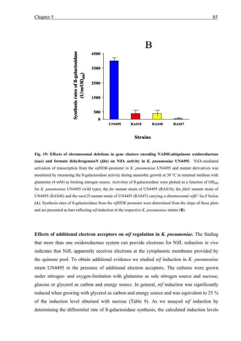
Chapter 5 85
Fig. 19: Effects of chromosomal deletions in gene clusters encoding NADH:ubiquinone oxidoreductase
(nuo) and formate dehydrogenaseN (fdn) on NifA activity in K. pneumoniae UN4495. NifA-mediated
activation of transcription from the nifHDK-promoter in K. pneumoniae UN4495 and mutant derivatives was
monitored by measuring the ß-galactosidase activity during anaerobic growth at 30 °C in minimal medium with
glutamine (4 mM) as limiting nitrogen source. Activities of ß-galactosidase were plotted as a function of OD600
for K. pneumoniae UN4495 (wild type), the fnr mutant strain of UN4495 (RAS18), the fdnG mutant strain of
UN4495 (RAS48) and the nuoCD mutant strain of UN4495 (RAS47) carrying a chromosomal nifK'-'lacZ fusion
(A). Synthesis rates of ß-galactosidase from the nifHDK promoter were determined from the slope of these plots
and are presented as bars reflecting nif-induction in the respective K. pneumoniae strains (B).
Effects of additional electron acceptors on nif regulation in K. pneumoniae. The finding
that more than one oxidoreductase system can provide electrons for NifL reduction in vivo
indicates that NifL apparently receives electrons at the cytoplasmic membrane provided by
the quinone pool. To obtain additional evidence we studied nif induction in K. pneumoniae
strain UN4495 in the presence of additional electron acceptors. The cultures were grown
under nitrogen- and oxygen-limitation with glutamine as sole nitrogen source and sucrose,
glucose or glycerol as carbon and energy source. In general, nif induction was significantly
reduced when growing with glycerol as carbon and energy source and was equivalent to 25 %
of the induction level obtained with sucrose (Table 9). As we assayed nif induction by
determining the differential rate of ß-galactosidase synthesis, the calculated induction levels
0
500
1500
2500
3500
4500
UN4495 RAS18 RAS48 RAS47
Synt
hesi
s rat
esof
ß-g
alac
tosi
dase
(U/m
/OD
600)
B
Strains
0
500
1500
2500
3500
4500
UN4495 RAS18 RAS48 RAS47
Synt
hesi
s rat
esof
ß-g
alac
tosi
dase
(U/m
/OD
600)
B
Strains
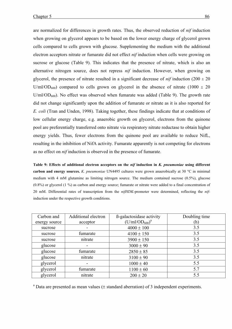
Chapter 5 86
are normalized for differences in growth rates. Thus, the observed reduction of nif induction
when growing on glycerol appears to be based on the lower energy charge of glycerol grown
cells compared to cells grown with glucose. Supplementing the medium with the additional
electron acceptors nitrate or fumarate did not effect nif induction when cells were growing on
sucrose or glucose (Table 9). This indicates that the presence of nitrate, which is also an
alternative nitrogen source, does not repress nif induction. However, when growing on
glycerol, the presence of nitrate resulted in a significant decrease of nif induction (200 ± 20
U/ml/OD600) compared to cells grown on glycerol in the absence of nitrate (1000 ± 20
U/ml/OD600). No effect was observed when fumarate was added (Table 9). The growth rate
did not change significantly upon the addition of fumarate or nitrate as it is also reported for
E. coli (Tran and Unden, 1998). Taking together, these findings indicate that at conditions of
low cellular energy charge, e.g. anaerobic growth on glycerol, electrons from the quinone
pool are preferentially transferred onto nitrate via respiratory nitrate reductase to obtain higher
energy yields. Thus, fewer electrons from the quinone pool are available to reduce NifL,
resulting in the inhibition of NifA activity. Fumarate apparently is not competing for electrons
as no effect on nif induction is observed in the presence of fumarate.
Table 9: Effects of additional electron acceptors on the nif induction in K. pneumoniae using different
carbon and energy sources. K. pneumoniae UN4495 cultures were grown anaerobically at 30 °C in minimal
medium with 4 mM glutamine as limiting nitrogen source. The medium contained sucrose (0.5%), glucose
(0.8%) or glycerol (1 %) as carbon and energy source; fumarate or nitrate were added to a final concentration of
20 mM. Differential rates of transcription from the nifHDK-promoter were determined, reflecting the nif-
induction under the respective growth conditions.
Carbon and energy source
Additional electron acceptor
ß-galactosidase activity (U/ml/OD600)a
Doubling time (h)
sucrose - 4000 ± 100 3.5 sucrose fumarate 4100 ± 150 3.5 sucrose nitrate 3900 ± 150 3.5 glucose - 3000 ± 90 3.5 glucose fumarate 2850 ± 85 3.5 glucose nitrate 3100 ± 90 3.5 glycerol - 1000 ± 40 5.5 glycerol fumarate 1100 ± 60 5.7 glycerol nitrate 200 ± 20 5.5
a Data are presented as mean values (± standard aberration) of 3 independent experiments.
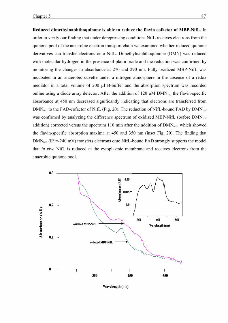
Chapter 5 87
Reduced dimethylnaphthoquinone is able to reduce the flavin cofactor of MBP-NifL. In
order to verify our finding that under derepressing conditions NifL receives electrons from the
quinone pool of the anaerobic electron transport chain we examined whether reduced quinone
derivatives can transfer electrons onto NifL. Dimethylnaphthoquinone (DMN) was reduced
with molecular hydrogen in the presence of platin oxide and the reduction was confirmed by
monitoring the changes in absorbance at 270 and 290 nm. Fully oxidized MBP-NifL was
incubated in an anaerobic cuvette under a nitrogen atmosphere in the absence of a redox
mediator in a total volume of 200 µl B-buffer and the absorption spectrum was recorded
online using a diode array detector. After the addition of 120 µM DMNred the flavin-specific
absorbance at 450 nm decreased significantly indicating that electrons are transferred from
DMNred to the FAD-cofactor of NifL (Fig. 20). The reduction of NifL-bound FAD by DMNred
was confirmed by analyzing the difference spectrum of oxidized MBP-NifL (before DMNred
addition) corrected versus the spectrum 110 min after the addition of DMNred, which showed
the flavin-specific absorption maxima at 450 and 350 nm (inset Fig. 20). The finding that
DMNred (E°'=-240 mV) transfers electrons onto NifL-bound FAD strongly supports the model
that in vivo NifL is reduced at the cytoplasmic membrane and receives electrons from the
anaerobic quinone pool.
0.0
0.025
0.05
Abs
orba
nce
(AU
)
350 450 550
Wavelength (nm)
Abs
orba
nce
(AU
)
0
0.1
0.2
0.3
350 450 550
Wavelength (nm)
oxidized MBP-NifL
reduced MBP-NifL
0.0
0.025
0.05
Abs
orba
nce
(AU
)
350 450 550
Wavelength (nm)
0.0
0.025
0.05
Abs
orba
nce
(AU
)
350 450 550
Wavelength (nm)
Abs
orba
nce
(AU
)
0
0.1
0.2
0.3
350 450 550
Wavelength (nm)
oxidized MBP-NifL
reduced MBP-NifL
Chapter 5 88
Fig. 20: Reduction of MBP-NifL using reduced dimethylnaphthoquinone as artificial electron donor. 40
µM fully oxidized MBP-NifL was incubated in B buffer under a nitrogen atmosphere at room temperature.
Reduced dimethylnaphthoquinone (DMNred) was added to a final concentration of 0.2 mM and the changes in
absorbance were recorded using a spectrophotometer with an integrated diode array detector (J & M Analytische
Mess- und Regeltechnik Aalen, Germany). Absorbance spectra of MBP-NifL before (oxidized MBP-NifL) and
40 min after the addition of 0.2 mM DMNred (reduced MBP-NifL) are shown. The corresponding difference
spectrum of oxidized MBP-NifL corrected versus the reduced spectrum after addition of DMNred is visualized in
the inset.
DISCUSSION
The NifL-bound FAD receives electrons from the reduced quinone pool at the
cytoplasmic membrane. We recently showed that in K. pneumoniae membrane-sequestration
of NifL under nitrogen- and oxygen-limited conditions seems to be the mechanism for nif
regulation in response to molecular oxygen and combined nitrogen (Klopprogge et al. 2002).
In order to verify our model that membrane-association of NifL is accompanied by the
reduction of the NifL-bound FAD cofactor, we studied the process of NifL reduction. Based
on our findings in this study we propose that the FAD cofactor of NifL is reduced by a
membrane-bound component of the anaerobic electron transport chain resulting in a reduced
non-inhibitory conformation of NifL, which is membrane-associated. A first line of evidence
was provided by biochemical analyses of the purified MBP-NifL protein. Spectral analysis
clearly showed that in the absence of a redox mediator no change in absorbance of the
flavoprotein was detectable upon the addition of NADH/H+. However, the presence of methyl
viologen or inside-out vesicles derived from K. pneumoniae cells grown under anaerobic
conditions allowed NifL reduction by NADH/H+ (Figs. 15 and 16). This strongly indicates
that in vivo NifL-bound FAD receives electrons from a membrane-bound oxidoreductase
system. Two other lines of evidence derived from in vivo studies of nif regulation support this
view. First, using K. pneumoniae or E. coli strains carrying null mutations of different
membrane-bound oxidoreductases we showed that the absence of formate dehydrogenaseN or
NADH:ubiquinone oxidoreductase in K. pneumoniae and the absence of NADH
dehydrogenaseII in the heterologous E. coli system affects nif regulation dramatically. In the
respective mutant strains nif induction was low, similar to induction levels under aerobic
conditions, even though cells were grown under oxygen- and nitrogen-limitation (Table 8 and
Fig. 19). These findings indicate that in the absence of the respective membrane-bound
oxidoreductases the FAD-cofactor of NifL was not reduced at the cytoplasmic membrane
Chapter 5 89
resulting in cytoplasmic NifL, which inhibits NifA activity. Second, additional studies of nif
induction in K. pneumoniae growing anaerobically on glycerol under nitrogen limitation
revealed that the presence of nitrate as additional electron acceptor resulted in a significant
decrease in nif induction (Table 9). It appears that under those energy-limited growth
conditions, electrons of the reduced quinone pool are preferentially transferred onto nitrate
allowing anaerobic respiration and energy conservation by the respiratory nitrate reductase
(Tran et al. 1997, Unden and Bongaerts 1997). Thus, a high percentage of NifL protein does
not receive electrons at the membrane and stays in its oxidized conformation in the cytoplasm
inhibiting NifA activity resulting in decreased nif induction. Taking together, these data
strongly indicate that more than one membrane-bound oxidoreductase system can provide
electrons for NifL reduction under anaerobic conditions and we propose that under anaerobic
conditions NifL receives electrons from the reduced quinone pool generated by different
membrane-bound oxidoreductase systems. Reduction of the NifL-bound cofactor finally
results in higher membrane-association of NifL, allowing cytoplasmic NifA to activate nif
induction. The demonstration that the reduced soluble quinone derivative
dimethylnaphthoquinone (E°'=-240 mV, Krafft et al. 1995) is able to reduce the FAD cofactor
of purified NifL in the absence of a redox mediator fully supports this model (Fig. 6). The
reduction of NifL under anaerobic conditions at the cytoplasmic membrane by electrons
derived from the reduced quinone pool rather than by a single specific membrane-bound
enzyme is a particularly attractive model for several reasons. It explains (i) that the absence of
different membrane-bound oxidoreductases in K. pneumoniae, which transfer electrons onto
the quinone pool, results in the oxidized inhibitory conformation of NifL; and (ii) that NADH
dehydrogenaseII in the heterologous E. coli system significantly affects nif regulation,
although a homologous oxidoreductase appears not to be present in K. pneumoniae. Finally,
reduction of NifL by the reduced quinone pool potentially allows the simultaneous signal
integration of the cell's energy status for nif regulation.
In contrast to K. pneumoniae NifL no membrane-association for A. vinelandii NifL has been
reported to date (Austin et al. 1994, Hill et al. 1996, Dixon, 1998). Thus, a different
mechanism for the transduction of the oxygen signal to NifL is expected. In in vitro
experiments A. vinelandii-NifL is reduced by NADH/H+ when catalyzed by the E. coli
cytoplasmic flavoheme protein (HMP), which is proposed to be a global oxygen sensor or an
oxidoreductase preventing cells from endogenous oxygen stress (Pool 1994, Macheroux et al.
1998, Stevanin et al. 2000;). However, the functional and physiological relevance of NifL
reduction by HMP has not been demonstrated in vivo. It is currently hypothesized, that the
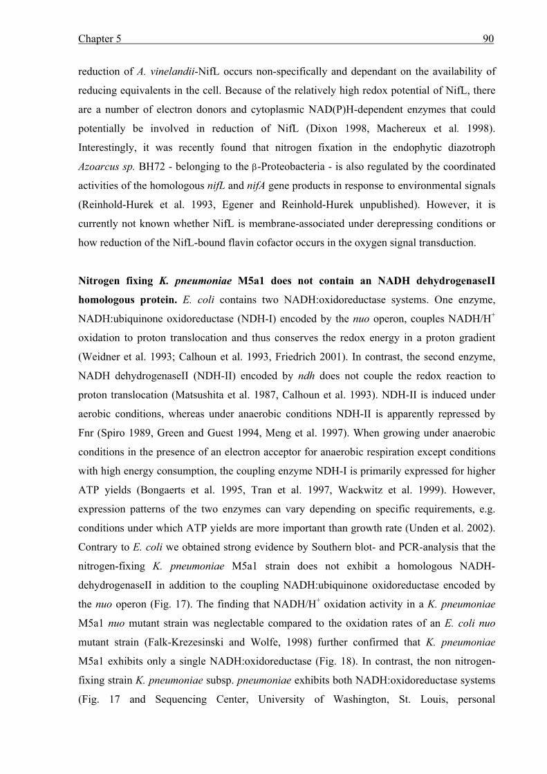
Chapter 5 90
reduction of A. vinelandii-NifL occurs non-specifically and dependant on the availability of
reducing equivalents in the cell. Because of the relatively high redox potential of NifL, there
are a number of electron donors and cytoplasmic NAD(P)H-dependent enzymes that could
potentially be involved in reduction of NifL (Dixon 1998, Machereux et al. 1998).
Interestingly, it was recently found that nitrogen fixation in the endophytic diazotroph
Azoarcus sp. BH72 - belonging to the β-Proteobacteria - is also regulated by the coordinated
activities of the homologous nifL and nifA gene products in response to environmental signals
(Reinhold-Hurek et al. 1993, Egener and Reinhold-Hurek unpublished). However, it is
currently not known whether NifL is membrane-associated under derepressing conditions or
how reduction of the NifL-bound flavin cofactor occurs in the oxygen signal transduction.
Nitrogen fixing K. pneumoniae M5a1 does not contain an NADH dehydrogenaseII
homologous protein. E. coli contains two NADH:oxidoreductase systems. One enzyme,
NADH:ubiquinone oxidoreductase (NDH-I) encoded by the nuo operon, couples NADH/H+
oxidation to proton translocation and thus conserves the redox energy in a proton gradient
(Weidner et al. 1993; Calhoun et al. 1993, Friedrich 2001). In contrast, the second enzyme,
NADH dehydrogenaseII (NDH-II) encoded by ndh does not couple the redox reaction to
proton translocation (Matsushita et al. 1987, Calhoun et al. 1993). NDH-II is induced under
aerobic conditions, whereas under anaerobic conditions NDH-II is apparently repressed by
Fnr (Spiro 1989, Green and Guest 1994, Meng et al. 1997). When growing under anaerobic
conditions in the presence of an electron acceptor for anaerobic respiration except conditions
with high energy consumption, the coupling enzyme NDH-I is primarily expressed for higher
ATP yields (Bongaerts et al. 1995, Tran et al. 1997, Wackwitz et al. 1999). However,
expression patterns of the two enzymes can vary depending on specific requirements, e.g.
conditions under which ATP yields are more important than growth rate (Unden et al. 2002).
Contrary to E. coli we obtained strong evidence by Southern blot- and PCR-analysis that the
nitrogen-fixing K. pneumoniae M5a1 strain does not exhibit a homologous NADH-
dehydrogenaseII in addition to the coupling NADH:ubiquinone oxidoreductase encoded by
the nuo operon (Fig. 17). The finding that NADH/H+ oxidation activity in a K. pneumoniae
M5a1 nuo mutant strain was neglectable compared to the oxidation rates of an E. coli nuo
mutant strain (Falk-Krezesinski and Wolfe, 1998) further confirmed that K. pneumoniae
M5a1 exhibits only a single NADH:oxidoreductase (Fig. 18). In contrast, the non nitrogen-
fixing strain K. pneumoniae subsp. pneumoniae exhibits both NADH:oxidoreductase systems
(Fig. 17 and Sequencing Center, University of Washington, St. Louis, personal
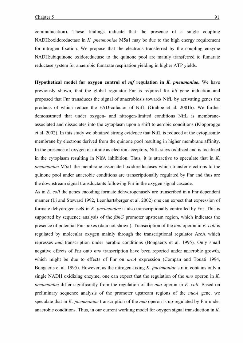
Chapter 5 91
communication). These findings indicate that the presence of a single coupling
NADH:oxidoreductase in K. pneumoniae M5a1 may be due to the high energy requirement
for nitrogen fixation. We propose that the electrons transferred by the coupling enzyme
NADH:ubiquinone oxidoreductase to the quinone pool are mainly transferred to fumarate
reductase system for anaerobic fumarate respiration yielding in higher ATP yields.
Hypothetical model for oxygen control of nif regulation in K. pneumoniae. We have
previously shown, that the global regulator Fnr is required for nif gene induction and
proposed that Fnr transduces the signal of anaerobiosis towards NifL by activating genes the
products of which reduce the FAD-cofactor of NifL (Grabbe et al. 2001b). We further
demonstrated that under oxygen- and nitrogen-limited conditions NifL is membrane-
associated and dissociates into the cytoplasm upon a shift to aerobic conditions (Klopprogge
et al. 2002). In this study we obtained strong evidence that NifL is reduced at the cytoplasmic
membrane by electrons derived from the quinone pool resulting in higher membrane affinity.
In the presence of oxygen or nitrate as electron acceptors, NifL stays oxidized and is localized
in the cytoplasm resulting in NifA inhibition. Thus, it is attractive to speculate that in K.
pneumoniae M5a1 the membrane-associated oxidoreductases which transfer electrons to the
quinone pool under anaerobic conditions are transcriptionally regulated by Fnr and thus are
the downstream signal transductants following Fnr in the oxygen signal cascade.
As in E. coli the genes encoding formate dehydrogenaseN are transcribed in a Fnr dependent
manner (Li and Steward 1992, Leonhartsberger et al. 2002) one can expect that expression of
formate dehydrogenaseN in K. pneumoniae is also transcriptionally controlled by Fnr. This is
supported by sequence analysis of the fdnG promoter upstream region, which indicates the
presence of potential Fnr-boxes (data not shown). Transcription of the nuo operon in E. coli is
regulated by molecular oxygen mainly through the transcriptional regulator ArcA which
represses nuo transcription under aerobic conditions (Bongaerts et al. 1995). Only small
negative effects of Fnr onto nuo transcription have been reported under anaerobic growth,
which might be due to effects of Fnr on arcA expression (Compan and Touati 1994,
Bongaerts et al. 1995). However, as the nitrogen-fixing K. pneumoniae strain contains only a
single NADH oxidizing enzyme, one can expect that the regulation of the nuo operon in K.
pneumoniae differ significantly from the regulation of the nuo operon in E. coli. Based on
preliminary sequence analysis of the promoter upstream regions of the nuoA gene, we
speculate that in K. pneumoniae transcription of the nuo operon is up-regulated by Fnr under
anaerobic conditions. Thus, in our current working model for oxygen signal transduction in K.
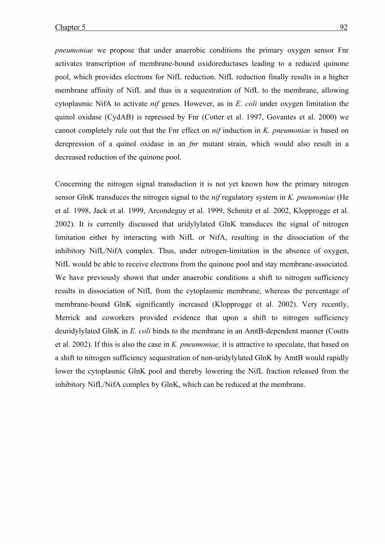
Chapter 5 92
pneumoniae we propose that under anaerobic conditions the primary oxygen sensor Fnr
activates transcription of membrane-bound oxidoreductases leading to a reduced quinone
pool, which provides electrons for NifL reduction. NifL reduction finally results in a higher
membrane affinity of NifL and thus in a sequestration of NifL to the membrane, allowing
cytoplasmic NifA to activate nif genes. However, as in E. coli under oxygen limitation the
quinol oxidase (CydAB) is repressed by Fnr (Cotter et al. 1997, Govantes et al. 2000) we
cannot completely rule out that the Fnr effect on nif induction in K. pneumoniae is based on
derepression of a quinol oxidase in an fnr mutant strain, which would also result in a
decreased reduction of the quinone pool.
Concerning the nitrogen signal transduction it is not yet known how the primary nitrogen
sensor GlnK transduces the nitrogen signal to the nif regulatory system in K. pneumoniae (He
et al. 1998, Jack et al. 1999, Arcondeguy et al. 1999, Schmitz et al. 2002, Klopprogge et al.
2002). It is currently discussed that uridylylated GlnK transduces the signal of nitrogen
limitation either by interacting with NifL or NifA, resulting in the dissociation of the
inhibitory NifL/NifA complex. Thus, under nitrogen-limitation in the absence of oxygen,
NifL would be able to receive electrons from the quinone pool and stay membrane-associated.
We have previously shown that under anaerobic conditions a shift to nitrogen sufficiency
results in dissociation of NifL from the cytoplasmic membrane, whereas the percentage of
membrane-bound GlnK significantly increased (Klopprogge et al. 2002). Very recently,
Merrick and coworkers provided evidence that upon a shift to nitrogen sufficiency
deuridylylated GlnK in E. coli binds to the membrane in an AmtB-dependent manner (Coutts
et al. 2002). If this is also the case in K. pneumoniae, it is attractive to speculate, that based on
a shift to nitrogen sufficiency sequestration of non-uridylylated GlnK by AmtB would rapidly
lower the cytoplasmic GlnK pool and thereby lowering the NifL fraction released from the
inhibitory NifL/NifA complex by GlnK, which can be reduced at the membrane.
Conclusions 93
Conclusions
In Klebsiella pneumoniae and Azotobacter vinelandii the nitrogen regulatory proteins NifL
and NifA tightly control synthesis of nitrogen fixation genes in response to the environmental
signals molecular oxygen and combined nitrogen. In this regulation the negative regulator
NifL inhibits NifA transcriptional activity. Immunological studies, co-chromatography and
complex formation analyses using the yeast two-hybrid system demonstrated that NifA
interacts directly with NifL by protein-protein interaction (Henderson et al., 1989; Money et
al. 1999 and 2001; Lei et al., 1999). This indicates that the signals of nitrogen sufficiency
and/or molecular oxygen finally result in complex formation between NifL and NifA, which
inhibits NifA activity and thus prevents unnecessary consumption of energy for nitrogen
fixation. The inhibitor NifL is a flavoprotein, which regulates NifA activity depending on the
reduction status of its N-terminally bound FAD-cofactor, allowing NifA transcriptional
activity only under anaerobic conditions. Thus, the redox-sensitive FAD-cofactor appears to
be involved in oxygen signal-transduction (Hill et al., 1996; Schmitz, 1997; Macheroux et al.,
1998; Klopprogge and Schmitz, 1999). Both proteins, K. pneumoniae NifL and A. vinelandii
NifL, were biochemically analyzed (Macheroux et al., 1998; Klopprogge and Schmitz, 1999),
however, the physiological electron donor for NifL reduction is still discussed to date. In A.
vinelandii, NifL reduction under anaerobic conditions apparently occurs unspecifically,
depending on reducing equivalents available in the cell (Macheroux et al., 1998; Dixon,
1998). In K. pneumoniae, the relief of NifL inhibition under anaerobic conditions requires the
presence of iron (Schmitz et al. 1996). As the NifL-protein does not contain iron or acid-labile
sulphur (Schmitz, 1997), this finding indicates the involvement of an iron-containing protein
in the oxygen-signaling mechanism, which is directly or indirectly required for NifL
reduction. In Fig. 21 a model for oxygen signal-transduction in K. pneumoniae is presented.
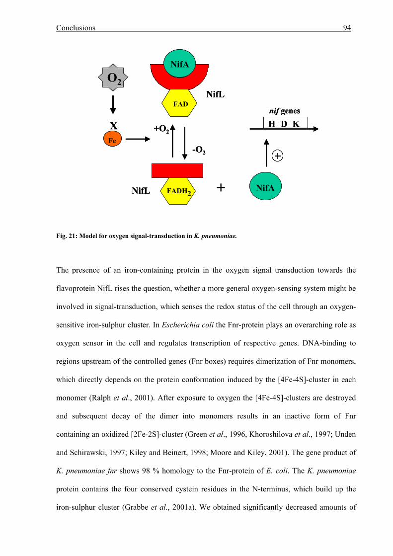
Conclusions 94
Fig. 21: Model for oxygen signal-transduction in K. pneumoniae.
The presence of an iron-containing protein in the oxygen signal transduction towards the
flavoprotein NifL rises the question, whether a more general oxygen-sensing system might be
involved in signal-transduction, which senses the redox status of the cell through an oxygen-
sensitive iron-sulphur cluster. In Escherichia coli the Fnr-protein plays an overarching role as
oxygen sensor in the cell and regulates transcription of respective genes. DNA-binding to
regions upstream of the controlled genes (Fnr boxes) requires dimerization of Fnr monomers,
which directly depends on the protein conformation induced by the [4Fe-4S]-cluster in each
monomer (Ralph et al., 2001). After exposure to oxygen the [4Fe-4S]-clusters are destroyed
and subsequent decay of the dimer into monomers results in an inactive form of Fnr
containing an oxidized [2Fe-2S]-cluster (Green et al., 1996, Khoroshilova et al., 1997; Unden
and Schirawski, 1997; Kiley and Beinert, 1998; Moore and Kiley, 2001). The gene product of
K. pneumoniae fnr shows 98 % homology to the Fnr-protein of E. coli. The K. pneumoniae
protein contains the four conserved cystein residues in the N-terminus, which build up the
iron-sulphur cluster (Grabbe et al., 2001a). We obtained significantly decreased amounts of
NifA
+O2
FADH2NifL
X
-O2
NifL
Fe
O2
FAD
NifA+
+
H D Knif genes
NifA
+O2
FADH2NifL
X
-O2
NifL
FeFe
O2
FAD
NifA+
++
H D Knif genes
Conclusions 95
iron in K. pneumoniae Fnr under aerobic conditions indicating that K. pneumoniae Fnr is
sensing the redox status of the cell via a redox-sensitive [4Fe-4S]-cluster (Chapter 2, Grabbe
et al., 2001a). The goal of this thesis was to analyze the potential role of Fnr in oxygen signal-
transduction to the nif regulatory system.
We obtained strong evidence that in K. pneumoniae Fnr influences the oxygen-dependent
nitrogen regulation by analyzing (i) NifA dependent nif induction in different fnr mutant
strains (Chapter 3, Grabbe et al., 2001b), (ii) membrane-association of NifL in K. pneumoniae
wild-type and fnr mutant strains (Chapter 4, Klopprogge et al., 2002), and finally (iii) by
studying the influence of different Fnr-dependent membrane-bound oxidoreductase systems
on NifL modulated NifA activity (Chapter 5, Grabbe and Schmitz, 2002). In the following,
each line of evidence is discussed in more detail.
(i) In the absence of Fnr, NifA mediated transcription of nif genes decreased significantly
under nitrogen- and oxygen-limitation (Grabbe et al., 2001b). This indicates that the FAD
moiety of NifL is not reduced, resulting in an inhibitory conformation of NifL. As we can rule
out that the transcriptional activator Fnr provides electrons to reduce NifL, we postulate that
Fnr transcriptionally controls genes, the products of which function to reduce the NifL-bound
FAD-cofactor, resulting in a non-inhibitory conformation of NifL. Attractive candidates for
the physiological electron donor are members of the anaerobic electron transport chain.
(ii) Localization of NifL in K. pneumoniae revealed that under anaerobic and nitrogen-limited
conditions NifL is highly membrane-associated. Based on a shift to oxygen or nitrogen
sufficiency in the absence of Fnr NifL is located mainly in the cytoplasm (Klopprogge et al.,
2002). This indicates that NifL is apparently reduced during membrane-association by a
membrane-bound oxidoreductase.
(iii) Studying the influence of different oxidoreductase systems on nif induction in K.
pneumoniae we observed a remarkable decrease in NifA activity under oxygen and nitrogen
limitation in the absence of a functional NADH:ubiquinone oxidoreductase or formate
Conclusions 96
dehydrogenaseN. Analyzing nif induction in a heterologous E. coli system showed that in a
ndh mutant background NifL inhibition was not relieved under nitrogen and oxygen limitation
(Grabbe and Schmitz, 2002). Based on these findings we conclude that under anaerobic
conditions in K. pneumoniae the oxidoreductase systems NADH:ubiquinone oxidoreductase
and formate dehydrogenaseN generate a reduced quinone pool in the cytoplasmic membrane.
In the absence of molecular oxygen, NifL contacts the membrane and receives electrons from
the reduced quinone pool resulting in a non-inhibitory conformation of NifL. Subsequent
analysis to verify the role of the reduced quinone pool towards the reduction of the NifL-
bound FAD-cofactor revealed, that reduced dimethylnaphthoquinone, a soluble quinone
derivative, is able to function as electron donor for NifL in vitro (Grabbe and Schmitz, 2002).
This further supports the model of NifL reduction by the reduced quinone pool, which is
generated by those oxidoreductase systems. We further hypothesize that transcription of
formate dehydrogenaseN and NADH:ubiquinone oxidoreductase is Fnr-dependent (Grabbe
and Schmitz, 2002).
Current working model for oxygen-dependent control of NifA in K. pneumoniae. The
results presented in this thesis are summarized in Fig. 22. Under anaerobic nitrogen-limiting
conditions the NifL-protein of K. pneumoniae contacts the cytoplasmic membrane. This
membrane-association is accompanied by reducing the FAD-cofactor with electrons from the
reduced quinone pool, which apparently results in a membrane-associated NifL protein. Upon
a shift to aerobiosis the FAD-cofactor is oxidized, NifL switches into an inhibitory
conformation and dissociates from the membrane. The increased amount of inhibitory NifL in
the cytoplasm interacts with NifA, resulting in a decrease of nif induction. Thus, we propose
that sequestration of reduced NifL to the membrane under oxygen- and nitrogen-limitation
creates a spatial gap between NifL and its target NifA, which is the regulatory mechanism for
oxygen dependent control of NifA activity in K. pneumoniae.
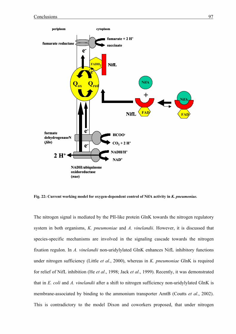
Conclusions 97
Fig. 22: Current working model for oxygen-dependent control of NifA activity in K. pneumoniae.
The nitrogen signal is mediated by the PII-like protein GlnK towards the nitrogen regulatory
system in both organisms, K. pneumoniae and A. vinelandii. However, it is discussed that
species-specific mechanisms are involved in the signaling cascade towards the nitrogen
fixation regulon. In A. vinelandii non-uridylylated GlnK enhances NifL inhibitory functions
under nitrogen sufficiency (Little et al., 2000), whereas in K. pneumoniae GlnK is required
for relief of NifL inhibition (He et al., 1998; Jack et al., 1999). Recently, it was demonstrated
that in E. coli and A. vinelandii after a shift to nitrogen sufficiency non-uridylylated GlnK is
membrane-associated by binding to the ammonium transporter AmtB (Coutts et al., 2002).
This is contradictory to the model Dixon and coworkers proposed, that under nitrogen
e-
NADH:ubiquinoneoxidoreductase(nuo)
formatedehydrogenaseN(fdn)
HCOO-
CO2 + 2 H+
NifA
Qred
e-
NADH/H+
NAD+2 H+
e-
e-
fumarate + 2 H+
succinate
NifA
fumarate reductase
Qox
FADH2 NifL
FAD
periplasm cytoplasm
FADNifL
+
e-
NADH:ubiquinoneoxidoreductase(nuo)
formatedehydrogenaseN(fdn)
HCOO-
CO2 + 2 H+
NifA
Qred
e-
NADH/H+
NAD+2 H+
e-
e-
fumarate + 2 H+
succinate
NifA
fumarate reductase
Qox
FADH2 NifLFADH2 NifL
FADFAD
periplasm cytoplasm
FADFADNifL
+
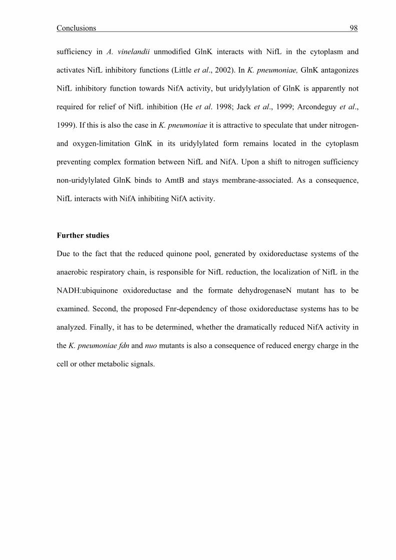
Conclusions 98
sufficiency in A. vinelandii unmodified GlnK interacts with NifL in the cytoplasm and
activates NifL inhibitory functions (Little et al., 2002). In K. pneumoniae, GlnK antagonizes
NifL inhibitory function towards NifA activity, but uridylylation of GlnK is apparently not
required for relief of NifL inhibition (He et al. 1998; Jack et al., 1999; Arcondeguy et al.,
1999). If this is also the case in K. pneumoniae it is attractive to speculate that under nitrogen-
and oxygen-limitation GlnK in its uridylylated form remains located in the cytoplasm
preventing complex formation between NifL and NifA. Upon a shift to nitrogen sufficiency
non-uridylylated GlnK binds to AmtB and stays membrane-associated. As a consequence,
NifL interacts with NifA inhibiting NifA activity.
Further studies
Due to the fact that the reduced quinone pool, generated by oxidoreductase systems of the
anaerobic respiratory chain, is responsible for NifL reduction, the localization of NifL in the
NADH:ubiquinone oxidoreductase and the formate dehydrogenaseN mutant has to be
examined. Second, the proposed Fnr-dependency of those oxidoreductase systems has to be
analyzed. Finally, it has to be determined, whether the dramatically reduced NifA activity in
the K. pneumoniae fdn and nuo mutants is also a consequence of reduced energy charge in the
cell or other metabolic signals.
References 99
References Ackrell, B.A. 2000. Progress in understanding structure-function relationships in respiratory
chain complex II. FEBS Lett. 466: 1-5.
Arcondeguy, T., Jack, R., and Merrick, M. 2001. The PII signal transduction proteins: pivotal
players in microbial nitrogen control. Microbiol. Mol. Biol. Rev. 65: 80-105.
Arcondeguy, T., Lawson, D., and Merrick, M. 2000. Two residues in the T-loop of Klebsiella
pneumoniae GlnK determine NifL-dependent nitrogen control of nif gene expression. J.
Biol. Chem. 275(49): 38452-6.
Arcondeguy, T., van Heeswijk, W.C., and Merrick, M. 1999. Studies on the roles of GlnK and
GlnB in regulating Klebsiella pneumoniae NifL-dependent nitrogen control. FEMS
Microbiol. Lett. 2: 263-70.
Arnott, M., Sidoti, C., Hill, S., and Merrick, M. 1989. Deletion analysis of the nitrogen
fixation regulatory gene nifL of Klebsiella pneumoniae. Arch. Microbiol. 151: 180-82.
Arsene, F., Kaminski, P.A., and Elmerich, C. 1999. Control of Azospirillum brasilense NifA
activity by P(II): effect of replacing Tyr residues of the NifA N-terminal domain on
NifA activity. FEMS Microbio. Lett. 2: 339-43.
Ausubel, F., Brent, R., Kingston, R.E., Moore, D.D., Seidmann, J.G., Smith, J.A., and Struhl,
K. 1987. Current protocols in molecular biology. John Wiley and Sons, New York.
Atkinson, M.R., and Ninfa, A.J. 1999. Characterization of the GlnK protein of Escherichia
coli. Mol. Microbiol. 32: 301-13.
Atkinson, M.R, and Ninfa, A.J. 1998. Role of the GlnK signal transduction protein in the
regulation of nitrogen assimilation in Escherichia coli. Mol. Microbiol. 29(2): 431-47.
Atkinson, M.R., Kamberov, E.S., Weiss, R.L. and Ninfa, A.J. 1994. Reversible uridylylation
of the Escherichia coli PII signal transduction protein regulates its ability to stimulate
the dephosphorylation of the transcription factor nitrogen regulator I (NRI or NtrC). J.
Biol. Chem. 269(45): 28288-93.
Austin, S., Buck, M., Cannon, W., Eydmann, T., and Dixon, R. 1994. Purification and in
vitro activities of the native nitrogen fixation control proteins NifA and NifL. J.
Bacteriol. 176: 3460-55.
Austin, S., Henderson, N. and Dixon R. 1990. Characterisation of the Klebsiella pneumoniae
nitrogen-fixation regulatory proteins NIFA and NIFL in vitro. Eur. J. Biochem. 187(2):
353-60.
References 100
Barrett, J., Ray, P., Sobczyk, A., Little, R., and Dixon, R. 2001. Concerted inhibition of the
transcriptional activation functions of the enhancer-binding protein NIFA by the anti-
activator NIFL. Mol. Microbiol. 39(2): 480-93.
Bauer, C.E., Elsen, S., and Bird, T.H. 1999. Mechanisms for redox control of gene
expression. Annu. Rev. Microbiol. 53: 495-523.
Beinert, H., and Kiley, P.J. 1998. Oxygen sensing by the global regulator, FNR: the role of
the iron-sulfur cluster. FEMS Microbiol. Rev. 22(5): 341-52.
Bergmayer, H.U.1983. Methods of enzymatic analysis Volume II. 3. Auflage, Verlag Chemie
Weinheim.
Bibikov, S.I., Biran, R., Rudd, K.E., and Parkinson, J.S. 1997. A signal transducer for
aerotaxis in Escherichia coli. J. Bacteriol. 179(12): 4075-9.
Blanco, G., Drummond, M., Woodley, P., and Kennedy, C. 1993. Sequence and molecular
analysis of the nifL gene of Azotobacter vinelandii. Mol. Microbiol. 9: 869-80.
Bongaerts, J., Zoske, S., Weidner, U., and Unden, G. 1995. Transcriptional regulation of the
proton translocating NADH dehydrogenase genes (nuoA-N) of Escherichia coli electron
acceptors, electron donors and gene regulators. Mol. Microbiol. 16(3): 521-34.
Bott, M., Meyer, M., and Dimroth, P. 1995. Regulation of anaerobic citrate metabolism in
Klebsiella pneumoniae. Mol. Microbiol. 18: 533-46.
Bradford, M.M. 1976. A rapid and sensitive method for the quantitation of microgram
quantities of protein utilizing the principle of protein-dye binding. Anal. Biochem. 72:
248-54.
Brown, E.D. and Wood, J.M. 1993. Nucleotide Conformational change and membrane
association of the PutA protein are coincident with reduction of its FAD cofactor by
proline. J. Biol. Chem. 268(12): 8972-9.
Buck, M., Cannon, W., and Woodcock, J. 1987. Transcriptional activation of the Klebsiella
pneumoniae nitrogenase promoter may involve DNA loop formation. Mol. Microbiol.
1: 243-49.
Burgess, B.K., and Lowe, D.J. 1996. Mechanism of molybdenum nitrogenase. Chem. Rev.
96: 2983-3011.
Calhoun, M.W., Oden, K.L., Gennis, R.B., de Mattos, M.J., and Neijssel, O.M. 1993.
Energetic efficiency of Escherichia coli: effects of mutations in components of the
aerobic respiratory chain. J, Bacteriol. 175(10): 3020-5.
Cline, J.D. 1969. Spectromphotometric determination of hydrogen sulfide in natural waters.
Limnol. Oceanogr. 14: 454-58.
References 101
Compan, I., and Touati, D. 1994. Anaerobic activation of arcA transcription in Escherichia
coli: roles of Fnr and ArcA. Mol, Microbiol, 11(5): 955-64.
Cotter, P.A., Melville, S.B., Albrecht, J.A., and Gunsalus, R.P. 1997. Aerobic regulation of
cytochrome d oxidase (cydAB) operon expression in Escherichia coli: roles of Fnr and
ArcA inrepression and activation. Mol, Microbiol, 25(3): 605-15.
Coutts, G., Thomas, G., Blakeley, D., and Merrick, M. 2002. Membrane sequestration of the
signal transduction protein GlnK by the ammónium transproter AmtB. EMBO Journal
21(4):536-45.
Cruz Ramos, H., Boursier, L., Moszer, I., Kunst, F., Danchin, A., and Glaser, P. 1995.
Anaerobic transcription activation in Bacillus subtilis: identification of distinct FNR-
dependent and -independent regulatory mechanisms. EMBO J. 14: 5984-94.
Daesch, G., and Mortenson, L.E. 1972. Effect of ammonia on the synthesis and function of
the N2-fixing enzyme systeme in Clostridium pasteurianum. J. Bacteriol. 110: 103-9.
Dean, D.R., and Jacobson, M.R. 1992. Biochemical genetics of nitrogenase In: Biological
nitrogen fixation. G. Stacey, R.H. Burris and H.J. Evans, eds. Chapman & Hall, New
York . p. 763-834.
DeLorenzo, V., Herrero, M., Jakubzik, U., and Timmis, K.N. 1990. Mini-Tn5 Transposon
Derivatives for Insertion Mutagenesis, Promoter Probing, and Chromosomal Insertion
of Cloned DNA in Gram-Negative Eubacteria. J. Bacteriol. 172(11): 6568-72.
Deppenmeier, U., Johann, A., Hartsch, T., Merkl, R., Schmitz, R.A., Lienard, T., Henne, A.,
Martinez-Arias, R., Wiezer, A., Jacobi, C., Brüggemann, H., Christmann, A., Bäumer,
S., Bömeke, M., Steckel, S., Bhattacharyya, A., Lykidis, A., Overbeek, R., Klenk, H.-P.,
Gunsalus, R.P., Fritz, H.-J., and Gottschalk, G. (2002). The genome of Methanosarcina
mazei: evidence for lateral gene transfer between Bacteria and Archaea. J. Mol.
Microbiol. Biotechnol. (ahead of print).
Devereux, J., Haeberli, P., and Smithies, O. 1984. A comprehensive set of sequence analysis
programs for the VAX. Nucleic Acids Res. 12: 387-95.
Dixon, R. 1998. The oxygen-responsive NifL-NifA complex: a novel two-component
regulatory system controlling nitrogenase synthesis in gamma-proteobacteria. Arch.
Microbiol. 169: 371-80.
Dixon, R.A., Cannon, F., and Kondorosi, A. 1976. Construction of a P plasmid carrying
nitrogen fixation genes from Klebsiella pneumoniae. Nature 260: 268-71.
References 102
Drummond, M.H., Contreras, A., and Mitchenall, L.A. 1990. The function of isolated
domains and chimaeric proteins constructed from the transcriptional activators NifA
and NtrC of Klebsiella pneumoniae. Mol. Microbiol. 4: 29-37.
Drummond, M.H., and Wootton, J.C. 1987. Sequence of nifL from Klebsiella pneumoniae:
mode of action and relationship to two families of regulatory proteins. Mol. Microbiol.
1: 37-44.
Falk-Krzesinski, H.J., and Wolfe, A. 1998. Genetic Analysis of the nuo Locus, Which
Encodes the Proton-Translocating NADH Dehydrogenase in Escherichia coli. J.
Bacteriol. 180(5): 1174-84.
Filser, M., Merrick, M., and Cannon, F. 1983, Cloning and characterization of nifLA
regulatory mutations from Klebsiella pneumoniae. Mol. Gen. Genet. 191: 485-91.
Fischer, H.M. 1994. Genetic regulation of nitrogen fixation in rhizobia. Microbiol. Reviews
58: 352-86.
Fischer, H.M. 1996. Environmental regulation of rhizobial symbiotic nitrogen fixation genes.
Trends Microbiol. 4: 317-20.
Fish, W.W. 1988. Rapid colorimetric micromethod for the quantitation of complexed iron in
biological samples. Methods Enzymol. 158: 357-64.
Flückiger, R., Paz, M.A. and Gallop, P.M. 1995. Redox-cycling detection of dialyzable
pyrroloquinoline quinone and quinoproteins. Methods in Enzymology 258: 140-49.
Forchhammer, K., and Hedler, A. 1997. Phosphoprotein PII from cyanobacteria--analysis of
functional conservation with the PII signal-transduction protein from Escherichia coli.
Eur. J. Biochem. 244(3): 869-75.
Friedrich, T. 2001. Complex I: a chimaera of a redox and conformation-driven proton pump?
J. Bioenerg. Biomembr. 33(3): 169-77.
Friedrich, T., Hofhaus, G., Ise, W., Nehls, U., Schmitz, B., and Weiss, H. 1989. A small
isoform of NADH:ubiquinone oxidoreductase (complex I) without mitochondrially
encoded subunits is made in chloramphenicol-treated Neurospora crassa. Eur J
Biochem. 180(1): 173-80.
Galagan, J.E., Nusbaum, C., Roy, A., Endrizzi, M.G., Macdonald, P., FitzHugh, W., Calvo,
S., Engels, R., Smirnov, S., Atnoor, D., Brown, A., Allen, N., Naylor, J., Stange-
Thomann, N., DeArellano, K., Johnson, R., Linton, L., McEwan, P., McKernan, K.,
Talamas, J., Tirrell, A., Ye, W., Zimmer, A., Barber, R.D., Cann, I., Graham, D.E.,
Grahame, D.A., Guss, A.M., Hedderich, R., Ingram-Smith, C., Kuettner, H.C., Krzycki,
J.A., Leigh, J.A., Li, W., Liu, J., Mukhopadhyay, B., Reeve, J.N., Smith, K., Springer,
References 103
T.A., Umayam, L.A., White, O., White, R.H., Conway de Macario, E., Ferry, J.G.,
Jarrell, K.F., Jing, H., Macario, A.J., Paulsen, I., Pritchett, M., Sowers, K.R., Swanson,
R.V., Zinder, S.H., Lander, E., Metcalf, W.W., and Birren, B. 2002. The genome of M.
acetivorans reveals extensive metabolic and physiological diversity. Genome Res.
12(4): 532-42.
Govantes, F., Orjalo, A.V., and Gunsalus, R.P. 2000. Interplay between three global
regulatory proteins mediates oxygen regulation of the Escherichia coli cytochrome
doxidase (cydAB) operon. Mol. Microbiol. 38(5): 1061-73.
Govantes, F., Andújar, E., and Santero, E. 1998. Mechanism of translational coupling in the
nifLA operon of Klebsiella pneumoniae. EMBO J. 17: 2368-77.
Gostick, D.O., Green, J., Irvine, A.S., Gasson, M.J., and Guest, J.R. 1998. A novel regulatory
switch mediated by the FNR-like protein of Lactobacillus casei. Microbiology 144: 705-
17.
Grabbe, R., and Schmitz, R.A. 2002. Oxygen Control of nif Gene Expression in Klebsiella
pneumoniae is dependent on NifL reduction at the cytoplasmic membrane by electrons
derived from the reduced quinone pool. submitted.
Grabbe, R., Kuhn, A., and Schmitz, R.A. 2001a. Cloning, sequencing and characterization of
Fnr from Klebsiella pneumoniae. Antonie Van Leeuwenhoek 79: 319-26.
Grabbe, R., Klopprogge, K., and Schmitz, R.A. 2001b. Fnr is Required for NifL-dependent
oxygen control of nif gene expression in Klebsiella pneumoniae. J. Bacteriol. 183:
1385-93.
Green, J., Bennett, B., Jordan, P., Ralph, E.T., Thomson, A.J., and J.R. Guest. 1996.
Reconstitution of the [4Fe-4S] cluster in FNR and demonstration of the aerobic-anaerobic
transcription switch in vitro. Biochem. J. 316: 887-92.
Green, J., and Guest, J.R. 1994. Regulation of transcription at the ndh promoter of
Escherichia coli by Fnr and novel factors. Mol. Microbiol. 12(3): 433-44.
Gutierrez, D., Hernando, Y., Palacios, J.M., Imperial, J., and Ruiz-Argueso, T. 1997. FnrN
controls symbiotic nitrogen fixation and hydrogenase activities in Rhizobium
leguminosarum biovar viciae UPM791. J. Bacteriol. 179: 5264-70.
Gunsalus, R.P., and Park, S.J. 1994. Aerobic-anaerobic gene regulation in Escherichia coli:
control by the ArcAB and Fnr regulons. Res. Microbiol. 145: 437-50.
Halbleib, C.M., and Ludden, P.W. 2000. Regulation of biological nitrogen fixation. J. Nutr.
130: 1081-84.
References 104
He, L., Soupene, E., Ninfa, A., and Kustu, S. 1998. Physiological role for the GlnK protein of
enteric bacteria: relief of NifL inhibition under nitrogen-limiting conditions. J.
Bacteriol. 180: 6661-67.
He, L., Soupene, E., and Kustu, S. 1997. NtrC is required for control of Klebsiella
pneumoniae NifL activity. J. Bacteriol. 179: 7446-55.
Henderson, N., Austin, S., and Dixon, R. A. 1989. Role of metal ions in negative regulation of
nitrogen fixation by the nifL gene produkt from Klebsiella pneumoniae. Mol. Gen.
Genet. 216: 484-91.
Hill, S., Austin, S., Eydmann, T., Jones, T., and Dixon, R. 1996. Azotobacter vinelandii NifL
is a flavoprotein that modulates transcriptional activation of nitrogen-fixation genes via
a redox-sensetive switch. Proc. Natl. Acad. Sci. USA 93: 2143-48.
Hill, S. 1985. Redox regulation of enteric nif expression is independent of the fnr gene
product. FEMS Microbiol. Lett. 29: 5-9.
Hill, S., Kennedy, C., Kavanagh, E., Goldberg, R., and Hanau, R. 1981. Nitrogen fixation
gene (nifL) involved in oxygen regulation of nitrogenase synthesis in Klebsiella
pneumoniae. Nature 290: 424-26.
Hoover, T.R., Santero, E., Porter, S., and Kustu, S. 1990. The integration host factor
stimulates interaction of RNA polymerase with NIFA, the transcriptional activator for
nitrogen fixation operons. Cell 63: 11-22.
Hoppert, M., Holzenburg, A. 1998. Electron Microscopy in Microbiology. Bios.. Scientific.
Publ., Oxford, UK.
Howard, J.B., and Rees, D.C. 1996. Structural basis of biological nitrogen fixation. Chem.
Rev. 96: 2965-82.
Imlay, J.A. 1995. A metabolic enzyme that rapidly produces superoxide, fumarate reductase
of Escherichia coli. J. Biol. Chem. 270(34): 19767-77.
Inoue, H., H. Nojima, and Okayama, H. 1990. High efficiency transformation of Escherichia
coli with plasmids. Gene 9: 23-28.
Jack, R., DeZamaroczy, M., and Merrick, M. 1999. The signal transduction protein GlnK is
required for NifL-dependent nitrogen control of nif gene expression in Klebsiella
pneumoniae. J. Bacteriol. 4: 1156-62.
Jayaraman, P.S., Gaston, K.L., Cole, J.A., and Busby, S.J.W. 1988. The nirB promoter of
Escherichia coli: location of nucleotide sequences essential for regulation by oxygen,
the FNR protein and nitrite. Mol. Microbiol. 2: 527-30.
References 105
Jiang, P., Peliska, J.A., and Ninfa, A.J. 1998. Enzymological characterization of the signal-
transducing uridylyltransferase/uridylyl-removing enzyme (EC2.7.7.59) of Escherichia
coli and its interaction with the PII protein. Biochemistry. 37: 12782-94.
Khoroshilova, N., Popescu, C., Munck, E., Beinert, H., and Kiley, P.J. 1997. Iron-sulfur
cluster disassembly in the FNR protein of Escherichia coli by O2: [4Fe-4S] to [2Fe-2S]
conversion with loss of biological activity. Proc. Natl. Acad. Sci. USA. 94: 6087-92.
Kiley, P.J., and Beinert, H. 1998. Oxygen sensing by the global regulator, FNR: the role of
the iron-sulfur cluster. FEMS Microbiol. Rev. 5: 341-52.
Klinger, A., Schirawski, J., Glaser, P., and Unden, G. 1998. The fnr gene of Bacillus
licheniformis and the cysteine ligands of the C-terminal FeS cluster. J. Bacteriol. 180:
3483-5.
Klopprogge, K., Grabbe, R., Hoppert, M., and Schmitz, R.A. 2002. Membrane association of
Klebsiella pneumoniae NifL is affected by molecular oxygen and combined nitrogen.
Arch. Microbiol. 177(3): 223-34.
Klopprogge, K., and Schmitz, R.A. 1999. NifL of Klebsiella pneumoniae: redox
characterization in relation to the nitrogen source. Biochim. Biophys. Acta 1431: 462-
70.
Krafft, T., Gross, R., and Kröger, A. 1995. The function of Wolinella succinogenes psr genes
in electron transport with polysulphide as the terminal electron acceptor. Eur. J.
Biochem. 230(2): 601-6.
Krebs, W., Steuber, J., Gemperli, A.C., and Dimroth, P. 1999. Na+ translocation by the
NADH:ubiquinone oxidoreductase (complex I) from Klebsiella pneumoniae. Mol.
Microbiol. 33(3): 590-8.
Laemmli, U.K. 1970. Cleavage of the structural proteins during the assembly of the head of
the Bacteriophage T4. Nature 227: 680-85.
Lazazzera, B.A., Beinert, H., Khoroshilova, N., Kennedy, M.C., and Kiley, P.J. 1996. DNA
binding and dimerization of the Fe-S-containing FNR protein from Escherichia coli are
regulated by oxygen. Biol. Chem. 271: 2762-68.
Lee, H.S., Berger, D.K., and Kustu, S. 1993a. Activity of purified NIFA, a transcriptional
activator of nitrogen fixation genes. Proc. Natl. Acad. Sci. U S A 90: 2266-70.
Lee, H. S., Narberhaus, F., and Kustu, S. 1993b. In vitro activity of NifL, a signal
transduction protein for biological nitrogen fixation. J. Bacteriol. 175: 7683-88.
References 106
Lei, S., Pulakat, L., and Gvani, N. 1999. Genetiv analysis of nif regulatory genes by utilizing
the yeast two-hybrid system detected formation of a NifL-NifA complex that is
implicated in regulated expression of nif genes. J. Bacteriol. 181: 6535-39.
Leonhartsberger, S., Korsa, I., and Boeck, A. 2002. The molecular biology of formate
metabolism in Enterobacteria. J. Mol. Microbiol. Biotechnol. 4(3): 269-76.
Li, J., and Steward, V. 1992. Localization of upstream sequence elements required for nitrate
and anaerobic induction of fdn (Formate Dehydrogenase-N) operon expression in
Escherichia coli K-12. J. Bacteriol. 174(15): 4935-42.
Little, R., Colombo, V., Leech, A., and Dixon, R. 2002. Direct interaction of the NifL
regulatory protein with the GlnK signal transducer enables the Azotobacter vinelandii
NifL-NifA regulatory system to respond to conditions replete for nitrogen. J. Biol.
Chem. 277(18): 15472-81.
Little, R., Reyes-Ramirez, F., Zhang, Y., van Heeswijk, W.C., and Dixon, R. 2000. Signal
transduction to the Azotobacter vinelandii NIFL-NIFA regulatory system is influenced
directly by interaction with 2-oxoglutarate and the PII regulatory protein. EMBO J. 19:
6041-50.
Lobo, A.L., and Zinder, S.H. 1992. Nitrogen fixation by methanogenic bacteria. In:
Biological nitrogen fixation. G. Stacey, R.H. Burris and H.J. Evans, eds. Chapman &
Hall, New York. p. 736-62.
Macheroux, P., Hill, S., Austin, S., Eydmann, T., Jones, T., Kim, S.O., Poole, R., and Dixon,
R. 1998. Electron donation to the flavoprotein NifL, a redox-rensing transcriptional
regulator. Biochem. J. 332: 413-19.
MacNeil, D., Zhu, J., and. Brill, W.J. 1981. Regulation of nitrogen fixation in Klebsiella
pneumoniae: isolation and characterization of strains with nif-lac fusions. J. Bacteriol.
145: 348-57.
Maloy, S. 1987. The proline utilization operon, in: Neidhardt, F.C., Ingraham, J.L, Low, K.B.,
Magasanik, B., Schaechter M. and Umbarger H.E. (Eds.), Salmonella typhimurium:
Cellular and molecular biology, American Society for Microbiology, Washington, DC:
1513-19.
Manodori, A., G. Cecchini, I. Schröder, R. P. Gunsalus, M. T. Werth, and M. K. Johnson.
1992. [3Fe-4S] to [4Fe-4S] cluster conversion in Escherichia coli fumarate reductase by
site-directed mutagenesis. Biochemistry 31: 2703-12.
Matsushita, K., Ohnishi, T., and Kaback, H.R. 1987. NADH-ubiquinone oxidoreductases of
the Escherichia coli aerobic respiratory chain. Biochemistry. 26(24): 7732-7.
References 107
Mayer, D., Schlensog, V., and Böck, A. 1995. Identification of the transcriptional activator
controlling the butanediol fermentation pathway in Klebsiella terrigena. J. Bacteriol.
177: 5261-69.
Meletzus, D., Rudnick, P., Doetsch, N., Green, A., and Kennedy, C. 1998. Characterization of
the glnK-amtB operon of Azotobacter vinelandii. J. Bacteriol. 12: 3260-64.
Melville, S.B., and Gunsalus, R.P. 1996. Isolation of an oxygen-sensitive FNR protein of
Escherichia coli: interaction at activator and repressor sites of FNR-controlled genes.
Proc. Natl. Acad. Sci. USA. 93: 1226-31.
Melville, S.B., and R.P. Gunsalus. 1990. Mutations in fnr that alter anaerobic regulation of
electron transport-associated genes in Escherichia coli. J. Biol.Chem. 265: 18733-36.
Meng, W., Green, J., and Guest, J.R. 1997. FNR-dependent repression of ndh gene expression
requires two upstream FNR-binding sites. Microbiology. 143 ( Pt 5): 1521-32.
Merrick, M., Hill, S. Hennecke, H., Hahn, M. Dixon, R., and Kennedy, C. 1982. Repressor
properties of the nifL gene product in Klebsiella pneumoniae. Mol. Gen. Genet. 185:
75-81.
Money, T., Barrett, J., Dixon, R., and Austin, S. 2001. Protein-protein interactions in the
complex between the enhancer binding protein NIFA and the sensor NIFL from
Azotobacter vinelandii. J. Bacteriol. 183(4): 1359-68.
Money, T., Jones, T., Dixon, R., and Austin, S. 1999. Isolation and properties of the complex
between the enhancer binding protein NifA and the sensor NifL. J. Bacteriol. 181: 4461-
68.
Monteiro, R.A., Souza, E.M., Yates, M.G., Pedrosa, F.O., and Chubatsu,
L.S. 1999a. In-trans regulation of the N-truncated-NIFA protein of Herbaspirillum
seropedicae by the N-terminal domain. FEMS Microbio. Lett. 2: 157-61.
Monteiro, R.A., Souza, E.M., Funayama, S., Yates, M.G., Pedrosa, F.O., and Chubatsu, L.S.
1999b. Expression and functional analysis of an N-truncated NifA protein of
Herbaspirillum seropedica. FEBS Lett. 447: 283-86.
Moore, L.J, and Kiley, P.J. 2001. Characterization of the dimerization domain in the FNR
transcription factor. J. Biol. Chem. 276(49): 45744-50.
Morett, E., and Segovia, L. 1993. The sigma 54 bacterial enhancer-binding protein family:
mechanism of action and phylogenetic relationship of their functional domains. J.
Bacteriol. 175: 6067-74.
References 108
Morett, E., and Buck, M. 1989. In vivo studies on the interaction of RNA polymerase-54
with the Klebsiella pneumoniae and Rhizobium meliloti nifH promoters. J. Mol. Biol.
210: 65-77.
Morett, E., and Buck, M. 1988. NifA-dependent in vivo protection demonstrates that the
upstream activator sequence of nif promoters is a protein binding site. Proc. Natl. Acad.
Sci. USA 85: 9401-05.
Mott, J.E., Grant, R.A., Ho, Y.S., and Platt, T. (1985) Maximizing gene expression from
plasmid vectors containing the lambda PL promoter: Strategies for overproducing
transcription termination factor-. Proc. Natl. Acad. Sci. USA 82, 88-92.
Muro-Pastor, A., Ostrovsky, P., and Maloy, J. 1997. Regulation of gene expression by
repressor localization: biochemical evidence that membrane and DNA binding by the
PutA protein are mutually exclusive. J. Bacteriol. 179(8): 2788-91.
Nakano, Y., Y. Yoshida, Y. Yamashita, and T. Koga. 1995. Construction of a series of
pACYC-derived plasmid vectors. Gene 162: 157-58.
Narberhaus, F., Lee, H.-S., Schmitz, R.A., He, L., and Kustu, S. 1995. The C-terminal domain
of NifL is sufficient to inhibit NifA activity. J. Bacteriol. 177: 5078-87.
Ostrovsky de Spicer, P., O'Brien, K. and Maloy, S. 1991. Regulation of proline utilization in
Salmonella typhimurium: a membrane-associated dehydrogenase binds DNA invitro. J.
Bacteriol. 173(1): 211-9.
Parkinson, J.S., and Kofoid, E.C. 1992. Communication modules in bacterial signaling
proteins. Annu. Rev. Genet. 26: 71-112.
Poole, R. K. 1994. Oxygen reactions with bacterial oxidases and globins: binding, reduction
and regulation. Antonie van Leuwenhoek 65: 289-310.
Prentki, P., and Kirsch, H. M. 1984. In vitro insertional mutagenesis with a selectable DNA
fragment. Gene 29: 303-13.
Ralph, E.T., Scott, C., Jordan, P.A., Thomson, A.J., Guest, J.R., and Green, J. 2001.
Anaerobic acquisition of [4FE 4S] clusters by the inactive FNR(C20S) variant and
restoration of activity by second-site amino acid substitutions. Mol. Microbiol. 39(5):
1199-211.
Rebbapragada, A., Johnson, M.S., Harding, G.P., Zuccarelli, A.J., Fletcher, H.M., Zhulin,
I.B., and Taylor, B.L. 1997. The Aer protein and the serine chemoreceptor Tsr
independently sense intracellular energy levels and transduceoxygen, redox, and energy
signals for Escherichia coli behavior. Proc. Natl. Acad. Sci. U S A. 94(20): 10541-6.

References 109
Rees, D.C., and Howard, J.B. 1999. Structural bioenergetics and energy transduction
mechanisms. J. Mol. Biol. 293: 343-50.
Reinhold-Hurek, B., Hurek, T., Gillis, M., Hoste, B., Vancanneyt, M., Kersters, K., and
Deley, J. 1993. Azoarcus gen. nov., nitrogen-fixing proteobacteria associated withroots
of kallar grass (Leptochloa fusca (L.) Kunth), and description of two species, Azoarcus
indigens sp. nov. and Azoarcus communis sp. nov. Int. J. Syst. Bacteriol. 43: 574-84.
Reyes-Ramirez, F., Little, R., and Dixon, R. 2001. Role of Escherichia coli nitrogen
regulatory genes in the nitrogen response of the Azotobacter vinelandii NifL-NifA
complex. J. Bacteriol. 183(10): 3076-82.
Reyes-Ramirez, F., Little, R., Hill, S., van Heeswijk, W., and Dixon, R. 2000. Regulation of
Azotobacter vinelandii NifA activity by NifL: role of PII-like proteins in nitrogen
sensing. In:Nitrogen fixation; from molecules to crop productivity. F.O. Pedrosa, M.
Hungria, M.G. Yates and W.E. Newton, eds.Kluwer Academic, Dordrecht. p. 97-98.
Roth, J., Bendayan, M., Carlemalm, E., Villinger, W., and Garavito, M. 1981. Enhancement
of structural preservation and immunocytochemical staining in low temperature
embedded pancreatic tissue. J. Histochem. Cytochem. 29: 6633-69.
Roth, J., Bendayan, M., and Orci, L. 1978. Ultrastructural localization of intracellular
antigens by the use of Protein A-gold complex. J. Histochem. Cytochem. 26: 1074-81.
Rudnick, P., Kunz, C., Gunatilaka, M.K., Hines, E.R., and Kennedy, C. 2002. Role of GlnK
in NifL-Mediated Regulation of NifA Activity in Azotobacter vinelandii. J. Bacteriol.
184(3): 812-20.
Sambrock, J., Fritsch, E. F., and Maniatis, T. 1989. Molecular Cloning: A Laboratory Manual,
2nd ed. Cold Spring Harbor, NY: Cold Spring Harbor Laboratory Press.
Saunders, N.F., Houben, E.N., Koefoed, S., de Weert, S., Reijnders, W.N., Westerhoff, H.V.,
De Boer, A.P., and Van Spanning, R.J. 1999. Transcription regulation of the nir gene
cluster encoding nitrite reductase of Paracoccus denitrificans involves NNR and NirI, a
novel type of membrane protein. Mol. Microbiol. 34(1): 24-36.
Sawers, G. 1993. Specific transcriptional requirements for positive regulation of the
anaerobically induceable ffl operon by ArcA and FNR proteins. J. Bacteriol. 174: 3474-
78.
Sawers, G., and Suppmann, B. 1992. Anaerobic induction of pyruvate formate-lyase gene
expression is mediated by the ArcA and FNR proteins. J. Bacteriol. 174: 3474-78.
References 110
Schmitz, R.A., Klopprogge, K. and Grabbe, R. 2002. Regulation of nitrogen fixation in
Klebsiella pneumoniae: NifL, transducing two environmental signals to the nif
transcriptional activator NifA. J. Mol. Microbiol. Biotech. 4(3): 235-42.
Schmitz, R.A. 2000. Internal glutamine and glutamate pools in Klebsiella pneumoniae grown
under different conditions of nitrogen availability. Curr. Microbiol. 41: 357-62.
Schmitz, R.A. 1997. NifL of Klebsiella pneumoniae carries an N-terminally bound FAD
cofactor, which is not directly required for the inhibitiory function of NifL. FEMS
Microbiol. Lett. 157: 313-18.
Schmitz, R.A, He, L., and Kustu, S. 1996. Iron is required to relieve inhibitory effects of NifL
on transcriptional activation by NifA in Klebsiella pneumoniae. J. Bacteriol. 178: 4679-
87.
Scott, C., Guest, J.R., and Green, J. 2000. Characterization of the Lactococcus lactis
transcription factor FlpA and demonstration of an in vitro switch. Mol. Microbiol. 35:
1383-93.
Shaw, D.J., and Guest, J.R. 1982. Nucleotide sequence of the fnr gene and primary structure
of the Fnr protein of Escherichia coli. Nucleic Acids Res. 10: 6119-30.
Sidoti, C., Harwood, G., Ackerman, R., Coppard, J. and Merrick, M. 1993, Characterization of
mutations in the Klebsiella pneumoniae nitrogen fixation regulatory gene nifL which
impair oxygen regulation. Arch. Microbiol. 159: 276-81.
Silhavy, T.J., M. Bermann, and L. W. Enquist. 1984. Experiments with Gene Fusions, p.107-
12. Cold Spring Harbor, NY: Cold Spring Harbor Laboratory Press.
Skorupsky, K., and Taylor, R.K. 1996. Positive selection vectors for allelic exchange. Gene
169: 47-52.
Skotnicki, M. L., and B. G. Rolfe. 1979. Pathways of energy metabolism required for
phenotypic expression of nif+Kp genes in Escherichia coli. Aust. J. Biol. Sci. 32: 637-49.
Söderbäck, E., Reyes-Ramirez, F., Eydmann, T., Austin, S., Hill, S., and Dixon R. 1998. The
redox and fixed nitrogen-responsive regulatory protein NifL from Azotobacter
vinelandii comprises discrete flavin and nucleotide-binding domains. Mol. Microbiol.
28: 179- 92.
Souza, E.M., Pedrosa, F.O., Drummond, M., Rigo, L.U., and Yates, M.G. 1999. Control of
Herbaspirillum seropedicae NifA activity by ammonium ions and oxygen. J. Bacteriol.
181: 681-84.
Spiro, S. 1994. The FNR family of transcriptional regulators. Antonie Van Leeuwenhoek 66:
23-36.
References 111
Spiro S., and Guest, J.R. 1990. FNR and its role in oxygen-regulated gene expression in
Escherichia coli. FEMS Microbiol. Rev. 6: 399-428.
Spiro, S., Roberts, R.E., and Guest, J.R. 1989. FNR-dependent repression of the ndh gene of
Escherichia coli and metal ion requirement for FNR-regulated gene expression. Mol.
Microbiol. 3(5): 601-8.
Spiro, S., and Guest, J.R. 1988. Inactivation of the FNR protein of Escherichia coli by
targeted mutagenesis in the N-terminal region. Mol. Microbiol. 2: 701-07.
Steenhoudt, O., and Vanderleyden, J. 2000. Azospirillum, a free-living nitrogen-fixing
bacterium closely associated with grasses: genetic, biochemical and ecological aspects.
FEMS Microbiol. Rev. 24: 487-506.
Stevanin, T.M., Ioannidis, N., Mills, C.E., Kim, S.O., Hughes, M.N., and Poole, R.K. 2000.
Flavohemoglobin Hmp affords inducible protection for Escherichia coli respiration,
catalyzed by cytochromes bo' orbd, from nitric oxide. J. Biol. Chem. 275(46): 35868-
75.
Taylor, B.L. and Zhulin, I.B. 1999. PAS domains: internal sensors of oxygen, redox potential,
and light. Microbiol. Mol. Biol. Rev. 63(2): 479-506.
Tran, Q.H., and Unden, G. 1998. Changes in the proton potential and the cellular energetics of
Escherichia coli during growth by aerobic and anaerobic respiration or by fermentation.
Eur. J. Biochem. 251: 538-43.
Tran, Q.H., Bongaerts, J., Vlad, D., and Unden, G. 1997. Requirement for the proton-
pumping NADH dehydrogenase I of Escherichia coli in respiration of NADH to
fumarate and its bioenergetic implications. Eur. J. Biochem. 244(1): 155-60.
Unden, G., Achebach, S., Holighaus, G., Tran, H.G., Wackwitz, B., Zeuner, Y. 2002. Control
of FNR function of Escherichia coli by O2 and reducing conditions. J. Mol. Microbiol.
Biotechnol. 4(3): 263-8.
Unden, G., and Bongaerts, J. 1997. Alternative respiratory pathways of Escherichia coli:
energetics and transcriptional regulation in response to electron acceptors. Biochim.
Biophys. Acta. 1320(3): 217-34.
Unden, G., and Schirawski, J. 1997. The oxygen-responsive transcriptional regulator FNR of
Escherichia coli: the search for signals and reactions. Mol. Microbiol. 25: 205-10.
Unden, G., Becker, S., Bongaerts, J., Holighaus, G., Schirawski, J., and Six, S. 1995. O2-
sensing and O2-dependent gene regulation in facultatively anaerobic bacteria. Arch.
Microbiol. 164: 81-90.
References 112
Upchurch, R.G., and Mortenson, L.E. 1980. In vivo energetics and control of nitrogen
fixation: Changes in the adenylate energy charge and adenosine 5´-
diphosphate/adenosine 5´-triphosphate ratio of cells during growth on dinitrogen versus
growth on ammonia. J. Bacteriol. 143: 274-84.
van Heeswijk, W.C., Hoving, S., Molenaar, D., Stegeman, B., Kahn, D., and Westerhoff,
H.V. 1996. An alternative PII protein in the regulation of glutamine synthetase in
Escherichia coli. Mol. Microbiol. 21(1): 133-46.
VanHellemond, J.J., and A.G. Tielens. 1994. Expression and functional properties of fumarate
reductase. Biochem. J. 304: 321-31.
Vollack, K.U., Hartig, E., Korner, H., and Zumft, W.G. 1999. Multiple transcription factors of
the FNR family in denitrifying Pseudomonas stutzeri: characterization of four fnr-
likegenes, regulatory responses and cognate metabolic processes. Mol. Microbiol. 31:
1681-94.
Wackwitz, B., Bongaerts, J., Goodman, S.D., and Unden, G. 1999. Growth phase-dependent
regulation of nuoA-N expression in Escherichia coli K-12 by the Fis protein: upstream
binding sites and bioenergetic significance. Mol. Gen. Genet. 262(4-5): 876-83.
Weidner, U., Geier, S., Ptock, A., Friedrich, T., Leif, H., and Weiss, H. 1993. The gene locus
of the proton-translocating NADH: ubiquinone oxidoreductase in Escherichia coli.
Organization of the 14 genes and relationship between the derived proteins and subunits
of mitochondrial complex I. J. Mol. Biol. 233(1): 109-22.
Whitby, L.G. 1953. A New Method for Preparing Flavin-adenine Dinucleotide. Biochem. J.
54: 437-42.
Wood, J.M. 1987. Membrane association of proline dehydrogenase in Escherichia coli is
redox dependent. Proc. Natl. Acad. Sci. U S A. 84(2): 373-7.
Woodley, P., and Drummond, M. 1994. Redundancy of the conserved His residue in
Azotobacter vinelandii NifL, a histidine protein kinase homologue which regulates
transcription of nitrogen fixation genes. Mol. Microbiol. 13: 619-26.
Xiao, H., Shen, S. and Zhu, J. 1998. NifL, an antagonistic regulator of NifA interacting with
NifA. Science in China (Series C) 41: 303 - 308.
Xu, Y., Cheah, E., Carr, P.D., van Heeswijk, W.C., Westerhoff, H.V., Vasdevan, S., G. and
Ollis, D.L. 1989. GlnK, a III-homologue: Structure reveals ATP binding site and
indicates how the T-loops may be involved in molecular recognition. J. Mol. Biol. 282:
149-65.

References 113
Young, J.P.W. 1992. Phylogenetic classification of nitrogen fixing organisms. In: Biological
nitrogen fixation. G. Stacey, R.H. Burris and H.J. Evans, eds. Chapman & Hall, New
York . p. 43-86.
Zhulin, I.B., Tayler, B.L., and Dixon, R. 1997. PAS domain S-boxes in Archaea, Bacteria and
sensors for oxygen and redox. Trends Biochem. Sci. 22: 331-33.

Curriculum vitae 114
Curriculum vitae
Roman Grabbe, Dipl. Biol. Born on December, 31th 1969 in Helmarshausen, Germany
Education:
1976-1980 Primary education in Oberweser
1980-1986 Secondary education at the Marie-Duran-Schule, Bad
Karlshafen
1986-1989 Secondary education at the Albert-Schweitzer-Gymnasium in
Hofgeismar
Scientific Background:
Oct. 1991-Aug. 1998 Study of Biology at the Georg-August University, Göttingen
Mar. 1997-Aug. 1998 Diploma thesis in Microbiology:“Regulation des
Isopropylbenzolabbaus in Rhodococcus erythropolis BD2.“
Jan. 1999-June 2002 Scientific assistant at the Institute of Microbiology and
Genetics, Georg-August University, Göttingen