structural characterization of the siderophore rhodochelin
TRANSCRIPT
Structural characterization of the siderophore rhodochelin from
Rhodococcus jostii RHA1 and elucidation of its biosynthetic machinery
Strukturelle Charakterisierung des Siderophors Rhodochelin aus
Rhodococcus jostii RHA1 und Untersuchung seiner biosynthetischen Maschinerie
Dissertation
zur
Erlangung des Doktorgrades
der Naturwissenschaften
(Dr. rer. nat.)
dem Fachbereich Biologie
der Philipps-Universität Marburg
vorgelegt von
Mattia Bosello
aus Bentivoglio, Italien
Marburg an der Lahn, 2012
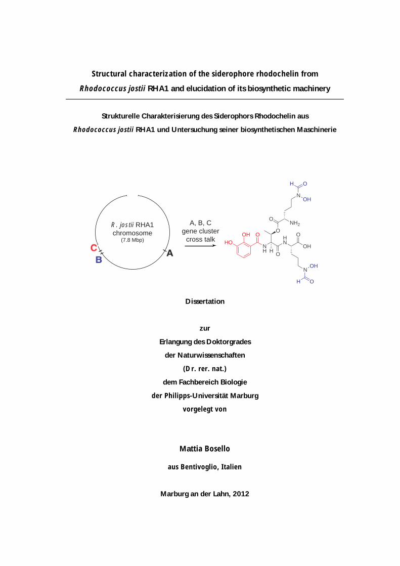
R. jostii RHA1chromosome
(7.8 Mbp)
A, B, Cgene cluster
cross talk HOOH O
OH
OH
OH
OH
NH
O
ONH2
N
HN
OOH
O
N
H
II
Die Untersuchungen zur vorliegenden Arbeit wurden am Fachbereich Chemie der
Philipps-Universität Marburg unter der Leitung von Herrn Prof. Dr. Mohamed A.
Marahiel durchgeführt.
Vom Fachbereich Biologie der Philipps-Universität Marburg als Dissertation
angenommen am 19 Juni 2012.
Erstgutachter: Prof. Dr. Mohamed A. Marahiel (Philipps-Universität Marburg)
Zweitgutachter: Prof. Dr. Michael Bölker (Philipps-Universität Marburg)
Tag der mündlichen Prüfung am: 5 Juli 2012
III
The majority of the work presented herein has been published:
Mattia Bosello, Lars Robbel, Uwe Linne, Xiulan Xie, and Mohamed A. Marahiel
Biosynthesis of the siderophore rhodochelin requires the coordinated expression
of three independent gene clusters in Rhodococcus jostii RHA1
Journal of the American Chemical Society 2011 133 (12), 4587-4595
Mattia Bosello, Andreas Mielcarek, Tobias W. Giessen, and Mohamed A. Marahiel
An enzymatic pathway for the biosynthesis of the formylhydroxyornithine
required for rhodochelin iron coordination
Biochemistry 2012 51 (14), 3059-3066
Additional publications:
Tobias W. Giessen, Kamila B. Franke, Thomas A. Knappe, Femke I. Kraas, Mattia
Bosello, Xiulan Xie, Uwe Linne, and Mohamed A. Marahiel
Isolation, structure elucidation, and biosynthesis of an unusual hydroxamic acid
ester-containing siderophore from Actinosynnema mirum
Journal of Natural Products 2012 75 (5), 905-914


V
dedicated to my parents
Table of contents
VI
Table of contents
Table of contents VI List of abbreviations IX Summary XII Zusammenfassung XIII Chapter 1 Introduction 1 1.1 Siderophore-based iron acquisition 2
1.1.1 The biological role of iron 2 1.1.2 Siderophore classification 2 1.1.3 Siderophore assembly strategies 3
1.2 The non-ribosomal assembly of peptides 6
1.2.1 The essential NRPS domains 7 1.2.1.1 The adenylation domain 7 1.2.1.2 The peptidyl-carrier-protein domain 8 1.2.1.3 The condensation domain 9 1.2.1.4 The thioesterase domain and the termination of non-ribosomal peptide assembly 9 1.2.2 Additional NRPS domains and related enzymes 10 1.2.2.1 In cis operating modification enzymes 10 1.2.2.1.1 The epimerization domain 10 1.2.2.1.2 The cyclization domain 11 1.2.2.1.3 The methylation domain 11 1.2.2.1.4 The formylation domain 11 1.2.2.2 Modifications through in trans acting tailoring enzymes 12 1.2.2.2.1 Methylation 12 1.2.2.2.2 Hydroxylation 13 1.2.2.2.3 Acetylation and formylation 14 1.2.2.3 NRPS repair mechanism: the type II thioesterase 14 1.2.3 Classification of non-ribosomal assembly line logic 15 1.2.3.1 Linear NRPS-assembly line logic 15 1.2.3.2 Iterative NRPS-assembly line logic 15 1.2.3.3 Non-linear NRPS assembly line logic 16
1.3 Rational strategies for natural product discovery via genome mining 18 1.4 Aim of the work 21 Chapter 2 Material 23 2.1 Equipment 24 2.2 Chemicals, enzymes and consumables 25 2.3 Oligonucleotides 26 2.4 Plasmids 28
2.4.1 pET28a(+) and pCB28a(+) 28 2.4.2 pK18mobsacB 28
2.5 Bacterial strains 30
2.5.1 Rhodococcus jostii RHA1 30 2.5.2 Escherichia coli TOP10 30 2.5.3 Escherichia coli BL21 (DE3) 30 2.5.4 Escherichia coli S17-1 30
2.6 Culture media 31
2.6.1 Lysogeny broth (LB-Miller) 31 2.6.2 M9 minimal medium 31
Table of contents
VII
Chapter 3 Methods 33 3.1 Molecular biology techniques 34
3.1.1 General strains maintenance 34 3.1.2 Preparation of genomic DNA 34 3.1.3 Preparation of plasmid DNA 34 3.1.4 PCR-based gene amplification 35 3.1.5 Vector construction 36 3.1.6 Construction of Rhodococcus mutants 36
3.2 Expression and purification of recombinant proteins 39
3.2.1 Gene expression 39 3.2.2 Protein purification 39 3.2.3 Protein quantification 39
3.3 Analytical methods 40
3.3.1 HPLC-MS 40 3.3.2 Peptide mass fingerprinting 40 3.3.3 HPLC-ESI-qTOF-MS 41 3.3.4 Natural product isolation 41 3.3.5 UV-vis spectroscopy 41 3.3.6 IR-spectroscopy 42 3.3.7 NMR-spectroscopy 42 3.3.8 Assignment of amino acid stereochemistry via FDAA-derivatization 42 3.3.9 Analytical size-exclusion chromatography 43
3.4 Chemical synthesis 44
3.4.1 Synthesis of L-δ-N-hydroxylornithine (L-hOrn) 44 3.4.2 Synthesis of L-δ-N-formylornithine (L-fOrn) 46 3.4.3 Synthesis of the formyl-donor cosubstrate intermediate N5,N10-methenylH4F 47
3.5 Biochemical methods 48
3.5.1 ATP/PPi exchange assay 48 3.5.2 Fluoresceinyl-CoA phospopantetheinylation assay 48 3.5.3 RhcE priming and coupled reaction with DhbE 49 3.5.4 Rmo-mediated L-Orn hydroxylation 50 3.5.5 In situ N10-fH4F conversion and L-hOrn formylation assay 50 3.5.6 Coupled L-Orn hydroxylation and formylation 51 3.5.7 HPLC-MS analysis of the L-Orn tailoring reactions 51
3.6 Bioinformatic Methods 52 Chapter 4 Results 53 4.1 Isolation and structural characterization of rhodochelin 54
4.1.1 Extraction and purification of rhodochelin 54 4.1.2 MSn analysis of rhodochelin composition 55 4.1.3 Structure elucidation of rhodochelin via NMR 56 4.1.4 Assignment of rhodochelin stereochemistry 57 4.1.5 Physico-chemical properties of rhodochelin 58
4.2 Identification of the rhodochelin biosynthetic gene clusters 60
4.2.1 Identification of the rhodochelin biosynthetic genes via genome mining 60 4.2.2 Construction of isogenic deletion mutants in R. jostii RHA1 and test for rhodochelin activity 63
Table of contents
VIII
4.3 Biochemical characterization of rhodochelin NRPS assembly-enzymes 65
4.3.1 DhbE ATP/PPi exchange 65 4.3.2 Coupled assay of DhbE and RhcE 65
4.4 Biochemical characterization of L-Orn tailoring enzymes 68
4.4.1 Biochemical characterization of Rmo L-Orn Monooxygenase 68 4.4.1.1 Bioinformatic analysis of the NMO Rmo 68 4.4.1.2 Recombinant production and purification of active apo-Rmo 68 4.4.1.3 Biochemical characterization of Rmo 71 4.4.2 Biochemical characterization of the Rft L-hOrn formylatransferase 73 4.4.2.1 Bioinformatic analysis of Rft and other homologous formyltransferases 73 4.4.2.2 Biochemical characterization of Rft and CchA L-hOrn formyltransferases 75 4.4.3 L-fhOrn coupled enzymatic biosynthesis 78
Chapter 5 Discussion 79 5.1 Isolation and structural characterization of the siderophore rhodochelin 80
5.1.1 Rhodococcus spp. as a new source for secondary metabolites 80 5.1.2 Isolation and structural characterization of the siderophore rhodochelin 81
5.2 The biosynthesis of rhodochelin requires NRPS cross-talk 84
5.2.1 Identification of the gene set associated with rhodochelin biosynthesis 84 5.2.2 Genome comparison between sequenced Rhodococcus strains 85 5.2.3 Genome cluster cross-talk associated with the production of microbial secondary metabolites 86
5.3 Biosynthesis of the non-proteinogenic amino acid L-fhOrn 89
5.3.1 Characterization of the L-Orn monooxygenase Rmo 89 5.3.2 Characterization of the L-hOrn formyltransferase Rft 91
5.4 Biochemical and genetic model for rhodochelin biosynthesis 95
5.4.1 A biosynthetic model for rhodochelin assembly 95 5.4.2 Putative regulation of the rhodochelin biosynthesis 96
5.5 Perspective and outlook 99 References 101 Supplementary section 111
Supporting tables 111 Supporting figures 114
Acknowledgements 119 Erklärung 121
Abbreviations
IX
List of abbreviations
A-domain adenylation domain aa amino acid Ac-CoA acetyl coenzyme A ArCP aryl carrier protein ACP acyl carrier protein α-KG α-ketoglutarate ACV δ-aminoadipyl-cysteinyl-D-valine ADP adenosine diphosphate AMP adenosine monophosphate AT acetyltransferase ATP adenosine-5'-triphosphate BLAST Basic Local Alignment Search Tool BSA bovine serum albumine Boc tert-butoxycarbonyl bp base pairs C-domain condensation domain Cy-domain heterocyclization domain CAS chromazurol S CDA calcium-dependent antibiotic CoA coenzyme A COSY correlation spectroscopy ddH2O double-distilled water DHB 2,3-dihydroxybenzoic acid DAD diode-array detector DMF dimethyl formamide DMSO dimethylsulfoxide DNA deoxyribonucleic acid DSS 4,4-dimethyl-4-silapentane sodium sulfonate DTT dithiothreitol E-domain epimerization domain EDTA ethylenediaminetetraacetic acid EIC extracted ion chromatogram ESI electron-spray ionization F-domain formylation domain Fl-CoA fluoresceinyl-CoA fhOrn δ-N-formyl-δ-N-hydroxyornithine fOrn δ-N-formylornithine FTICR fourier transform ion cyclotron resonance FA fatty acid FAS fatty acid synthase FAD flavin adenine dinucleotide FDAA N-α-(2,4-dinitro-5-fluorophenyl)-L-alaninamide FMN flavin mononucleotide Fmoc fluorenylmethyloxycarbonyl FT-IR Fourier transform infrared spectroscopy FPLC fast protein liquid chromatography GARF glycinamide ribonucleotide formyltransferase H4F tetrahydrofolate haOrn δ-N-acetyl-δ-N-hydroxyornithine haLys ε-N-acetyl-ε-N-hydroxylysine HEPES 4-(2-hydroxyethyl)-1-piperazine ethanesulfonic acid HMBC heteronuclear multiple bond coherence hLys ε-N-hydroxylysine hOrn δ-N-hydroxyornithine HPLC high performance liquid chromatography HR-MS high-resolution mass spectrometry HSQC heteronuclear single-quantum correlation spectroscopy ICL isochorismate lyase, isochorismatase IMAC immobilized metal affinity chromatography IPTG isopropyl-β-D-thiogalactopyranoside LTQ linear triple quadrupole MCS multiple cloning site MT-domain methyltransferase domain
Abbreviations
X
mRNA messenger ribonucleic acid MS mass spectrometry N5-fH4F N5-formyl-tetrahydrofolate N5,N10-methenylH4F N5,N10-methenyl-tetrahydrofolate N5-methylH4F N5-methyl-tetrahydrofolate N5,N10-methyleneH4F N5,N10-methylene-tetrahydrofolate N10-fH4F N10-formyl-tetrahydrofolate; n.a. not applicable NAD(P)H nicotinamide adenine dinucleotide (phosphate) NFPA nonafluoropentanoic acid, n-perfluoropentanoic acid NIS NRPS independent siderophore NMO N-hydroxylating flavoprotein monooxygenases NMR nuclear magnetic resonance NOE nuclear Overhauser effect NOESY nuclear Overhauser effect spectroscopy NRP non-ribosomal peptide NRPS non-ribosomal peptide synthetase NTA nitrilotriacetic acid NTP nucleoside triphosphate Ox-domain oxidation domain OD optical density ORF open reading frame Orn ornithine p.a. per analysis PCP peptidyl-carrier-protein PCR polymerase chain reaction PDB protein data bank PK polyketide PKS polyketide synthase ppan 4'-phosphopantetheine PPi inorganic pyrophosphate PPTase 4'-phosphopantetheine transferase qTOF quadrupole time-of-flight R-domain reductase domain RNA ribonucleic acid ROESY rotating frame nuclear Overhauser effect spectroscopy ROS radical oxygen species RP reversed-phase RT room temperature SAM S-denosylmethionine SDS sodium dodecyl sulfate SDS-PAGE sodium dodecyl sulfate polyacrylamide gel electrophoresis SIC selected (single) ion chromatogram SIM single ion mode SOE splicing overlap extension spp. species T-domain thiolation domain TA transaminase TE thioesterase domain TEII type II thioesterase TFA trifluoroacetic acid THF tetrahydrofuran TIC total ion chromatogram TOCSY total correlation spectroscopy tR retention time TRIS tris-(hydroxymethyl)-aminomethane tRNA transfer ribonucleic acid UDP uridine diphosphate v/v volume/volume w/ with w/o without w/v weight/volume w.t. wild-type
Abbreviations
XI
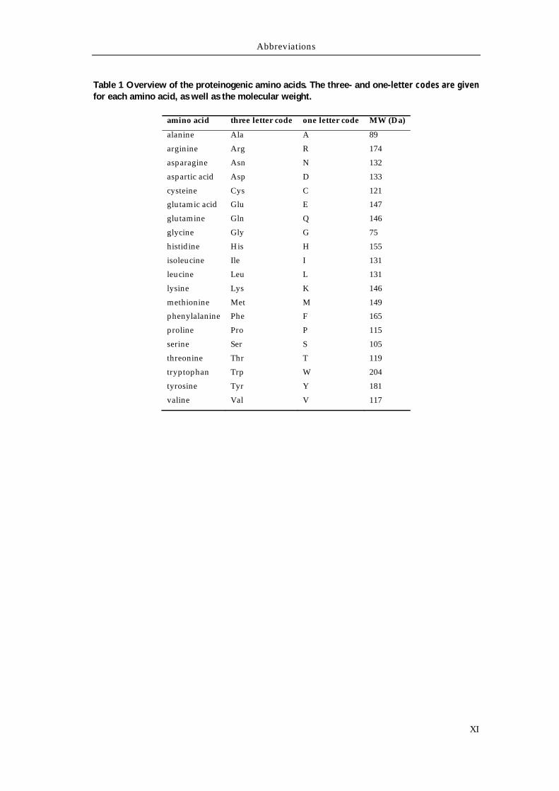
Table 1 Overview of the proteinogenic amino acids. The three- and one-letter codes are given for each amino acid, as well as the molecular weight.
amino acid three letter code one letter code MW (Da)
alanine Ala A 89
arginine Arg R 174
asparagine Asn N 132
aspartic acid Asp D 133
cysteine Cys C 121
glutamic acid Glu E 147
glutamine Gln Q 146
glycine Gly G 75
histidine His H 155
isoleucine Ile I 131
leucine Leu L 131
lysine Lys K 146
methionine Met M 149
phenylalanine Phe F 165
proline Pro P 115
serine Ser S 105
threonine Thr T 119
tryptophan Trp W 204
tyrosine Tyr Y 181
valine Val V 117
Summary
XII
Summary
Rhodococci represent an important genus of industrial interest, both because of
their role in bioremediation and biocatalysis, as well as for their potential as
producers of natural products. In this context, the genome sequencing of the
biphenyl-degrading soil bacterium Rhodococcus jostii RHA1 represents the first
attempt to harness the biosynthetic metabolic potential of the genus Rhodococcus, by
enabling the systematic exploration of its natural product-producing capabilities.
The genome of R. jostii RHA1 contains 23 secondary metabolite gene clusters, all
considered to be orphan with respect to their product, including two clusters
putatively involved in siderophore biosynthesis. In this study, the isolation,
structural characterization and genetic analysis of the biosynthetic origin of
rhodochelin, a unique mixed-type catecholate-hydroxamate siderophore isolated
from R. jostii RHA1, which represents the first characterized NRPS-derived natural
product of the strain, is reported. Structure elucidation of rhodochelin was
accomplished via MSn- and NMR-analysis and revealed the tetrapeptide to contain
an unusual ester bond between an L-δ-N-formyl-δ-N-hydroxyornithine (L-fhOrn)
moiety and the side chain of a threonine residue. Bioinformatic analysis of the R.
jostii RHA1 genome revealed the enzymes responsible for siderophore biosynthesis
to be encoded in three distant NRPS gene clusters. Single gene deletions within the
three putative biosynthetic gene clusters abolished rhodochelin production, proving
that the ORFs responsible for rhodochelin biosynthesis are located in different
chromosomal loci. Biochemical characterization of the monooxygenase Rmo and the
formyltransferase Rft established a route for the biosynthesis of the non-
proteinogenic amino acid L-fhOrn, prior to its incorporation into the peptide
scaffold by the NRPS-assembly line. The insights gained from the structural and
functional characterization of rhodochelin, together with the genetic and
biochemical characterization of the respective biosynthetic gene clusters, allowed
the proposal of a biosynthetic model for rhodochelin assembly. Finally, the efficient
and, in this work, first reported cross-talk between three distantly located secondary
metabolite gene clusters provides deep insights into natural product biosynthesis
that may facilitate future attempts to isolate new natural products.
Zusammenfassung
XIII
Zusammenfassung
Bei den Rhodococci handelt es sich um ein bakterielles Genus von industrieller
Relevanz, welches bei der biologischen Dekontaminierung und Biokatalyse zum
Einsatz kommt und dessen Mitglieder großes Potential als Produzenten neuer
Naturstoffe zeigen. In diesem Zusammenhang stellt die Genomsequenzierung des
Biphenyl-abbauenden Bodenbakteriums Rhodococcus jostii RHA1 den ersten Versuch
dar das biosynthetische Potential des Genus Rhodococcus auszuloten, da das
Vorliegen der kompletten Genomsequenz die systematische Erforschung der
Naturstoffproduktion erlaubt. Das Genom von R. jostii RHA1 enthält 23
Sekundärmetabolit-Gencluster, darunter zwei putative Siderophor-
Biosynthesecluster, wobei alle als „orphan“ zu bezeichnen sind, da ihnen kein
konkretes Produkt zugeordnet werden kann. In der vorliegenden Arbeit soll die
Isolierung, strukturelle Charakterisierung und genetische sowie biochemische
Analyse des biosynthetischen Ursprungs von Rhodochelin, dem ersten aus R. jostii
RHA1 isolierten „mixed-type“ Catechol-Hydroxamat Siderophore, welches das
erste charakterisierte NRPS-abhängige Naturprodukt dieses Stamms darstellt,
behandelt werden. Zur Strukturaufklärung von Rhodochelin wurden sowohl MSn-
als auch NMR-Studien durchgeführt, welche ergaben, dass es sich um ein
Tetrapeptid handelt, das eine ungewöhnliche Esterbindung zwischen einem L-δ-N-
Formyl-δ-N-hydroxy-Ornithinrest (L-fhOrn) und einer Threoninseitenkette enthält.
Eine bioinformatische Analyse des R. jostii RHA1 Genoms zeigte, dass die für die
Biosynthese verantwortlichen Gene in drei unterschiedlichen, voneinander weit
entfernten NRPS-Genclustern lokalisiert sind. Durch Einzelgendeletionen in den
jeweiligen Clustern, durch welche die Rhodochelinproduktion komplett
aufgehoben wurde, konnte eindeutig gezeigt werden, dass die für die Rhodochelin-
Biosynthese verantwortlichen Gencluster in drei unterschiedlichen Loci auf dem R.
jostii RHA1 Chromosom vorliegen. Durch die biochemische Charakterisierung der
Monooxygenase Rmo und der Formyltransferase Rft konnte ein Biosyntheseweg für
die nicht-proteinogene Aminosäure L-fhOrn etabliert werden, welche anschließend
durch die NRPS-Maschinerie in das Peptidgerüst eingebaut wird. Mit Hilfe der
durch die strukturelle und funktionelle Charakterisierung von Rhodochelin, sowie
der genetischen und biochemischen Analysen der verantwortlichen Biosynthese-
Gencluster gewonnen Einsichten, konnte eine Biosyntheseroute für das Siderophor
Rhodochelin postuliert werden. Die aus der vorliegenden Arbeit gewonnenen
Erkenntnisse bezüglich des effizienten und vormals unbekannten „cross-talks“
zwischen drei weit voneinander entfernten Sekundärmetabolit-Genclustern
Zusammenfassung
XIV
erlauben neue Einblicke in die Organisation des bakteriellen
Sekundärmetabolismus und tragen zu einem besseren Verständnis der Biosynthese
von Naturstoffen bei. Des Weiteren können die gewonnenen Ergebnisse als
Ausgangspunkt für eine zukünftige Isolierung neuer Naturstoffe dienen.
Chapter 1
Introduction
Introduction
2
1.1 Siderophore-based iron acquisition
1.1.1 The biological role of iron
Under physiological conditions, iron exists in two redox forms: Fe2+ (ferrous iron)
and Fe3+ (ferric iron), easily convertible into each other under acidic or basic
conditions [E0acid (Fe2+/Fe3+) = 0.771 V, E0
basic (Fe2+/Fe3+) = -0.690 V]. These redox
properties put the element in a central and extremely versatile position for almost
the entire spectrum of biological processes needing a redox potential between -0.5
and 0.6 V.1 In fact, being the fourth most common element on Earth (and the second
most common metal), iron takes part as an essential cofactor in many enzymes of
cellular metabolism (photosynthesis, Krebs cycle, respiratory chain, nitrogen
fixation and methanogenesis among others) since the early days of anaerobic life on
the planet. Under aerobic conditions, soluble Fe(II) spontaneously oxidizes to Fe(III)
which, in the presence of oxygen, water and at neutral pH, forms insoluble ferric
oxide hydrate complexes, leading to a free Fe(III) concentration of only up to 10-18 M.2
In addition, in the presence of molecular oxygen, iron can react to form extremely
toxic reactive oxygen species (ROS), in the well-known Fenton reaction:
Fe2+ + H2O2 → Fe3+ + OH + OH-
Fe3+ + H2O2 → Fe2+ + OOH + H+
These radical species are subsequently able to damage cellular structures, e.g.
nucleic acids and membranes, severely impairing cellular functions.3 Thus, under
oxidative conditions, iron is both rarely available and extremely toxic.
The development of high-affinity iron uptake systems and the precise regulation of
the iron homeostasis is therefore an essential process to sustain cellular life. In
particular, in order to cope with iron-limiting conditions, microbes have developed
mechanisms for highly selective metal uptake.4 The secretion of low-molecular
weight organic chelators called siderophores is one of the main iron-mobilizing
strategies used by both environmental and pathogenic strains to support their life
under strict iron-liming conditions.5
1.1.2 Siderophore classification
The production and secretion of siderophores is the most efficient and widespread
iron-scavenging strategy used by microorganisms (bacteria and fungi) to mobilize
iron from iron-depleted environments.6 Most siderophores display a molecular
mass below 1 kDa, coordinate the ferric iron via six donor atoms as an octahedral
Introduction
3
complex (in an ferric iron:siderophore ratio of 1:1) and have an extremely high
affinity (Kf = 1022 - 1049 M-1).2 After secretion to the extracellular space and
complexation of iron, the ferric iron-siderophore complex is subsequently
selectively and actively imported into the intracellular space, the iron is released
from the chelator-complex and channeled to the intracellular targets.3,7,8 Although
they perform the same biological function, siderophores are structurally diverse
natural products and display great chemical diversity, in both iron coordination and
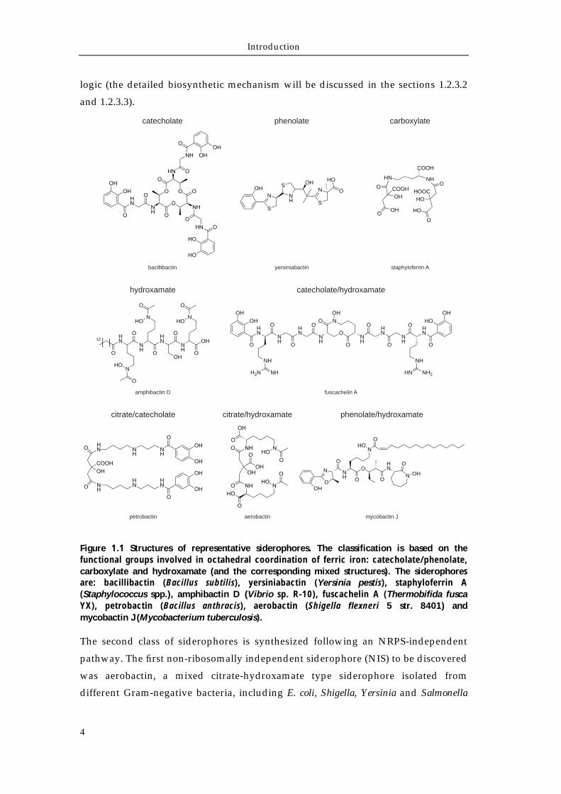
their biosynthesis.9 Siderophores may have a linear, exocyclic, endocyclic or tripodal
structure.10 On the other hand, on the basis of the chemical nature of the moieties
involved in ferric iron coordination, they are usually classified into three main
classes: catecholates (better termed as “aryl caps”), hydroxamates, and (α-hydroxy)-
carboxylates. The most commonly known catecholate siderophores are the tris-
cathecolates enterobactin11 (Escherichia coli) and bacillibactin12 (Bacillus subtilis)
which possess the highest known affinity constants for the siderophore:metal
complex, with (Fe:enterobactin)3- being 1049 M-1 and (Fe:bacillibactin)3- being 1047 M-1,
respectively. Tris-hydroxamate siderophores coelichelin13 [Streptomyces coelicolor
A3(2)] and erythrochelin14 (Saccharopolyspora erythraea) contain iron-coordinating
hydroxamic acid moieties, deriving from the subsequent tailoring of hydroxylated
side chain amino groups of lysine or ornithine residues, via acetylation or
formylation.15 Moreover, amphibactins16 and aquachelins17 are additional examples
of tris-hydroxamate amphiphilic siderophores isolated from the marine
environment. Finally, carboxylate-type siderophores generally utilize α-
hydroxycarboxylic acids as bidentate iron-chelating group. In the case of
staphyloferrin A,18 two citrate groups provide the iron-coordinating moieties.
However, the continuous discovery of new structures led to a more complex
classification, due to the presence of at least two different coordinating groups
within one molecule, resulting in “mixed-type” siderophores. A representative
overview of the structural diversity of siderophores is shown in Figure 1.1.
1.1.3 Siderophore assembly strategies
In addition to the classification relying on the different iron-coordination functions,
siderophores diversity can be further organized on the basis of their biosynthetic
origin in NRPS-dependent (NRPS, non-ribosomal peptide synthetase) and NRPS-
independent.19,20 NRPS-dependent is the most prominent and extensively studied
assembly strategy. Notable examples are the tris-catecholate enterobactin11 and the
tris-hydroxamate coelichelin,13 whose genetic and biochemical analysis has allowed
for the elucidation of alternatives strategies to the canonical linear NRPS assembly
Introduction
4
logic (the detailed biosynthetic mechanism will be discussed in the sections 1.2.3.2
and 1.2.3.3).
Figure 1.1 Structures of representative siderophores. The classification is based on the functional groups involved in octahedral coordination of ferric iron: catecholate/phenolate, carboxylate and hydroxamate (and the corresponding mixed structures). The siderophores are: bacillibactin (Bacillus subtilis), yersiniabactin (Yersinia pestis), staphyloferrin A (Staphylococcus spp.), amphibactin D (Vibrio sp. R-10), fuscachelin A (Thermobifida fusca YX), petrobactin (Bacillus anthracis), aerobactin (Shigella flexneri 5 str. 8401) and mycobactin J (Mycobacterium tuberculosis).
The second class of siderophores is synthesized following an NRPS-independent
pathway. The first non-ribosomally independent siderophore (NIS) to be discovered
was aerobactin, a mixed citrate-hydroxamate type siderophore isolated from
different Gram-negative bacteria, including E. coli, Shigella, Yersinia and Salmonella
bacillibactin
catecholate
OH
S
N NH
S OH
S
N O
HO
yersiniabactin
phenolate
HN
O
OHCOOH
OHO
NH
COOH
O
HOHOOC
O
HO
staphyloferrin A
carboxylate
HN
NH
HN
NH
O
NHO
O
O
NHO
O
OOH
O
NHO
O
O
OH12
OHOH
HN
O
O
NH
HN
ONH
ON
O
OHO
ONH
OHN
ONH
OHN
NH
NH2HN
NH
NHH2N
O
HOOH
amphibactin D fuscachelin A
hydroxamate catecholate/hydroxamate
N
O
NH
OHN
OH
O
ON
O
OHO
NHO
O
petrobactin aerobactin mycobactin J
citrate/catecholate citrate/hydroxamate phenolate/hydroxamate
Introduction
5
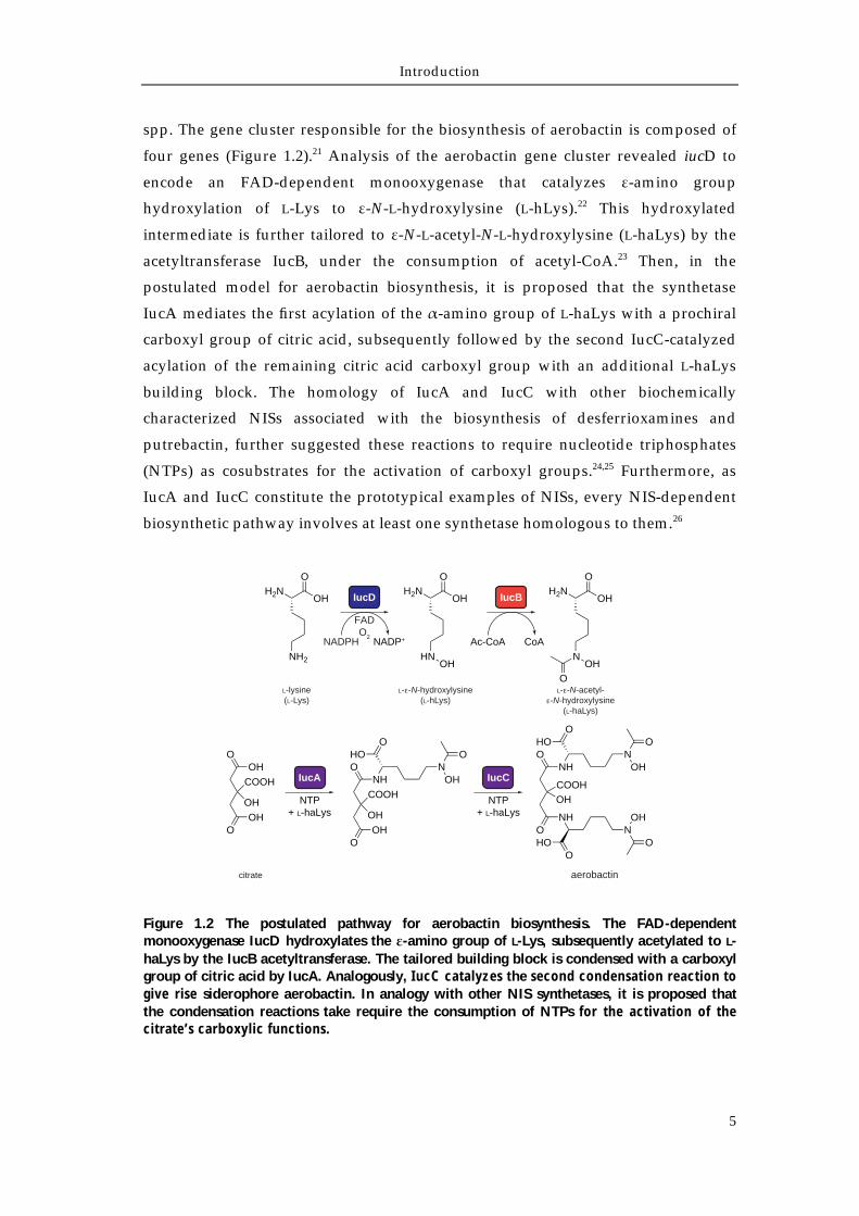
spp. The gene cluster responsible for the biosynthesis of aerobactin is composed of
four genes (Figure 1.2).21 Analysis of the aerobactin gene cluster revealed iucD to
encode an FAD-dependent monooxygenase that catalyzes ε-amino group
hydroxylation of L-Lys to ε-N-L-hydroxylysine (L-hLys).22 This hydroxylated
intermediate is further tailored to ε-N-L-acetyl-N-L-hydroxylysine (L-haLys) by the
acetyltransferase IucB, under the consumption of acetyl-CoA.23 Then, in the
postulated model for aerobactin biosynthesis, it is proposed that the synthetase
IucA mediates the first acylation of the α-amino group of L-haLys with a prochiral
carboxyl group of citric acid, subsequently followed by the second IucC-catalyzed
acylation of the remaining citric acid carboxyl group with an additional L-haLys
building block. The homology of IucA and IucC with other biochemically
characterized NISs associated with the biosynthesis of desferrioxamines and
putrebactin, further suggested these reactions to require nucleotide triphosphates
(NTPs) as cosubstrates for the activation of carboxyl groups.24,25 Furthermore, as
IucA and IucC constitute the prototypical examples of NISs, every NIS-dependent
biosynthetic pathway involves at least one synthetase homologous to them.26
Figure 1.2 The postulated pathway for aerobactin biosynthesis. The FAD-dependent monooxygenase IucD hydroxylates the ε-amino group of L-Lys, subsequently acetylated to L-haLys by the IucB acetyltransferase. The tailored building block is condensed with a carboxyl group of citric acid by IucA. Analogously, IucC catalyzes the second condensation reaction to give rise siderophore aerobactin. In analogy with other NIS synthetases, it is proposed that the condensation reactions take require the consumption of NTPs for the activation of the citrate’s carboxylic functions.
IucB
Ac-CoA CoA
IucD
NADPH NADP+
FADO2
L-lysine(L-Lys)
L- -N-hydroxylysine(L-hLys)
L- -N-acetyl--N-hydroxylysine
(L-haLys)
H2NOH
O
NH2
H2NOH
O
HNOH
H2NOH
O
NOH
O
OHO
COOH
OH
OHO
NHO
COOH
OH
OHO
OHO
NOH
ONH
O
COOHOH
NHO
OHO
NOH
O
N
OHO
OH
O
IucA
NTP+ L-haLys
IucC
NTP+ L-haLys
citrate aerobactin
Introduction
6
1.2 The non-ribosomal assembly of peptides
Non-ribosomal peptide synthetases (NRPSs) are large multimodular megaenzymes
that catalyze the biosynthesis of biologically active peptides.27 In contrast to
ribosomal peptide synthesis, the assembly of the oligopeptide is carried out in an
mRNA-independent function.28 NRPS encoding genes are widely spread in nature,
being mainly found in bacteria and fungi.29 The best examined producing species of
NRPS-assembling natural products are soil actinobacteria, who have already proved
to be a rich source of new potent and pharmacologically-relevant molecules.30,31
Figure 1.3 The catalytic steps of an elongation NRPS module are shown: (1) Substrate recognition and activation by the A-domain under ATP-consumption, (2) Substrate transfer onto the 4'-phosphopantetheine (ppan) cofactor covalently attached to an invariant Ser of a PCP-domain, (3) trapping of the thioesterified amino-acid in the acceptor site of the C-domain, followed by condensation with the incoming building block trapped in the donor-site, (4) trapping of the peptidyl-S-ppan moiety in the donor-site of the downstream C-domain.
The genetic and biochemical characterization of the NRPS assembly machinery
revealed a multimodular organization, which can be further dissected into single
catalytic domains.32 Each module is responsible for the incorporation of a building
block, catalyzing the elongation of the oligopeptide chain by one unit. In particular,
a minimal module (Figure 1.3) contains all the essential units required for the
recognition and activation of the monomer [adenylation (A)-domain], the formation
of the peptide bond [condensation (C)-domain] and the translocation of the peptidyl
intermediates to the subsequent module [peptidyl-carrier-protein (PCP)-domain]. A
fourth catalytic NRPS domain, the thioesterase (TE)-domain is responsible for the
release of the product from the NRPS machinery. Recently the crystal structure of
SrfA-C, the termination module of the surfactin synthetase, was determined,
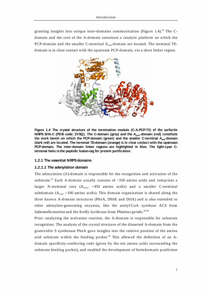
Introduction
7
granting insights into unique inter-domains communication (Figure 1.4).33 The C-
domain and the core of the A-domain constitute a catalytic platform on which the
PCP-domain and the smaller C-terminal Asub-domain are located. The terminal TE-
domain is in close contact with the upstream PCP-domain, via a short linker region.
Figure 1.4 The crystal structure of the termination module (C-A-PCP-TE) of the surfactin NRPS SrfA-C (PDB code: 2VSQ). The C-domain (grey) and the Acore-domain (red) constitute the work bench on which the PCP-domain (green) and the smaller C-terminal Asub-domain (dark red) are located. The terminal TE-domain (orange) is in close contact with the upstream PCP-domain. The inter-domain linker regions are highlighted in blue. The light-cyan C-terminal helix is the peptidic fusion-tag for protein purification.
1.2.1 The essential NRPS domains
1.2.1.1 The adenylation domain
The adenylation (A)-domain is responsible for the recognition and activation of the
substrate.27 Each A-domain usually consists of ~550 amino acids and comprises a
larger N-terminal core (Acore, ~450 amino acids) and a smaller C-terminal
subdomain (Asub, ~100 amino acids). This domain organization is shared along the
three known A-domain structures (PheA, DhbE and DtlA) and is also extended to
other adenylate-generating enzymes, like the acetyl-CoA synthase ACS from
Salmonella enterica and the firefly luciferase from Photinus pyralis.34-38
Prior catalyzing the activation reaction, the A-domain is responsible for substrate
recognition. The analysis of the crystal structure of the dissected A-domain from the
gramicidin S synthetase PheA gave insights into the relative position of the amino
acid substrate within the binding pocket.34 This allowed the definition of an A-
domain specificity-conferring code (given by the ten amino acids surrounding the
substrate binding pocket), and enabled the development of bioinformatic prediction
Introduction
8
tools which facilitated the identification and the isolation of NRPS-derived natural
products via genome mining approaches.39-44
After the recognition of the proper building block, in the presence of Mg2+ and ATP,
the A-domain activates the amino acid substrate as an adenylate, with the
subsequent release of pyrophosphate. Although this reaction is analogous to the one
carried out by aminoacyl-tRNA synthetases during ribosomal protein synthesis, the
enzymes do not share any sequential or structural homology. Furhtermore, A-
domains lack a proof-reading mechanism that, combined with relaxed substrate
specificity, often results in the synthesis of NRPs with different amino acid
composition by one synthetase.45
1.2.1.2 The peptidyl-carrier-protein domain
The peptidyl-carrier-protein (PCP)-domain, also known as thiolation (T)-domain, is
responsible for the covalent tethering of the monomeric building blocks and the
translocation of the peptidyl intermediates.46 It is usually located at the C-terminus
of an A-domain and, despite its size of ~80 amino acids, it is of the greatest
importance for the functionality of the NRPS assembly line. PCP-domains are post-
translationally modified at a highly-conserved Ser redidue embedded in the core-T
motif (GGxS) with a 4'-phospopantetheine (ppan) cofactor. In fact, during non-
ribosomal peptide synthesis, the mobile ppan-arm delivers all the substrates and
peptidyl intermediates as thioesters to the adjacent NRPS domains for the formation
of the peptide bond, for modification of the PCP-bound substrate or for product
release.
The conversion from the inactive apo-PCP to the active holo-PCP is mediated by
phosphopantetheinyl-transferases (PPTases, e.g. Sfp), which catalyze the
nucleophilic attack of the hydroxyl-group of the conserved Ser residue onto the β-
phosphate of a donor coenzyme A molecule (and the subsequent release of 3',5'-
adenosinediphosphate).47,48
PCPs share a high degree of sequential and structural homology with acyl-carrier-
proteins (ACP) of fatty acid (FAS) and polyketide synthases (PKS).49 NMR-based
studies showed that the PCP-domain adopts a four-helix bundle structure and exists
in three different conformational states: the apo (A), the holo (H) and the A/H form.50
When the PCP is in the apo-state, both A and A/H coexist. On the other hand, when
the PCP is in the holo-state, it slowly interconverts between the H and A/H states.
These extensive conformational changes evidence the dynamic nature of the PCP
carrying the ppan-bound substrates and intermediates to the adjacent domains. In
particular, the terminal sulfhydryl-group of the ppan-arm is able to move
Introduction
9
approximately 16 Å, confirming the long-proposed swinging mode of the ppan
prostetic group during the non-ribosomal peptide synthesis.
1.2.1.3 The condensation domain
The condensation (C)-domain is the last essential domain of an NRPS module and
carries out the peptide bond formation.51 The C-domain contains an acceptor and a
donor site, which harbor the nucleophilic aminoacyl-S-PCP substrate and the
electrophilic peptidyl-S-PCP electrophilic substrate, respectively.52,53 The formation
of the peptide bond is initiated by the nucleophilic attack of the α-amino group of
the aminoacyl-S-PCP substrate onto the thioester bond of the peptidyl-S-PCP. Upon
the amid bond formation, the elongated peptide is transferred onto the downstream
PCP-domain and serves as a donor substrate in the downstream condensation step.
All C-domains have been found to operate unidirectionally, translocating the
growing peptide chain towards the C-terminus.53,54 In addition, following to the
prototypical co-linearity assembly rule of non-ribosomal peptides, the number of C-
domains is in agreement with the number of peptide bonds found in the mature
NRP.
C-domains contain approximately 450 amino acids and are composed of two big
similar subdomains arranged in a V-shaped canyon-like structure, of which the N-
terminal one shares high sequence and structural homology with the
chloramphenicol acetyltransferases.55-57 This characteristic V-shaped structure allows
the correct positioning of the up- and downstream PCP-domains at each opening
(acceptor and donor site), with respect to the highly conserved catalytic His residue
of the HHxxxDG motif, which remains at the bottom of the canyon. Althought the
exact reaction mechanism has not yet been elucidated, it is suggested that the
second His residue takes part in the deprotonation of the α-amino group of the
aminoacyl-S-PCP substrate, enhancing the electron-donor character of the
nucleophilic PCP-bound substrate, and therefore facilitating the reaction.
1.2.1.4 The thioesterase domain and the termination of non-ribosomal peptide
assembly
The thioesterase (TE)-domain is the fourth essential domain of NRPSs and is usually
located in the termination module of the assembly line. TE-domains catalyze the
product release from the NRPS, resulting in a linear, cyclic or branched cyclic
peptide.58,59 These independently working domains contain approximately 230-270
amino acids arranged in an α/β hydrolase fold, similarly to serine proteases and
lipases.60-62 The catalytic Ser-His-Asp triad is located in a deep pocket, shielded from
Introduction
10
solvent by an α-helical lid region, or by the peptide itself. The hydrolytic release of
the template-bound NRP occurs in a two-step mechanism: first the formation of an
acyl-O-intermediate with the active Ser, which is subsequently cleaved by the attack
of a nucleophile.63 The different nature of the nucleophilic group results in the
different topology of the molecule: if an intramolecular attack occurs, a cyclic (or a
branched cyclic) molecule is released. On the other hand, if a water molecule cleaves
the intermediate, a linear peptide is released.64-66
The release of the peptide chain from the NRPS assembly machinery can also occur
via different alternative strategies, for example via the reduction of the C-terminal
carboxyl group to the corresponding aldehyde or alcohol, in an NAD(P)H
dependent manner.67,68 This reaction is catalyzed by a C-terminal reductase (R)-
domain, which takes the place of the TE-domain. In addition, in several NRPSs,
product release is carried out by a C-terminal C-domain, proposed to mediate the
cyclorelease of the peptide. Furthermore, the existence of in trans acting TE-domains
has been postulated for NRPSs that lack any C-terminal domain for the release of
the NRP product.13,69
1.2.2 Additional NRPS domains and related enzymes
The structural and functional diversity of NRPS-derived natural products is usually
extended by the addition of auxiliary domains to the essential core functions. in cis
operating NRPS domains are responsible for the on-line modification of the peptidic
backbone, whereas other stand-alone domains modify and tailor NRPS building
blocks prior to the incorporation into the assembly line. Finally, a repair mechanism
of the assembly machinery carries out in the regeneration of misprimed PCP-
domains.
1.2.2.1 In cis operating modification enzymes
1.2.2.1.1 The epimerization domain
The presence of D-configured amino acids can be observed in numerous NRPS-
derived compounds.64,70 The incorporation of D-configured amino acids in the
peptidic backbone is usually mediated by epimerization (E)-domains, located
directly downstream of the adjacent PCPs, and represents one of the major
differences between NRP and ribosomal peptide biosynthesis. E-domains epimerize
L-configured amino acids immediately after their immobilization as aminoacyl-S-
PCP intermediates. The E-domains of initiation modules generate a mixture of PCP-
S-L/D-monomers, later correctly selected by the downstream C-domain prior to
peptide bond formation. On the other hand, for E-domains embedded in elongation
Introduction
11
modules, the epimerization occurs before the transfer to the subsequent module of
the assembly line. Also in this case the downstream C-domains ensure the correct D-
configured substrate specificity.71,72
1.2.2.1.2 The cyclization domain
The heterocyclization of cysteine, serine or threonine side chains resulting in the
corresponding five-membered thiazoline, oxazoline or methyloxazoline
heterocycles is a structural feature found in several NRPs. These modifications
increase the diversity of the natural product, rigidifing the peptide backbone and
providing either metal-chelating or intercalating properties.73-76 The cyclization (Cy)-
domains are responsible of these structural modifications and are variants of C-
domains. First, Cy-domains catalyze the usual peptide bond formation and then
carry out cyclization of the nucleophilic sidechain of cysteine or the hydroxyl
sidechain of serine or threonine onto the newly formed peptide bond. The newly
formed heterocycles are often associated with oxidation (Ox)-domains, that catalyze
the FMN-dependent two-electron oxidation of the thiazoline or oxazoline ring
structures to the thermodynamically more stable corresponding thiazoles or
oxazoles.73,77 Conversely, the reduction of thiazoline or oxazolines structures is
mediated by in trans operating NAD(P)H-dependent reductases that recognize and
directly reduce the PCP-bound substrate.78
1.2.2.1.3 The methylation domain
Methylation (MT)-domains catalyze the in cis transfer of a methyl group from a S-
adenosylmethionine (SAM) donor to a carbon, nitrogen or oxygen atom of the NRP
chain. Therefore, on the basis of the different site of methylation, MT-domains are
classified as C-MT, N-MT or O-MT, respectively.79 MT-domains share a bidomain
structure, with the first subdomain containing the binding site for methyl group
donor, while the second subdomain harbors the pocket for the acceptor substrate. In
contrast to the domains described so far, MT-domains are usually embedded within
the corresponding A-domains, between the core A8- and A9-motifs, separating the
Acore, and Asub,subunits.
1.2.2.1.4 The formylation domain
The N-formylation of the N-terminal α-amino group of a non-ribosomal peptide
chain is catalyzed by the formylation (F)-domain, through the transfer of a formyl
group from the N10-fH4F or N5-fH4F donor cosubstrate to the α-amino function of the
activated aminoacyl-S-PCP substrate. The linear gramicidin NRPS LgrA contains a
Introduction
12
F-domain in the initiation module, upstream of the corresponding A-domain
(Figure 1.5).67 The F-domain catalyzes the reaction on the PCP-bound amino acid (L-
Val) and its N-formylation is essential for the subsequent elongation of the
peptide.80
Figure 1.5 The F-domain of the linear gramicidin NRPS LgrA is located at the N-terminus of the initiation module. Upon activation of the L-Val substrate, the F-domain catalyzes the transfer of a formyl moiety from the N10-fH4F or N5-fH4F cosubstrate to the α-amino function of the activated L-Val-S-PCP. This reaction is required for the subsequent elongation step. In the picture, the dissected NRPS LgrA lacks the terminal E-domain.
1.2.2.2 Modifications through in trans acting tailoring enzymes
1.2.2.2.1 Methylation
Similar to in cis acting methyltransferases, stand-alone methyltransferases catalyze
the in trans SAM-dependent (or N5-methylH4F-dependent) methylation of carbon,
nitrogen, oxygen or sulphur atoms of building blocks, prior to their incorporation
by the NRPS assembly line.81 Interestingly, these enzymes generate the methylated
amino acid by a two-step mechanism, by first methylating the corresponding α-
ketoacid, followed by the transamination of the corresponding α-keto group,
resulting in the correct building block (Figure 1.6).82-84
Figure 1.6 Side chain C-methylated amino acids are generated in a two-step reaction via the SAM-dependent modification of the corresponding α-ketoacid, followed by the transamination of the corresponding α-keto group. (A) GlmT, from the CDA-biosynthetic
PCPL-ValF C PCPL-Gly
OS
H2N
OS
HN
HO O
HN
HO
NH
SOO
S
H2N
PCPL-ValF C PCPL-Gly PCPL-ValF C PCPL-Gly
SHO
S
NH2
N10-fH4F H4F
LgrA1-2
GlmT
SAM AdoHCy
-ketoglutarate (3R)-3-methyl-2-oxoglutarate
O
OHO
O OH
O
OHO
O OH
TAO
OH
O OH
H2N
3-methyl-L-glutamate
MppJ
SAM AdoHCy
phenylpyruvate (R,S)-methyl-phenylpyruvate
TA
(R,S)-methyl-L-phenylalanine
OO
OH
OO
OH
O
OHH2N
A
B
Introduction
13
gene cluster [S. coelicolor A3(2), A], catalyzes the stereospecific methylation of α-KG to (3R)-3-methyl-2-oxoglutarate followed by the subsequent conversion to 3-methyl-L-glutamate.82,83 (B) MppJ (mannopeptimycin gene cluster, Streptomyces hygroscopicus) catalyzes the analogous reaction on phenylpyruvate but is not stereospecific.84,85 The NRPS machinery solely incorporates the (2S,3S)-3-methyl-phenylalanine precursor, found in mannopeptimycin.
1.2.2.2.2 Hydroxylation
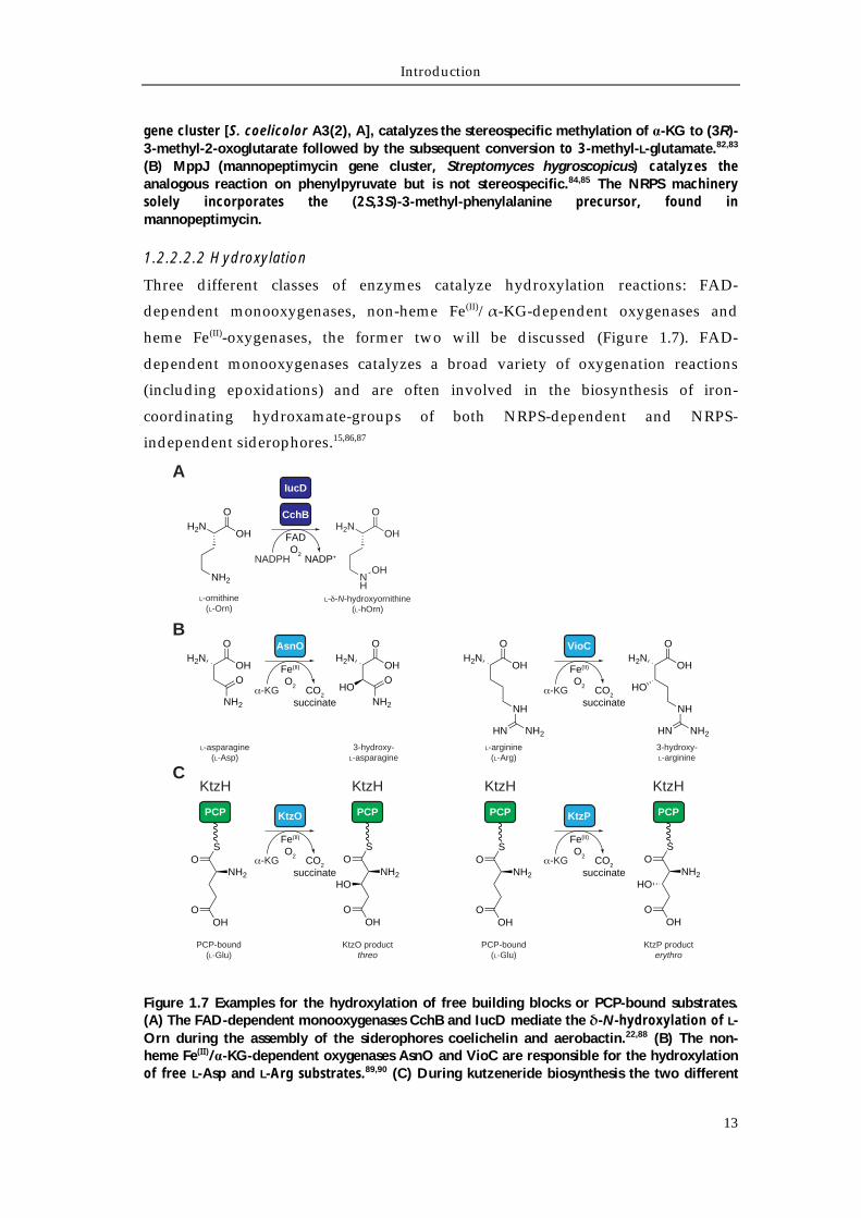
Three different classes of enzymes catalyze hydroxylation reactions: FAD-
dependent monooxygenases, non-heme Fe(II)/α-KG-dependent oxygenases and
heme Fe(II)-oxygenases, the former two will be discussed (Figure 1.7). FAD-
dependent monooxygenases catalyzes a broad variety of oxygenation reactions
(including epoxidations) and are often involved in the biosynthesis of iron-
coordinating hydroxamate-groups of both NRPS-dependent and NRPS-
independent siderophores.15,86,87
Figure 1.7 Examples for the hydroxylation of free building blocks or PCP-bound substrates. (A) The FAD-dependent monooxygenases CchB and IucD mediate the δ-N-hydroxylation of L-Orn during the assembly of the siderophores coelichelin and aerobactin.22,88 (B) The non-heme Fe(II)/α-KG-dependent oxygenases AsnO and VioC are responsible for the hydroxylation of free L-Asp and L-Arg substrates.89,90 (C) During kutzeneride biosynthesis the two different
H2NOH
O
NH2
L-ornithine(L-Orn)
H2NOH
O
NH
OH
L- -N-hydroxyornithine(L-hOrn)
CchB
NADPH NADP+
FADO2
IucD
OH
O
NH2
OHO
H2NVioC
-KG CO2
succinate
Fe(II)
O2
AsnO
-KG CO2
succinate
Fe(II)
O2
OH
O
NH2
O
H2N
L-asparagine(L-Asp)
3-hydroxy-L-asparagine
OH
OH2N
NH
NH2HN
OH
OH2N
NH
NH2HN
HO
A
B
PCP
OS
OHO
NH2
KtzO
-KG CO2
succinate
Fe(II)
O2
PCP
OS
OHO
NH2HO
PCP
OS
OHO
NH2HO
PCP
OS
OHO
NH2
KtzH KtzH
KtzP
-KG CO2
succinate
Fe(II)
O2
KtzH KtzH
PCP-bound(L-Glu)
KtzO productthreo
PCP-bound(L-Glu)
KtzP producterythro
C
L-arginine(L-Arg)
3-hydroxy-L-arginine
Introduction
14
non-heme Fe(II)/α-KG-dependent oxygenases KtzO and KtzP hydroxylate PCP-bound L-Glu generating the corresponding threo or erythro products, respectively.91
These enzymes require the reduction of the FAD-cofactor, with the needed electrons
usually supplied by an NAD(P)H cosubstrate. Fe(II)/α-KG-dependent oxygenases
couple the oxidative conversion of the substrate with the decarboxylation of the
cosubstrate α-ketoglutarate to succinate and carbon dioxide. Both enzyme classes
acts as in trans hydroxylating catalysts, prior to the incorporation of the modified
building block by the NRPS machinery.89,90,92,93 In addition, Fe(II)/α-KG-dependent
oxygenases can either hydroxylate free or PCP-bound substrates.91
1.2.2.2.3 Acetylation and formylation
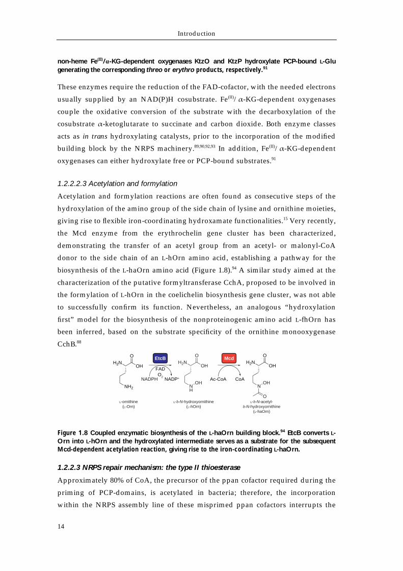
Acetylation and formylation reactions are often found as consecutive steps of the
hydroxylation of the amino group of the side chain of lysine and ornithine moieties,
giving rise to flexible iron-coordinating hydroxamate functionalities.15 Very recently,
the Mcd enzyme from the erythrochelin gene cluster has been characterized,
demonstrating the transfer of an acetyl group from an acetyl- or malonyl-CoA
donor to the side chain of an L-hOrn amino acid, establishing a pathway for the
biosynthesis of the L-haOrn amino acid (Figure 1.8).94 A similar study aimed at the
characterization of the putative formyltransferase CchA, proposed to be involved in
the formylation of L-hOrn in the coelichelin biosynthesis gene cluster, was not able
to successfully confirm its function. Nevertheless, an analogous “hydroxylation
first” model for the biosynthesis of the nonproteinogenic amino acid L-fhOrn has
been inferred, based on the substrate specificity of the ornithine monooxygenase
CchB.88
Figure 1.8 Coupled enzymatic biosynthesis of the L-haOrn building block.94 EtcB converts L-Orn into L-hOrn and the hydroxylated intermediate serves as a substrate for the subsequent Mcd-dependent acetylation reaction, giving rise to the iron-coordinating L-haOrn.
1.2.2.3 NRPS repair mechanism: the type II thioesterase
Approximately 80% of CoA, the precursor of the ppan cofactor required during the
priming of PCP-domains, is acetylated in bacteria; therefore, the incorporation
within the NRPS assembly line of these misprimed ppan cofactors interrupts the
Mcd
Ac-CoA CoA
H2NOH
O
NOH
O
EtcB
NADPH NADP+
FADO2
H2NOH
O
NH2
H2NOH
O
NH
OH
L-ornithine(L-Orn)
L- -N-hydroxyornithine(L-hOrn)
L- -N-acetyl--N-hydroxyornithine
(L-haOrn)
Introduction
15
NRP biosynthesis. Consequently, in order to overcome this critical step, a second
type of thioesterase (TEII) is often found in NRP gene clusters that ensures the
deacylation of the misprimed PCP-domain.95-97 These stand-alone domains display
structural homology with canonical NRPS termination domains, but the overall
greater accessibility of the catalytic pocket ensures the promiscuity of the TEII
enzymes towards a broad range of short chain acyl-misprimed PCP-domain
substrates.98 For the same reason, TEIIs are not able to release peptidyl-S-PCP bound
substrates.
1.2.3 Classification of non-ribosomal assembly line logic
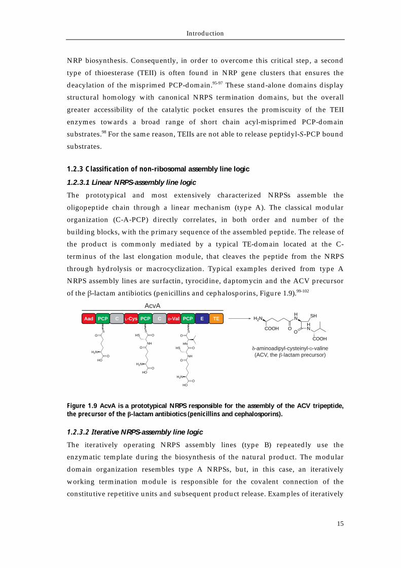
1.2.3.1 Linear NRPS-assembly line logic
The prototypical and most extensively characterized NRPSs assemble the
oligopeptide chain through a linear mechanism (type A). The classical modular
organization (C-A-PCP) directly correlates, in both order and number of the
building blocks, with the primary sequence of the assembled peptide. The release of
the product is commonly mediated by a typical TE-domain located at the C-
terminus of the last elongation module, that cleaves the peptide from the NRPS
through hydrolysis or macrocyclization. Typical examples derived from type A
NRPS assembly lines are surfactin, tyrocidine, daptomycin and the ACV precursor
of the β-lactam antibiotics (penicillins and cephalosporins, Figure 1.9).99-102
Figure 1.9 AcvA is a prototypical NRPS responsible for the assembly of the ACV tripeptide, the precursor of the β-lactam antibiotics (penicillins and cephalosporins).
1.2.3.2 Iterative NRPS-assembly line logic
The iteratively operating NRPS assembly lines (type B) repeatedly use the
enzymatic template during the biosynthesis of the natural product. The modular
domain organization resembles type A NRPSs, but, in this case, an iteratively
working termination module is responsible for the covalent connection of the
constitutive repetitive units and subsequent product release. Examples of iteratively
PCPL-Cys C PCPD-ValPCPAad TEE H2N
COOH
HN
OO
HN
SH
COOHO
S
H2N
HOO
ONH
H2N
HOO
HSS
O
ONH
H2N
HOO
HS OHN
SO
AcvA
-aminoadipyl-cysteinyl-D-valine(ACV, the -lactam precursor)
C
Introduction
16
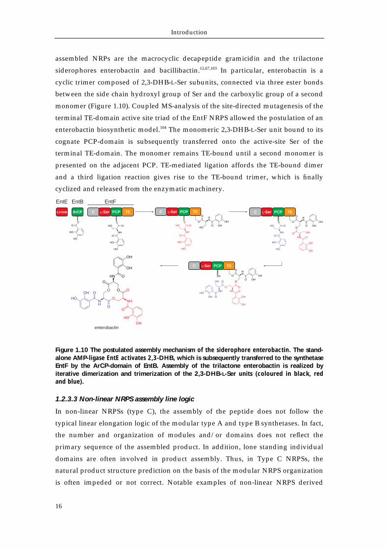
assembled NRPs are the macrocyclic decapeptide gramicidin and the trilactone
siderophores enterobactin and bacillibactin.12,67,103 In particular, enterobactin is a
cyclic trimer composed of 2,3-DHB-L-Ser subunits, connected via three ester bonds
between the side chain hydroxyl group of Ser and the carboxylic group of a second
monomer (Figure 1.10). Coupled MS-analysis of the site-directed mutagenesis of the
terminal TE-domain active site triad of the EntF NRPS allowed the postulation of an
enterobactin biosynthetic model.104 The monomeric 2,3-DHB-L-Ser unit bound to its
cognate PCP-domain is subsequently transferred onto the active-site Ser of the
terminal TE-domain. The monomer remains TE-bound until a second monomer is
presented on the adjacent PCP. TE-mediated ligation affords the TE-bound dimer
and a third ligation reaction gives rise to the TE-bound trimer, which is finally
cyclized and released from the enzymatic machinery.
Figure 1.10 The postulated assembly mechanism of the siderophore enterobactin. The stand-alone AMP-ligase EntE activates 2,3-DHB, which is subsequently transferred to the synthetase EntF by the ArCP-domain of EntB. Assembly of the trilactone enterobactin is realized by iterative dimerization and trimerization of the 2,3-DHB-L-Ser units (coloured in black, red and blue).
1.2.3.3 Non-linear NRPS assembly line logic
In non-linear NRPSs (type C), the assembly of the peptide does not follow the
typical linear elongation logic of the modular type A and type B synthetases. In fact,
the number and organization of modules and/or domains does not reflect the
primary sequence of the assembled product. In addition, lone standing individual
domains are often involved in product assembly. Thus, in Type C NRPSs, the
natural product structure prediction on the basis of the modular NRPS organization
is often impeded or not correct. Notable examples of non-linear NRPS derived
C PCPL-Ser TE
EntF
enterobactin
2,3-DHB ArCP
OS
HO
HO
O
O
HO
HN
O OHOH
O
O
O
HN
O OHOH
OHN O
OH
OH
O
O
NH
OH
OOHHO
O
O
O
HN
O OHOH
OHN O
OH
OH
OH
O O
O
O
O
O
NHNH
HN O
O
HOOH
OH
OH
OOHHO
EntBEntE
ONH
HO
HO
HOS
O
C PCPL-Ser TE
ONH
HO
HO
HOS
O
C PCPL-Ser TE
ONH
HO
HO
HOS
O
C PCPL-Ser TE
SH
Introduction
17
natural products are the antibiotic congocidin (Streptomyces ambofaciens) and the tris-
hydroxamate siderophore coelichelin.13,105 In this latter case, a trimodular NRPS is
responsible of the assembly of a tetrapeptide and the suggestion of the so-called
“module skipping” mechanism allowed the postulation of a biosynthetic model
(Figure 1.11).13,106 Before the final release, with the corresponding tripeptide bound
to the terminal PCP-domain, the initiation module of the CchH NRPS activates a
second L-fhOrn building block that is subsequently incorporated into the peptide
chain, to give rise to the final tetrapetide.
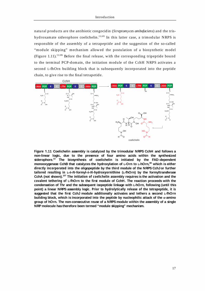
Figure 1.11 Coelichelin assembly is catalyzed by the trimodular NRPS CchH and follows a non-linear logic, due to the presence of four amino acids within the synthesized siderophore.13 The biosynthesis of coelichelin is initiated by the FAD-dependent monooxygenase CchB that catalyzes the hydroxylation of L-Orn to L-hOrn,88 which is either directly incorporated into the oligopeptide by the third module of the NRPS CchJ or further tailored resulting in L-δ-N-formyl-δ-N-hydroxyornithine (L-fhOrn) by the formyltransferase CchA (not shown).107 The initiation of coelichelin assembly requires is the activation and the covalent tethering of L-fhOrn to the first module of CchH. The reaction proceeds with the condensation of Thr and the subsequent isopeptide linkage with L-hOrn, following (until this point) a linear NRPS assembly logic. Prior to hydrolytically release of the tetrapeptide, it is suggested that the first CchJ module additionally activates and tethers a second L-fhOrn building block, which is incorporated into the peptide by nucleophilic attack of the α-amino group of hOrn. The non-consecutive reuse of a NRPS module within the assembly of a single NRP molecule has therefore been termed “module skipping” mechanism.
PCPL-fhOrn E C PCPL-Thr E C PCPL-hOrn
CchH
OS
N
H2N
OHO
HO
N
H2N
OHO
H
HNH
OS
OH
O
N
H2N
OHO
H
HNH
ON
OHOH
SO
NH2
PCPL-fhOrn E C PCPL-Thr E C PCPL-hOrn
O
N
H2N
HO
H
HNH
ON
OHOH
SO
NH2
SHO
S
N
H2N
OHO
H
O
NH
H2N
NOH
H O
NOH
H
OH
O
OHO
NH
ONH2
NHO
HO
coelichelin
Introduction
18
1.3 Rational strategies for natural product discovery via genome mining
The discovery of natural products often relies on bioassay-guided fractionation of
extracts from different natural sources and the subsequent isolation of the bioactive
compound. This employed strategy has historically led to the discovery of many
bioactive compounds, used in clinical therapy as antifungal, anticancer and
immunosuppressive agents.30 Lately, the discovery that the systematic cultivation of
one species under several conditions strongly influences secondary metabolite
production (OSMAC, One Strain - MAny Compounds approach) allowed the
discovery of different natural products.108 In recent years, with the increasing
amount of information derived by huge advances in sequencing technologies, a
plethora of sequenced microbial genomes has revealed a multitude of gene clusters
associated with the biosynthesis of secondary metabolites.109,110 This quantity of
information, in combination with a substantial increase in the understanding of
natural product biosynthesis has paved the way for the mining of genomes for
bioactive compounds.32
Since many microbial natural products are assembled by multimodular synthases
and synthetases (e.g. polyketides and non-ribosomal peptides), the development of
bioinformatic tools for the analysis and the prediction of the modular organization
and the substrate specificity of these assembly machineries has formed the basis for
the subsequent development of genome mining approaches, allowing the isolation
of new natural products solely on the basis of the genome sequence of the target
organism.32,40,111 The first microbial organism to be extensively analyzed for the
production of unknown secondary metabolites was S. coelicolor A3(2). Within its
genome, several gene clusters were identified to encode for new biosynthetic
pathways, later associated to newly isolated natural products. Besides, the gene
clusters that still remain “orphan” with respect to their cognate metabolites, were
renamed “cryptic”.13,112-115
During the past years, several approaches for microbial genome mining have been
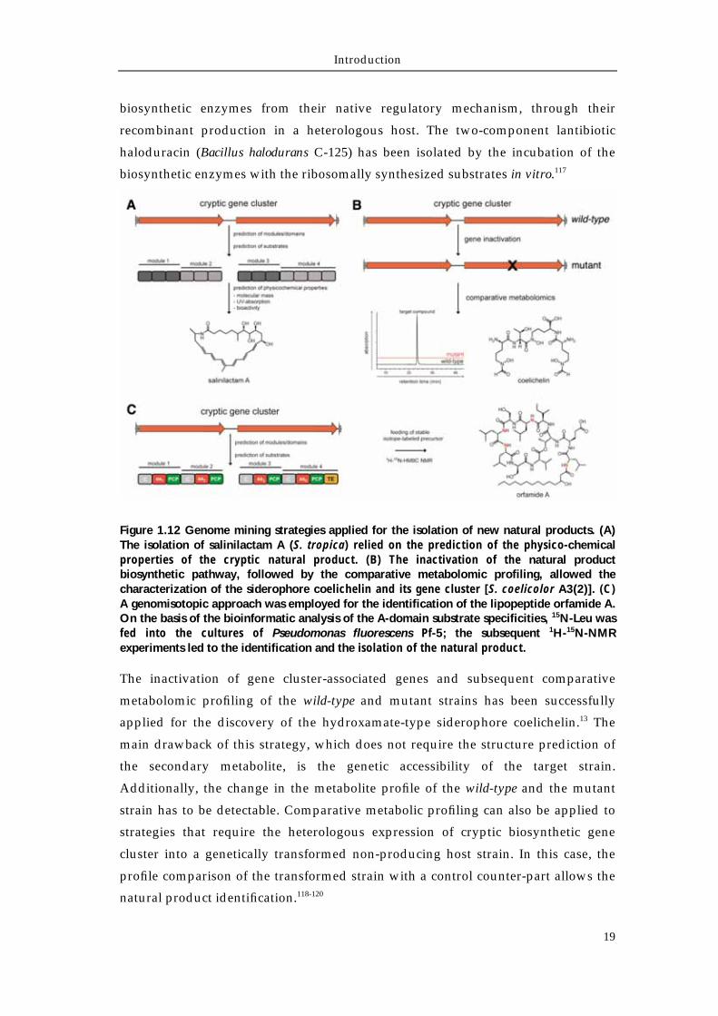
developed, leading to a successful characterization of new natural products (Figure
1.12). The first approach solely relies on the prediction of the physico-chemical
properties of the target compound, and has led to the identification of salinilactam
A (Salinispora tropica).116 The sequence analysis of a modular PKS gene cluster
putatively encoding a lysine-primed polyene macrolactam allowed the isolation and
the structural characterization of salinilactam A solely on the basis of polyene UV-
absorption properties. An alternative approach is represented by the in vitro
reconstitution of natural product biosynthesis. This strategy uncouples the
Introduction
19
biosynthetic enzymes from their native regulatory mechanism, through their
recombinant production in a heterologous host. The two-component lantibiotic
haloduracin (Bacillus halodurans C-125) has been isolated by the incubation of the
biosynthetic enzymes with the ribosomally synthesized substrates in vitro.117
Figure 1.12 Genome mining strategies applied for the isolation of new natural products. (A) The isolation of salinilactam A (S. tropica) relied on the prediction of the physico-chemical properties of the cryptic natural product. (B) The inactivation of the natural product biosynthetic pathway, followed by the comparative metabolomic profiling, allowed the characterization of the siderophore coelichelin and its gene cluster [S. coelicolor A3(2)]. (C) A genomisotopic approach was employed for the identification of the lipopeptide orfamide A. On the basis of the bioinformatic analysis of the A-domain substrate specificities, 15N-Leu was fed into the cultures of Pseudomonas fluorescens Pf-5; the subsequent 1H-15N-NMR experiments led to the identification and the isolation of the natural product.
The inactivation of gene cluster-associated genes and subsequent comparative
metabolomic profiling of the wild-type and mutant strains has been successfully
applied for the discovery of the hydroxamate-type siderophore coelichelin.13 The
main drawback of this strategy, which does not require the structure prediction of
the secondary metabolite, is the genetic accessibility of the target strain.
Additionally, the change in the metabolite profile of the wild-type and the mutant
strain has to be detectable. Comparative metabolic profiling can also be applied to
strategies that require the heterologous expression of cryptic biosynthetic gene
cluster into a genetically transformed non-producing host strain. In this case, the
profile comparison of the transformed strain with a control counter-part allows the
natural product identification.118-120
Introduction
20
The last more general strategy described for natural product discovery is the
genomisotopic approach, which has been successfully applied for the
characterization of the NRPS-derived cyclolipopeptide orfamide A (Pseudomonas
fluorescens Pf-5).121 This technique combines the bioinformatic predictions of the
modular assembly machinery with the incorporation of a stable-isotope precursor
added to the growing culture, allowing the labeling, detection and structural
characterization of the assembled product. In the case of orfamide A, 15N-labeled L-
Leu was fed to cultures of P. fluorescens Pf-5 and HPLC fractions of the culture
extracts were analyzed via 1H-15N-NMR experiments for the successful
identification of isotope-labeled metabolites.
In conclusion, the choice of the best strategy for the successful characterization of
new natural products via genome mining must take several aspects into account,
among them, the culturing conditions and the genetic accessibility of the producing
strain and the availability of the proper instrumentation for the isolation and
structural characterization of the natural product.
Introduction
21
1.4 Aim of the work
Rhodococcus spp. are extensively studied as extraordinary biocatalysts for steroid
production and fossil fuel biodesulfurization and as tools suitable for
bioremediation purposes.122,123 This widespread biotechnological and industrial
interest derive from their diverse metabolic capabilities. Furthermore, genome
sequencing information revealed Rhodococcus spp. to possess a vast genetic potential
for secondary metabolite production.124 However, only very few natural products
have been isolated from this genus, among them two siderophores, heterobactin A
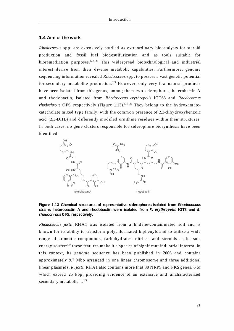
and rhodobactin, isolated from Rhodococcus erythropolis IGTS8 and Rhodococcus
rhodochrous OFS, respectively (Figure 1.13).125,126 They belong to the hydroxamate-
catecholate mixed type family, with the common presence of 2,3-dihydroxybenzoic
acid (2,3-DHB) and differently modified ornithine residues within their structures.
In both cases, no gene clusters responsible for siderophore biosynthesis have been
identified.
Figure 1.13 Chemical structures of representative siderophores isolated from Rhodococcus strains: heterobactin A and rhodobactin were isolated from R. erythropolis IGT8 and R. rhodochrous OFS, respectively.
Rhodococcus jostii RHA1 was isolated from a lindane-contaminated soil and is
known for its ability to transform polychlorinated biphenyls and to utilize a wide
range of aromatic compounds, carbohydrates, nitriles, and steroids as its sole
energy source:127 these features make it a species of significant industrial interest. In
this context, its genome sequence has been published in 2006 and contains
approximately 9.7 Mbp arranged in one linear chromosome and three additional
linear plasmids. R. jostii RHA1 also contains more that 30 NRPS and PKS genes, 6 of
which exceed 25 kbp, providing evidence of an extensive and uncharacterized
secondary metabolism.124
N
OH
O
HN
O
NH
HN
HN
O NH2
O
NH
OH2N
O
HN O
OH
OH
HN
O OH
OH
rhodobactin
N O
OH
NH
OHN O
HN
HN
O OHOH
O
OH
ON
heterobactin A
Introduction
22
On the basis of this knowledge, the isolation and the structural characterization of
the endogenous siderophore of R. jostii RHA1 will confirm the metabolic capacity of
the strain to produce secondary metabolites. Furthermore, the genome mining
identification of the gene cluster responsible for the biosynthesis of the molecule
will permit the rational construction of isogenic deletion mutant strains.
Subsequently, the metabolic profile comparison between the wild-type and the
mutant strains will undoubtedly connect the biosynthesis of the natural product
with the corresponding genes. Finally, the biochemical characterization of the
recombinantly-produced enzymes associated with the biosynthesis of the
siderophore will integrate the genetic results and will allow the postulation of a
model for the biosynthesis of the newly-discovered iron-scavenging compound.
Chapter 2
Material
Material
24
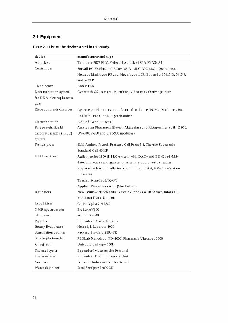
2.1 Equipment
Table 2.1 List of the devices used in this study.
device manufacturer and type
Autoclave Tuttnauer 5075 ELV, Fedegari Autoclavi SPA FVA3/A1
Centrifuges Sorvall RC 5B Plus and RC6+ (SS‐34, SLC‐300, SLC‐4000 rotors),
Heraeus Minifugue RF and Megafugue 1.0R, Eppendorf 5415 D, 5415 R
and 5702 R
Clean bench Antair BSK
Documentation system
for DNA‐electrophoresis
gels
Cybertech CS1 camera, Mitsubishi video copy thermo printer
Electrophoresis chamber Agarose gel chambers manufactured in‐house (PUMa, Marburg), Bio‐Rad Mini‐PROTEAN 3 gel chamber
Electroporation Bio-Rad Gene-Pulser II
Fast protein liquid
chromatography (FPLC)
system
Amersham Pharmacia Biotech Äktaprime and Äktapurifier: (pH/C-900,
UV-900, P-900 and Frac-900 modules)
French-press SLM Aminco French-Pressure Cell Press 5.1, Thermo Spectronic
Standard Cell 40 KP
HPLC-systems Agilent series 1100 (HPLC‐system with DAD‐ and ESI‐Quad‐MS‐
detection, vacuum degasser, quarternary pump, auto sampler,
preparative fraction collector, column thermostat, HP‐ChemStation
software)
Thermo Scientific LTQ‐FT
Applied Biosystems API QStar Pulsar i
Incubators New Brunswick Scientific Series 25, Innova 4300 Shaker, Infors HT
Multitron II and Unitron
Lyophilizer Christ Alpha 2‐4 LSC
NMR-spectrometer Bruker AV600
pH meter Schott CG 840
Pipettes Eppendorf Research series
Rotary Evaporator Heidolph Laborota 4000
Scintillation counter Packard Tri-Carb 2100-TR
Spectrophotometer PEQLab Nanodrop ND‐1000; Pharmacia Ultrospec 3000
Speed‐Vac Uniequip Univapo 150H
Thermal cycler Eppendorf Mastercycler Personal
Thermomixer Eppendorf Thermomixer comfort
Vortexer Scientific Industries VortexGenie2
Water deionizer Seral Seralpur Pro90CN
Material
25
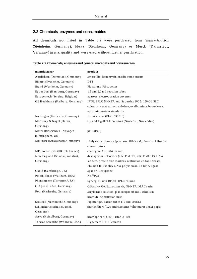
2.2 Chemicals, enzymes and consumables
All chemicals not listed in Table 2.2 were purchased from Sigma-Aldrich
(Steinheim, Germany), Fluka (Steinheim, Germany) or Merck (Darmstadt,
Germany) in p.a. quality and were used without further purification.
Table 2.2 Chemicals, enzymes and general materials and consumables.
manufacturer product
Applichem (Darmstadt, Germany) ampicillin, kanamycin, media components
Biomol (Ilvesheim, Germany) DTT
Brand (Wertheim, Germany) Plastbrand PS cuvettes
Eppendorf (Hamburg, Germany) 1.5 and 2.0 mL reaction tubes
Eurogentech (Seraing, Belgium) agarose, electroporation cuvettes
GE Healthcare (Freiburg, Germany) IPTG, FPLC Ni-NTA and Superdex 200 5/150 GL SEC
columns, yeast extract, aldolase, ovalbumin, ribonuclease,
aprotinin protein standards
Invitrogen (Karlsruhe, Germany) E. coli strains (BL21, TOP10)
Macherey & Nagel (Düren,
Germany)
C8- and C18-HPLC columns (Nucleosil, Nucleodur)
Merck4Biosciences - Novagen
(Nottingham, UK)
pET28a(+)
Millipore (Schwalbach, Germany) Dialysis membranes (pore size: 0.025 μM), Amicon Ultra‐15
concentrators
MP Biomedicals (Illkirch, France) coenzyme A trilithium salt
New England Biolabs (Frankfurt,
Germany)
desoxyribonucleotides (dATP, dTTP, dGTP, dCTP), DNA
ladders, protein size markers, restriction endonucleases,
Phusion Hi-Fidelity DNA polymerase, T4 DNA ligase
Oxoid (Cambridge, UK) agar nr. 1, tryptone
Perkin Elmer (Waltham, USA) Na432P2O7
Phenomenex (Torrance, USA) Synergi Fusion RP‐80 HPLC column
QIAgen (Hilden, Germany) QIAquick Gel Extraction kit, Ni‐NTA IMAC resin
Roth (Karlsruhe, Germany) acrylamide solution, β‐mercaptoethanol, ethidium
bromide, scintillation fluid
Sarstedt (Nümbrecht, Germany) Pipette tips, Falcon tubes (15 and 50 mL)
Schleicher & Schüll (Dassel,
Germany)
Sterile filters (0.20 and 0.45 μm), Whatmann-3MM paper
Serva (Heidelberg, Germany) bromophenol blue, Triton X‐100
Thermo Scientific (Waltham, USA) Hypercarb HPLC column
Material
26
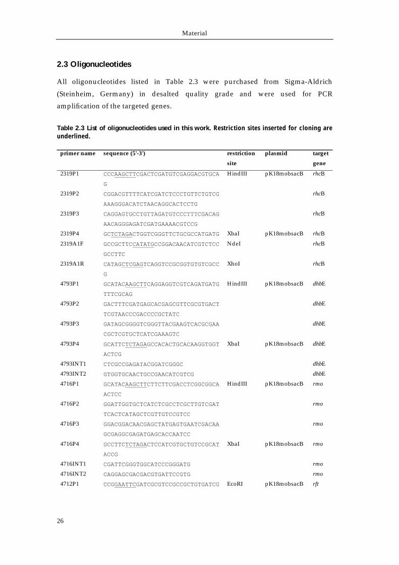
2.3 Oligonucleotides
All oligonucleotides listed in Table 2.3 were purchased from Sigma-Aldrich
(Steinheim, Germany) in desalted quality grade and were used for PCR
amplification of the targeted genes.
Table 2.3 List of oligonucleotides used in this work. Restriction sites inserted for cloning are underlined.
primer name sequence (5'-3') restriction
site
plasmid target
gene
2319P1 CCCAAGCTTCGACTCGATGTCGAGGACGTGCA
G
HindIII pK18mobsacB rhcB
2319P2 CGGACGTTTTCATCGATCTCCCTGTTCTGTCG
AAAGGGACATCTAACAGGCACTCCTG
rhcB
2319P3 CAGGAGTGCCTGTTAGATGTCCCTTTCGACAG
AACAGGGAGATCGATGAAAACGTCCG
rhcB
2319P4 GCTCTAGACTGGTCGGGTTCTGCGCCATGATG XbaI pK18mobsacB rhcB
2319A1F GCCGCTTCCATATGCCGGACAACATCGTCTCC
GCCTTC
NdeI rhcB
2319A1R CATAGCTCGAGTCAGGTCCGCGGTGTGTCGCC
G
XhoI rhcB
4793P1 GCATACAAGCTTCAGGAGGTCGTCAGATGATG
TTTCGCAG
HindIII pK18mobsacB dhbE
4793P2 GACTTTCGATGAGCACGAGCGTTCGCGTGACT
TCGTAACCCGACCCCGCTATC
dhbE
4793P3 GATAGCGGGGTCGGGTTACGAAGTCACGCGAA
CGCTCGTGCTCATCGAAAGTC
dhbE
4793P4 GCATTCTCTAGAGCCACACTGCACAAGGTGGT
ACTCG
XbaI pK18mobsacB dhbE
4793INT1 CTCGCCGAGATACGGATCGGGC dhbE
4793INT2 GTGGTGCAACTGCCGAACATCGTCG dhbE
4716P1 GCATACAAGCTTCTTCTTCGACCTCGGCGGCA
ACTCC
HindIII pK18mobsacB rmo
4716P2 GGATTGGTGCTCATCTCGCCTCGCTTGTCGAT
TCACTCATAGCTCGTTGTCCGTCC
rmo
4716P3 GGACGGACAACGAGCTATGAGTGAATCGACAA
GCGAGGCGAGATGAGCACCAATCC
rmo
4716P4 GCCTTCTCTAGACTCCATCGTGCTGTCCGCAT
ACCG
XbaI pK18mobsacB rmo
4716INT1 CGATTCGGGTGGCATCCCGGGATG rmo
4716INT2 CAGGAGCGACGACGTGATTCCGTG rmo
4712P1 CCGGAATTCGATCGCGTCCGCCGCTGTGATCG EcoRI pK18mobsacB rft
Material
27
4712P2 GTCCAGGAGGACCGCGTTGAGAGTCTGACGGT
CCCGCGCCGACACCAT
rft
4712P3 ATGGTGTCGGCGCGGGACCGTCAGACTCTCAA
CGCGGTCCTCCTGGAC
rft
4712P4 GCTCTAGACGTCCCGGAAATGCACGACCAGCG XbaI pK18mobsacB rft
4712INT1 CGATGACCATTCCGTCGCCTTCGTG rft
4712INT2 GACCATGAGGTCGTCCTCGCGATC rft
KANF ATGGATTGCACGCAGGTTCTC kanR
KANR CGATAGAAGGCGATGCGCT kanR
4793F GGGAATTCCATATGAGCACGAGCGTTCGCGCT NdeI pET28a(+) dhbE
4793R CCCCAAGCTTTTACGAAGTCACGAACGTCTTC
TCC
HindIII pET28a(+) dhbE
4712F GGAATTCCATATGAGAGTCGCCACACTCGGAT
ATC
NdeI pET28a(+) rft
4712R ATAAGAATGCGGCCGCTCAGCTGAGGTAGCCG
CCG
NotI pET28a(+) rft
4716F GGAATTCCATATGAGTGAATCGCCGGAAACGG
TCG
NdeI pET28a(+) rmo
4716R ATAAGAATGCGGCCGCTCATCTCGCCTCGCTT
GTCGCATAC
NotI pET28a(+) rmo
2322F AAAAAACCATGGCTAGTTCCGCTTCCAGCACA
GTTCC
NcoI pET28a(+) rhcE
2322R AAAAAAAAGCTTGCGTGCTGCCGTCACCTCGA
A
HindIII pET28a(+) rhcE
CchApCB128 AAAAAAGAATTCATGCGGGTCGTCATGTTCGG
CT
EcoRI pCB28a(+) cchA
CchApCB128 AAAAAACTCGAGTCAGGGGCGGGCGGTCAG XhoI pCB28a(+) cchA
Material
28
2.4 Plasmids
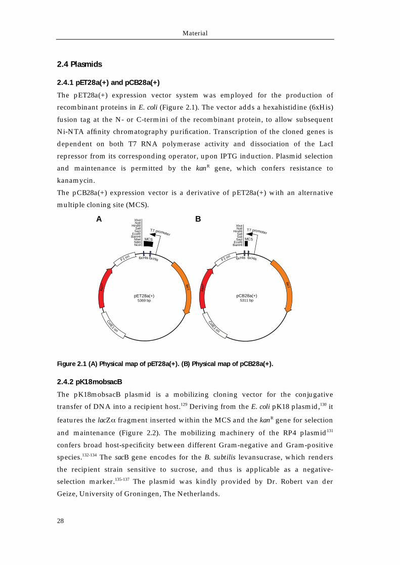
2.4.1 pET28a(+) and pCB28a(+)
The pET28a(+) expression vector system was employed for the production of
recombinant proteins in E. coli (Figure 2.1). The vector adds a hexahistidine (6xHis)
fusion tag at the N- or C-termini of the recombinant protein, to allow subsequent
Ni-NTA affinity chromatography purification. Transcription of the cloned genes is
dependent on both T7 RNA polymerase activity and dissociation of the LacI
repressor from its corresponding operator, upon IPTG induction. Plasmid selection
and maintenance is permitted by the kanR gene, which confers resistance to
kanamycin.
The pCB28a(+) expression vector is a derivative of pET28a(+) with an alternative
multiple cloning site (MCS).
Figure 2.1 (A) Physical map of pET28a(+). (B) Physical map of pCB28a(+).
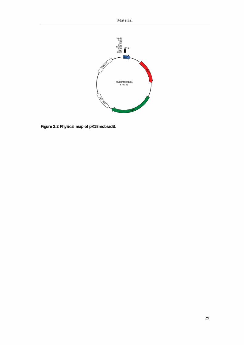
2.4.2 pK18mobsacB
The pK18mobsacB plasmid is a mobilizing cloning vector for the conjugative
transfer of DNA into a recipient host.129 Deriving from the E. coli pK18 plasmid,130 it
features the lacZα fragment inserted within the MCS and the kanR gene for selection
and maintenance (Figure 2.2). The mobilizing machinery of the RP4 plasmid131
confers broad host-specificity between different Gram-negative and Gram-positive
species.132-134 The sacB gene encodes for the B. subtilis levansucrase, which renders
the recipient strain sensitive to sucrose, and thus is applicable as a negative-
selection marker.135-137 The plasmid was kindly provided by Dr. Robert van der
Geize, University of Groningen, The Netherlands.
lacIkan
R
ColE1 ori
F1 ori 6xHis6xHis
MCS
T7 promoter
pET28a(+)5369 bp
XhoINotI
HindIIISalISacI
EcoRIBamHI
NheINdeINcoI
lacI
kan
R
ColE1 ori
F1 ori 6xHis6xHis
MCS
T7 promoter
pCB28a(+)5311 bp
XhoINotI
HindIIISalISstI
SacIEcoRI
BamHI
A B
Material
29
Figure 2.2 Physical map of pK18mobsacB.
sacB
kanR
ColE1 ori
oriTRP4
lacZ
MCS
HindIIISphIPstISalI
XbaIBamHI
SmaISacI
EcoRI
pK18mobsacB5702 bp
Material
30
2.5 Bacterial strains
2.5.1 Rhodococcus jostii RHA1
R. jostii RHA1 is a strain originally isolated from an insecticide-polluted soil sample.
It is classified as a Gram-positive, non-sporulating and non-motile microorganism
belonging to the Actinomicetales order.127 Its completely sequenced genome
comprises one linear chromosome and three linear plasmids, covering
approximately 9.2 Mbp in total.124 The strain has been cultured for the isolation of
the siderophore rhodochelin, for the isolation of genomic DNA and for the
construction of isogenic deletion mutants. The strain was kindly provided by Prof.
Linday E. Eltis of the University of British Columbia, Vancouver, Canada.
2.5.2 Escherichia coli TOP10
The E. coli TOP10 strain was used as a natural host for cloning and plasmid
propagation procedures. The genotype is: F- mcrA Δ(mrr-hsdRMS-mcrBC)
φ80lacZΔM15 ΔlacX74 recA1 araD139 Δ(ara-leu)7697 galU galK rpsL (StrR) endA1
nupG.
2.5.3 Escherichia coli BL21 (DE3)
The E. coli BL21 (DE3) strain was used as a general host for the heterologous
expression of recombinant proteins using T7-promoter driven expression plasmids.
It derives from the E. coli B strain and its genotype is: F– ompT hsdSB(rB–, mB
–) gal dcm
λ(DE3). The strain lacks the proteases Ion and OmpT to increase the half-life of the
overproduced recombinant protein. In addition, the insertion of the λ prophage DE3
carrying the IPTG-inducible T7 RNA polymerase (under the control of the lacUV5
promoter) is essential for recombinant protein expression.138
2.5.4 Escherichia coli S17-1
The E. coli S17-1 strain (genotype recA pro hsdR RP4-2-Tc::Mu-Km::Tn7) was used
as mobilizing strain for the transfer of the pK18mobsacB plasmid into the recipient
R. jostii RHA1, through bacterial conjugation. The mobilizing elements of the RP4
plasmid are stably integrated within the chromosome, in order to avoid self-transfer
of the original conjugating vector.139. The strain was kindly provided by Dr. Robert
van der Geize, University of Groningen, The Netherlands.
Material
31
2.6 Culture media
All media listed below were used for the cultivation and fermentation of the
bacterial strains indicated above. If solid agar plates were required for the
cultivation of the microorganism, agar nr. 1 was added to a final concentration of
1.5 % (m/w). All media were sterilized by autoclavation, (121 °C, 1.5 bar, 20 min).
Antibiotics and other additional temperature-labile components were added after
sterile-filtration, prior to use.
2.6.1 Lysogeny broth (LB-Miller)
LB broth (buffered at pH 7) has been routinely used for the cultivation and the
maintenance of R. jostii RHA1 and E. coli strains.
component quantity (1 L)
yeast extract 5 g
tryptone 10 g
NaCl 5 g
For the selection of Rhodococcus transconjugants sucrose was added to a final
concentration of 10% (m/w). Where required, antibiotics were added at the
following concentrations: nalidixic acid 30 μg/mL, kanamycin 50 μg/mL for
derivatives of pET28a(+), pCB28a(+) and pK18mobsacB integrants in Rhodococcus
host, and kanamycin 25 μg/mL for derivatives of pK18mobsacB in E. coli.
2.6.2 M9 minimal medium
M9 minimal medium was used for the isolation and the fermentative production of
rhodochelin.
component quantity (1 L)
Na2HPO4 · 2 H2O 8.5 g
KH2PO4 10 g
NH4Cl 1 g
NaCl 0.5 g
Prior to use, a concentrated trace element mix (100xMg/Ca/B1/Goodies) was
prepared as follows and added to a final 1x concentration. Sterile glucose solution
was used as a carbon source (final concentration: 4 g/L). Where required, Fe3+ was
added from a sterile-filtered FeCl3 solution.
Material
32
100x Mg/Ca/B1/Goodies
component quantity (100 mL)
MgSO4 1 M 20 mL
CaCl2 1 M 1 mL
thiamine 10 mM 1 mL
Conc. goodies 25 mM
ddH2O top up to volume
Concentrated goodies Stock salts solution140
component quantity (100 mL) component quantity (1 L)
Stock salts sol. 50 mL MgCl2 · 6 H2O 22.94 g
MgSO4 · 7 H2O 3 g CaCO3 2.0 g
ddH2O top up to volume ZnSO4 · 7 H2O 1.44 g
MnSO4 · H2O 0.85 g
CuSO4 · 5 H2O 0.25 g
CoCl2 · 6 H2O 0.24 g
H3BO3 0.06 g
HCl (conc.) 51.3 mL
ddH2O top up to volume
Chapter 3
Methods
Methods
34
3.1 Molecular biology techniques
General molecular biology material and methods (e.g. DNA agarose-gel
electrophoresis, protein SDS-PAGE, the composition of routine buffers solutions,
etc.) are not described in this section and were carried out according to established
protocols.141
3.1.1 General strains maintenance
All E. coli strains were routinely maintained on LB-agar plates and incubated at
37 °C. R. jostii RHA1 and derivative strains were maintained on LB-agar plates at
30 °C. Liquid cultures were incubated under continuous shaking at 180 rpm.
Antibiotics were added where required. For long-term storage, sterile glycerol was
added to a liquid culture [final concentration: 20% (v/v)] and the resulting stocks
were stored at -80 °C.
3.1.2 Preparation of genomic DNA
For genomic DNA isolation, 5 mL of liquid culture were harvested by centrifugation
and the resulting pellet was washed with 1 mL of water. The cell pellet was
resuspended in 500 μL lysis buffer [100 mM TRIS, 50 mM EDTA, 1% (w/v) SDS, pH
8] and acid-washed glass beads were added to a final volume of 1.25 mL. The
mixture was vortexed for two minutes and the recovered supernatant was
transferred into a new microfuge tube. 275 μL of 7 M ammonium acetate pH 7 was
added and the solution was incubated for 5 min at 65 °C, and then for 5 min on ice.
500 μL of chloroform were added and the mixture was vortexed for 2 min.
Following a centrifugation step at 13,000 rpm for 5 min, the recovered aqueous
phase was added to 800 μL of ice-cold isopropanol. The genomic DNA was
precipitated by centrifugation (5 min, 13,000 rpm), prior to 5 min incubation on ice.
Subsequently, the pellet was washed with 500 μL of ice-cold 70% ethanol solution,
dried and resuspended in 50 μL of EB buffer (10 mM TRIS, pH 8.5).
3.1.3 Preparation of plasmid DNA
Routine plasmid preparations were carried out according to the alkaline lysis with
SDS procedure.141 5 mL of LB medium (supplemented with the respective antibiotic)
was inoculated with a single colony and grown overnight. 2 mL of the stationary-
phase culture were harvested by centrifugation (13,000 rpm, 1 min) and the cell
pellet was resuspended in 300 μL of P1 buffer (50 mM TRIS, 10 mM EDTA, pH 8,
RNAse A 100 μg/mL). 300 μL of P2 buffer [0.2 M NaOH, 1% (w/v) SDS] were
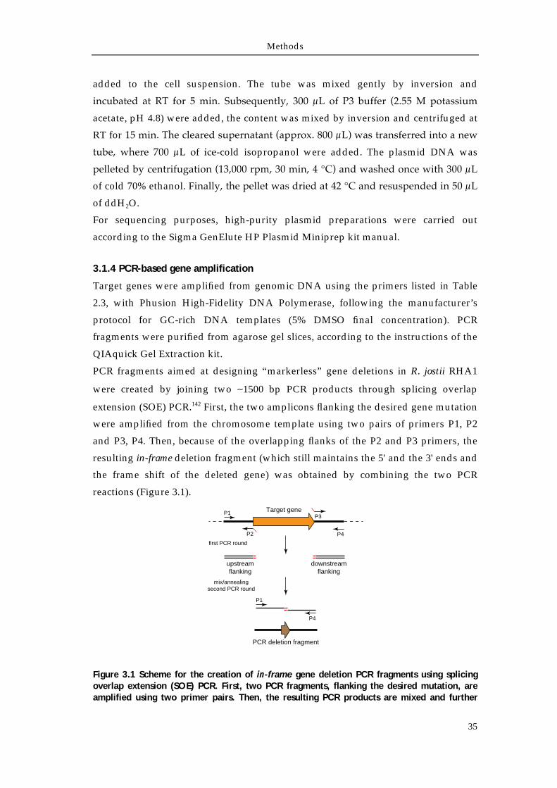
Methods
35
added to the cell suspension. The tube was mixed gently by inversion and
incubated at RT for 5 min. Subsequently, 300 μL of P3 buffer (2.55 M potassium
acetate, pH 4.8) were added, the content was mixed by inversion and centrifuged at
RT for 15 min. The cleared supernatant (approx. 800 μL) was transferred into a new
tube, where 700 μL of ice-cold isopropanol were added. The plasmid DNA was
pelleted by centrifugation (13,000 rpm, 30 min, 4 °C) and washed once with 300 μL
of cold 70% ethanol. Finally, the pellet was dried at 42 °C and resuspended in 50 μL
of ddH2O.
For sequencing purposes, high-purity plasmid preparations were carried out
according to the Sigma GenElute HP Plasmid Miniprep kit manual.
3.1.4 PCR-based gene amplification
Target genes were amplified from genomic DNA using the primers listed in Table
2.3, with Phusion High-Fidelity DNA Polymerase, following the manufacturer’s
protocol for GC-rich DNA templates (5% DMSO final concentration). PCR
fragments were purified from agarose gel slices, according to the instructions of the
QIAquick Gel Extraction kit.
PCR fragments aimed at designing “markerless” gene deletions in R. jostii RHA1
were created by joining two ∼1500 bp PCR products through splicing overlap
extension (SOE) PCR.142 First, the two amplicons flanking the desired gene mutation
were amplified from the chromosome template using two pairs of primers P1, P2
and P3, P4. Then, because of the overlapping flanks of the P2 and P3 primers, the
resulting in-frame deletion fragment (which still maintains the 5' and the 3' ends and
the frame shift of the deleted gene) was obtained by combining the two PCR
reactions (Figure 3.1).
Figure 3.1 Scheme for the creation of in-frame gene deletion PCR fragments using splicing overlap extension (SOE) PCR. First, two PCR fragments, flanking the desired mutation, are amplified using two primer pairs. Then, the resulting PCR products are mixed and further
Target geneP1
P2
P3
P4
first PCR round
P1
P4
upstreamflanking
downstreamflanking
mix/annealingsecond PCR round
PCR deletion fragment
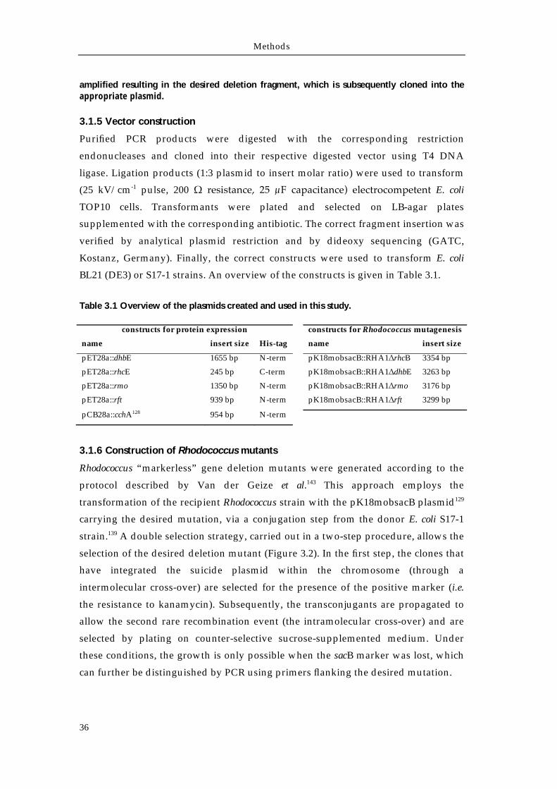
Methods
36
amplified resulting in the desired deletion fragment, which is subsequently cloned into the appropriate plasmid.
3.1.5 Vector construction
Purified PCR products were digested with the corresponding restriction
endonucleases and cloned into their respective digested vector using T4 DNA
ligase. Ligation products (1:3 plasmid to insert molar ratio) were used to transform
(25 kV/cm-1 pulse, 200 Ω resistance, 25 μF capacitance) electrocompetent E. coli
TOP10 cells. Transformants were plated and selected on LB-agar plates
supplemented with the corresponding antibiotic. The correct fragment insertion was
verified by analytical plasmid restriction and by dideoxy sequencing (GATC,
Kostanz, Germany). Finally, the correct constructs were used to transform E. coli
BL21 (DE3) or S17-1 strains. An overview of the constructs is given in Table 3.1.
Table 3.1 Overview of the plasmids created and used in this study.
constructs for protein expression constructs for Rhodococcus mutagenesis
name insert size His-tag name insert size
pET28a::dhbE 1655 bp N-term pK18mobsacB::RHA1∆rhcB 3354 bp
pET28a::rhcE 245 bp C-term pK18mobsacB::RHA1∆dhbE 3263 bp
pET28a::rmo 1350 bp N-term pK18mobsacB::RHA1∆rmo 3176 bp
pET28a::rft 939 bp N-term pK18mobsacB::RHA1∆rft 3299 bp
pCB28a::cchA128 954 bp N-term
3.1.6 Construction of Rhodococcus mutants
Rhodococcus “markerless” gene deletion mutants were generated according to the
protocol described by Van der Geize et al.143 This approach employs the
transformation of the recipient Rhodococcus strain with the pK18mobsacB plasmid129
carrying the desired mutation, via a conjugation step from the donor E. coli S17-1
strain.139 A double selection strategy, carried out in a two-step procedure, allows the
selection of the desired deletion mutant (Figure 3.2). In the first step, the clones that
have integrated the suicide plasmid within the chromosome (through a
intermolecular cross-over) are selected for the presence of the positive marker (i.e.
the resistance to kanamycin). Subsequently, the transconjugants are propagated to
allow the second rare recombination event (the intramolecular cross-over) and are
selected by plating on counter-selective sucrose-supplemented medium. Under
these conditions, the growth is only possible when the sacB marker was lost, which
can further be distinguished by PCR using primers flanking the desired mutation.
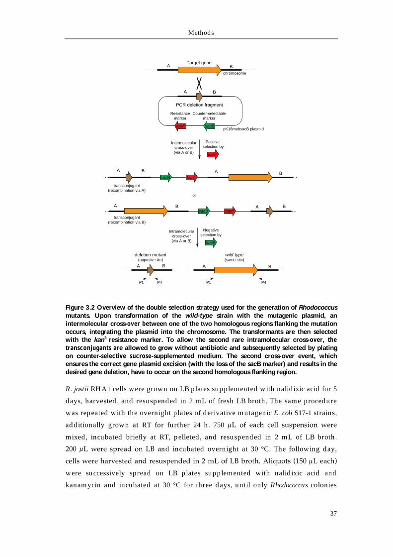
Methods
37
Figure 3.2 Overview of the double selection strategy used for the generation of Rhodococcus mutants. Upon transformation of the wild-type strain with the mutagenic plasmid, an intermolecular cross-over between one of the two homologous regions flanking the mutation occurs, integrating the plasmid into the chromosome. The transformants are then selected with the kanR resistance marker. To allow the second rare intramolecular cross-over, the transconjugants are allowed to grow without antibiotic and subsequently selected by plating on counter-selective sucrose-supplemented medium. The second cross-over event, which ensures the correct gene plasmid excision (with the loss of the sacB marker) and results in the desired gene deletion, have to occur on the second homologous flanking region.
R. jostii RHA1 cells were grown on LB plates supplemented with nalidixic acid for 5
days, harvested, and resuspended in 2 mL of fresh LB broth. The same procedure
was repeated with the overnight plates of derivative mutagenic E. coli S17-1 strains,
additionally grown at RT for further 24 h. 750 μL of each cell suspension were
mixed, incubated briefly at RT, pelleted, and resuspended in 2 mL of LB broth.
200 μL were spread on LB and incubated overnight at 30 °C. The following day,
cells were harvested and resuspended in 2 mL of LB broth. Aliquots (150 μL each)
were successively spread on LB plates supplemented with nalidixic acid and
kanamycin and incubated at 30 °C for three days, until only Rhodococcus colonies
A B
A
A
B
B
BA
kanRsacB
sacB kanR
pK18mobsacB plasmid
transconjugant(recombination via A)
transconjugant(recombination via B)
Target gene
Counter-selectablemarker
A B
Resistancemarker
XA B
kanR sacB
chromosome
PCR deletion fragment
Intermolecularcross-over(via A or B)
Positive selection by
kanR
or
A B
P1 P4
wild-type(same site)
A B
deletion mutant(opposite site)
P1 P4
Intramolecularcross-over(via A or B)
Negativeselection by
sacB
Methods
38
appeared. Transconjugants were grown in liquid medium and vector integration
was checked by PCR of the kanamycin cassette and by replica plating on LB
supplemented with kanamycin, or kanamycin and 10% sucrose. To force plasmid
excision, single integrant colonies were inoculated in LB broth and subsequently
plated on LB supplemented with 10% sucrose and grown at 30 °C until new colonies
appeared. To confirm correct plasmid excision, single clones were tested for
kanamycin sensitivity by replica plating and by PCR using different primer pairs:
for the kanamycin cassette, for a flanking region and for the deleted gene.
Methods
39
3.2 Expression and purification of recombinant proteins
3.2.1 Gene expression
5 mL of an overnight culture of E. coli BL21 (DE3) carrying the desired expression
construct was diluted inside a 2 L baffled flask containing 500 mL of fresh LB
medium supplemented with 50 μg/mL kanamycin. Cells were grown at 25 °C,
under continuous shaking at 230 rpm, until OD600 ~0.5 was reached. Gene
expression was induced with IPTG (50 μM), followed by incubation at 25 °C for 4 h.
Cells were harvested by centrifugation (6000 rpm, 15 min, 4 °C), resuspended in
HEPES A buffer (50 mM HEPES, 300 mM NaCl, pH 8) and stored at -20 °C until
further processing.
3.2.2 Protein purification
The frozen cell pellet was thawed on ice and lysed via French press. After a
centrifugation step (17,000 rpm, 4 °C, 30 min), the cleared lysate was sterile-filtered
and applied to a Ni-NTA IMAC column (equilibrated with HEPES A buffer) using a
ÄktaPrime system with a flow rate of 0.8 mL/min. The elution was carried out
using a linear gradient from 3 to 50% HEPES B buffer (50 mM HEPES, 250 mM
imidazole, 300 mM NaCl, pH 8) over 30 min, followed by a linear increase to 100% B
in 10 min at a flow rate of 1 mL/min, harvesting 2 mL fractions. Elution was
monitored at 280 nm and protein-containing fractions were checked by qualitative
Bradford assay144 and further analyzed by SDS-PAGE. Pooled fractions were
dialyzed against 25 mM HEPES, 100 mM NaCl, pH 7.5 buffer, and concentrated
with Amicon Ultra-15 concentrators.
3.2.3 Protein quantification
The final concentration of the overproduced and purified protein was determined
by Bradford colorimetric assay using a BSA calibration curve. Protein aliquots were
flash-frozen in liquid nitrogen and stored at -80 °C.
Methods
40
3.3 Analytical methods
3.3.1 HPLC-MS
High performance liquid chromatography (HPLC) was used as a standard
methodology for the characterization of compounds based on the retention time on
a chromatography column. Reversed-phase (RP) chromatography relies on the
hydrophobic interaction between compounds and the non-polar stationary phase of
the column (porous graphitic carbon or C8 or C18 coated silica gel). The elution of the
compounds is mediated employing a gradient with methanol or acetonitrile, which
shifts the interaction of the analyte for the column towards the mobile phase. The
retention time of the compounds is monitored by UV-vis detection. The mobile
phases were routinely supplied with either 0.1% TFA, or 0.05% formic acid, or
20 mM NFPA as ion pairing reagents to improve chromatographic separation and
electrospray mass ionization (ESI-MS) of the liquid compounds at atmospheric
pressure. Routine mass-spectrometry (MS) analysis were carried out on an Agilent
1100 MSD system. Acquisition of high resolution MS data and CID fragmentation
analysis of rhodochelin were carried out on a Thermo Electron LTQ-MS Fourier
transform ion cyclotron resonance mass spectrometer (FTICR-MS). Protein mass
fingerprinting of tryptic digests and measurements of whole proteins were carried
out on a Applied Biosystem API QStar Pulsar i system.
3.3.2 Peptide mass fingerprinting
The identity of recombinant proteins was verified by peptide mass fingerprinting.
Gel bands of the correct molecular mass were excised after SDS-PAGE and
incubated with 200 μL of wash solution [200 mM NH4HCO3, 50% (v/v) acetonitrile]
for 30 min at 37 °C. The solvent was subsequently removed in a speed-vac manifold
to complete dryness (37 °C, 30 min). In-band tryptic digestion was carried out by
addition of 20 μL of a trypsin solution (0.02 μg/μL trypsin, 10% NH4HCO3, 10%
acetonitrile, pH 8.1) at 37 °C for 45 min. The excess of trypsin was removed,
followed by an additional incubation period of 16 h. Peptide fragments were eluted
with 25 μL of a diffusion solution [1% (v/v) TFA, 10% acetonitrile, pH 8.1] and
sonicated at RT for 45 min. The samples were analyzed on a nanospray-ESI-qTOF-
MS system and the subsequent comparison of the peptide mass fingerprint with the
MASCOT database allowed the correct protein identification. 145,146
Methods
41
3.3.3 HPLC-ESI-qTOF-MS
The apo-, holo-, and the 2,3-DHB-loaded ArCP RhcE was analyzed by HPLC‐MS
using a QStar Pulsar i qTOF‐MS coupled to a HPLC system equipped with a C4
Nucleosil guard column (Macherey & Nagel, 10 x 3 mm, pore diameter of 300 Å,
particle size 5 μm) with the following conditions: solvent A (water + 0.45% formic
acid), solvent B (acetonitrile + 0.45% formic acid), flow rate 0.2 mL/min,
temperature 45 °C with a gradient of 10‐95% solvent B over 10 min, the gradient
was then held for 7 min.
3.3.4 Natural product isolation
For the isolation of rhodochelin, R. jostii RHA1 was grown for two days in LB
medium. Cells were harvested, washed once and resuspended in an equal amount
of M9 medium. A 1/100 aliquot was used to inoculate fresh minimal medium (in
polycarbonate flasks), and cultures were grown for two days, until a CAS positive
reaction of the supernatant was observed.147 The culture supernatant was extracted
with 5 g/L of XAD-16 resin for 2 h, and after a washing step, the adsorbed
compounds were eluted with methanol and immediately concentrated under
vacuum to dryness. The eluate was resuspended in 2 mL of water and analyzed on
an Agilent 1100 HPLC system equipped with a Nucleodur C8(ec) column 125 × 2
mm, and coupled to an ESI-MS detector, utilizing the solvent gradient water +
0.05% formic acid (solvent A) and methanol + 0.04% formic acid (solvent B), with a
linear gradient from 0% to 20% B within 40 min, followed by a linear increase to
95% B in 5 min and holding B for an additional 5 min. The flow rate was set to 0.3
mL/min and the column temperature to 40 °C. The gradient was also used to
analyze comparative extractions of R. jostii RHA1 mutants. Large-scale purification
was carried out by scaling-up the described protocol for 5 L cultures. The dried
eluate was dissolved in 10 mL of water and separated on a preparative Agilent 1100
preparative HPLC system equipped with a Nucleodur C8(ec) 250 × 21. Elution was
performed with the same gradient described previously, using 215 and 280 nm as
wavelengths for detection and a flow rate of 16 mL/min. Siderophore-containing
fractions were analyzed via CAS assay and ESI-MS. Positive fractions were pooled
according to their respective m/z, lyophilized, and subjected to further analysis.
3.3.5 UV-vis spectroscopy
UV-vis spectra were recorded on an Ultrospec 3000 (Pharmacia) spectrophotometer.
Wavescan measurements were performed within a wavelength range of 200-800 nm
and a scan rate of 750 nm/min. Absorption spectra of rhodochelin and holo-
Methods
42
rhodochelin as well as the apo- and holo-rhodochelin tripeptide were recorded in
water at a final concentration of 400 μM. holo-complexes were obtained by
incubating rhodochelin or the tripeptide (400 μM) with aqueous FeCl3 (400 μM) for
10 min at RT prior to the scan. Extinction coefficients were derived from the UV-vis
spectra. The following values were obtained. holo-rhodochelin: λmax 330 nm, ε =
3842.5 M-1 cm-1, λmax 395 nm, ε = 2205.0 M-1 cm-1, λmax 525 nm, ε = 1567.5 M-1 cm-1.
holo-rhodochelin tripeptide: λmax 327 nm, ε = 3372.5 M-1 cm-1, λmax 397 nm, ε = 1430.0
M-1 cm-1, λmax 580 nm, ε = 1567.5 M-1 cm-1.
3.3.6 IR-spectroscopy
Purified rhodochelin in a KBr disk was subjected to FT-IR-spectroscopy on a
Magna-IR 750 spectrometer (Nicolet). Main signals are the following: ν = 3367.2,
1749.5, 1660.5, 1586.1, 1534.1, 1448.0, 1381.8, 1204.0, 1137.4, 1066.9, 878.9, and
748.0 cm-1.
3.3.7 NMR-spectroscopy
NMR-spectroscopic structure elucidation of rhodochelin was carried out in
collaboration with Dr. Xiulan Xie (Department of chemistry - NMR facility,
Philipps-Universität Marburg). About 8 mg of rhodochelin were dissolved in 0.7 mL
of H2O/D2O (9:1). Measurements were carried out on a Bruker AV600 spectrometer
with an inverse broadband probe installed with z gradient. The one-dimensional
spectra 1H and 13C, the homonuclear two-dimensional spectra DQF-COSY, TOCSY,
NOESY, and ROESY, the 1H-13C HSQC and HMBC, and the 1H-15N HSQC spectra
were recorded with standard pulse programs at 283 K. The TOCSY spectrum was
recorded with a mixing time of 80 ms, whereas NOESY and ROESY spectra were
taken at 150 and 300 ms mixing times. The 1D spectra were acquired with 65,536
data points, whereas 2D spectra were collected using 4096 points in the F2
dimension and 512 increments in the F1 dimension. For 2D spectra, 16-32 transients
were used, while the 13C spectrum was recorded with 12 K scans. The relaxation
delay was 2.5 s. The 1H chemical shifts were referenced to 4,4-dimethyl-4-
silapentane sodium sulfonate (DSS) in H2O/D2O (9:1) at 283 K externally, whereas
those of 13C and 15N were referenced with spectrometer default calibration. The
spectra were processed with Bruker Topspin 2.1.
3.3.8 Assignment of amino acid stereochemistry via FDAA-derivatization
Assignment of the amino acid stereochemistry was achieved through rhodochelin
total acid hydrolysis followed by derivatization with Nα-(2,4-dinitro-5-
Methods
43
fluorophenyl)-L-alaninamide (FDAA, Marfey’s reagent).148 Purified rhodochelin
(3.76 mg) was hydrolyzed in 400 μL of 6 M HCl at 99 °C and 1000 rpm for 24 h. The
lyophilized hydrolysate was resuspended in 50 μL of 1 M NaHCO3, and 10 μL of
this solution was added to 170 μL of 1% FDAA solution in acetone. The
derivatization reaction was carried out for 1 h at 37 °C and terminated by addition
of 20 μL of 1 M HCl. FDAA standard derivatives of amino acids (L/D-Thr, L/D-Orn,
L-hOrn) were prepared by incubation of 25 μL of 50 mM amino acid solution with
50 μL of 1% FDAA solution and 10 μL of 1 M NaHCO3 for 1 h. After lyophilization,
all the products were resuspended in 200 μL of 1:1 water:acetonitrile solution prior
to injection (10 μL) into a HPLC-MS system equipped with a Synergi Fusion RP-80
250 × 2.0 mm column utilizing the following solvent gradient: 0-30 min, 0-30%
buffer A (10 mM ammonium formate, 1% methanol, 5% acetonitrile, pH 5.2) into
buffer B (10 mM ammonium formate, 1% methanol, 60% acetonitrile, pH 5.2),
followed by a linear increase to 95% buffer B in 2 min and holding 95% buffer B for
an additional 5 min. The flow rate was set at 0.3 mL/min and the column
temperature at 25 °C. Elution was monitored in single ion mode.
3.3.9 Analytical size-exclusion chromatography
Size-exclusion chromatography was employed to determine the quaternary
structure of the recombinant Rmo and Rft. 25 μL of a 50 μM Rmo or Rft solution was
analyzed using a Superdex 200 5/150 GL column combined with an ÄktaPurifier
system (Amersham Pharmacia Biotechnology) equilibrated with 25 mM TRIS 150
mM NaCl pH 8 buffer. Protein elution was monitored at 280 nm. Aldolase (158
kDa), ovalbumin (43 kDa), ribonuclease (13.7 kDa) and aprotinin (6.5 kDa) were
used as standards to determine the molecular weight of the proteins in solution.
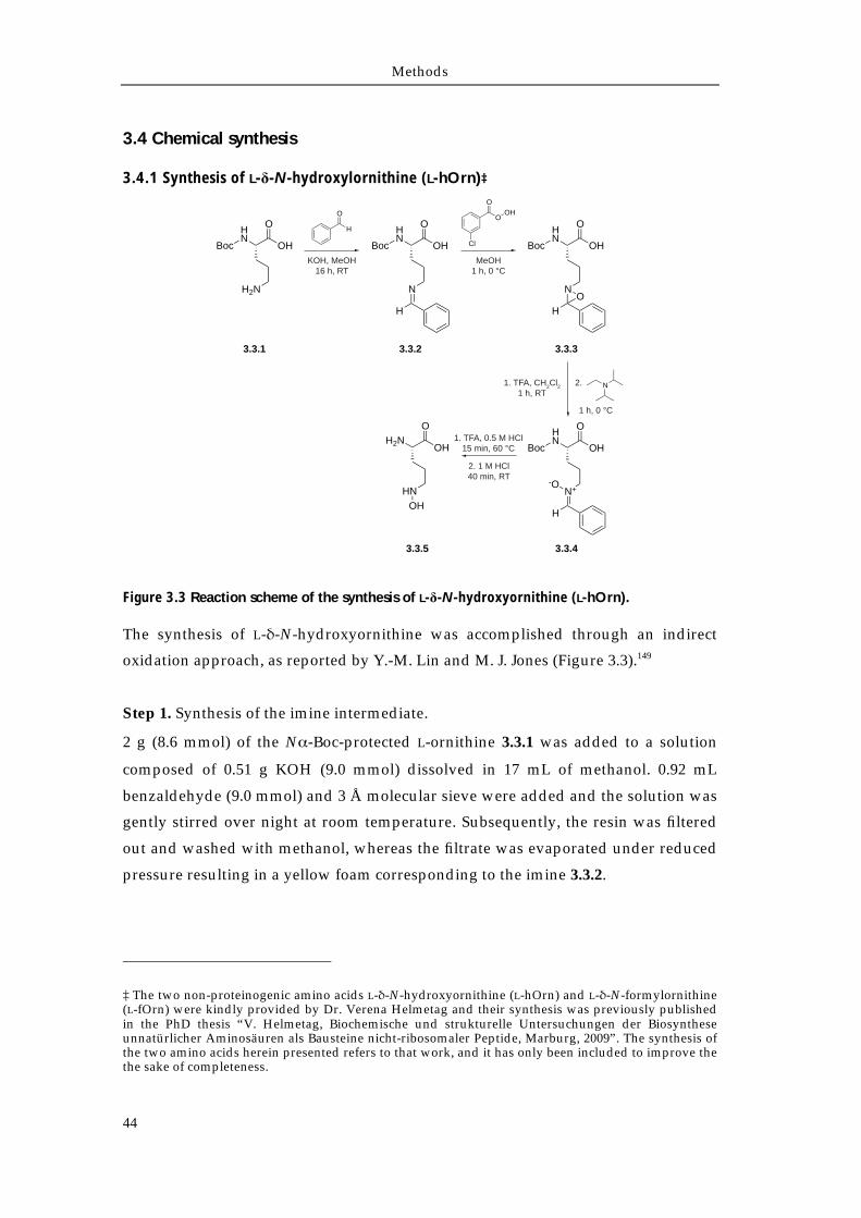
Methods
44
3.4 Chemical synthesis
3.4.1 Synthesis of L-δ-N-hydroxylornithine (L-hOrn)‡
Figure 3.3 Reaction scheme of the synthesis of L-δ-N-hydroxyornithine (L-hOrn).
The synthesis of L-δ-N-hydroxyornithine was accomplished through an indirect
oxidation approach, as reported by Y.-M. Lin and M. J. Jones (Figure 3.3).149
Step 1. Synthesis of the imine intermediate.
2 g (8.6 mmol) of the Nα-Boc-protected L-ornithine 3.3.1 was added to a solution
composed of 0.51 g KOH (9.0 mmol) dissolved in 17 mL of methanol. 0.92 mL
benzaldehyde (9.0 mmol) and 3 Å molecular sieve were added and the solution was
gently stirred over night at room temperature. Subsequently, the resin was filtered
out and washed with methanol, whereas the filtrate was evaporated under reduced
pressure resulting in a yellow foam corresponding to the imine 3.3.2.
‡ The two non-proteinogenic amino acids L-δ-N-hydroxyornithine (L-hOrn) and L-δ-N-formylornithine (L-fOrn) were kindly provided by Dr. Verena Helmetag and their synthesis was previously published in the PhD thesis “V. Helmetag, Biochemische und strukturelle Untersuchungen der Biosynthese unnatürlicher Aminosäuren als Bausteine nicht-ribosomaler Peptide, Marburg, 2009”. The synthesis of the two amino acids herein presented refers to that work, and it has only been included to improve the the sake of completeness.
Boc
HN
O
OH
H2N
Boc
HN
O
OH
N
H
Boc
HN
O
OH
N
HO
Boc
HN
O
OH
N+
H
-O
H2NO
OH
HNOH
H
O
KOH, MeOH16 h, RT
Cl
O
OOH
MeOH1 h, 0 °C
1. TFA, CH2Cl21 h, RT
1 h, 0 °C
N2.
1. TFA, 0.5 M HCl15 min, 60 °C
2. 1 M HCl40 min, RT
3.3.1 3.3.2 3.3.3
3.3.43.3.5
Methods
45
Step 2. Oxidation of the imine 3.3.2 to oxaziridine.
2.2 g of the imine 3.3.2 (7.0 mmol) was dissolved in 13 mL of methanol and the
solution was cooled to 0 °C. Then, a solution of 1.2 g of meta-chloroperbenzoic acid
(7.0 mmol) in 5 mL of methanol was added over 15 min. The reaction was kept at 0
°C and allowed to proceed for 1 h, under continuous stirring. The resulting
precipitate was filtered, washed with methanol and dried under reduced pressure.
Subsequently, the solid compound was dissolved in 34 mL of a 1:1 ddH2O:ethyl
acetate solution, acidified to pH 2 with HCl 1 M and extracted with ethyl acetate.
The recovered organic phases were washed with saturated NaCl solution, dried
with Na2SO4 and concentrated under reduced pressure, resulting in the
corresponding oxaziridine 3.3.3.
Step 3. Synthesis of the stable nitrone intermediate 3.3.3.
The unpurified oxaziridine precursor 3.3.3 was dissolved in 10 mL of trifluoroacetic
acid and, after the addition of 10 mL of CH2Cl2, the reaction mixture was stirred for
1 h at room temperature. The volatile compounds were removed by evaporation
and the resulting residue was dissolved in 30 mL of ethyl acetate. A solution of
diisopropyethylamine in THF was added until pH 8-9 was reached. The reaction
was cooled at 0 °C and allowed to proceed for 1 h, resulting in a pale-yellow
precipitate which was filtered and freeze-dried. The crude product was dissolved in
10 mL of ddH2O and purified by HPLC using a preparative C18-column equilibrated
with ddH2O (0.1% TFA, solvent A). The compounds were eluted with a 20 mL/min
flow rate, performing the following acetonitrile (0.1% TFA, solvent B) gradient: 0 %
B for 5 min, 0-30 % B in 30 min and 30-95 % in 3 min. The elution was monitored at
215 nm and the resulting fractions were identified by ESI-MS, pooled and
lyophilized, resulting in 945 mg of the corresponding nitrone 3.3.4.
Step 4. Conversion of the nitrone 3.3.4 to the hydroxylamine 3.3.5.
The final stage of the synthesis of L-hOrn was carried out from small quantities of
the stable nitrone, as this intermediate is more stable than the final product. 0.1 g of
the nitrone 3.3.4 (0.4 mmol) was diluted in 0.55 mL of hexane. Subsequently, 1.1 mL
of 0.5 M HCl and 0.27 mL of TFA were added. The mixture was heated at 60 °C
under reflux for 15 min. The volatile compounds were removed under reduced
pressure and the resulting yellowish oil was mixed with 1.1 mL of CH2Cl2 and 1.62
mL of HCl 1 M. The mixture was heated at 40 °C to allow the compounds to
dissolve and mixed for 40 min at RT. The organic phase was removed, whereas the
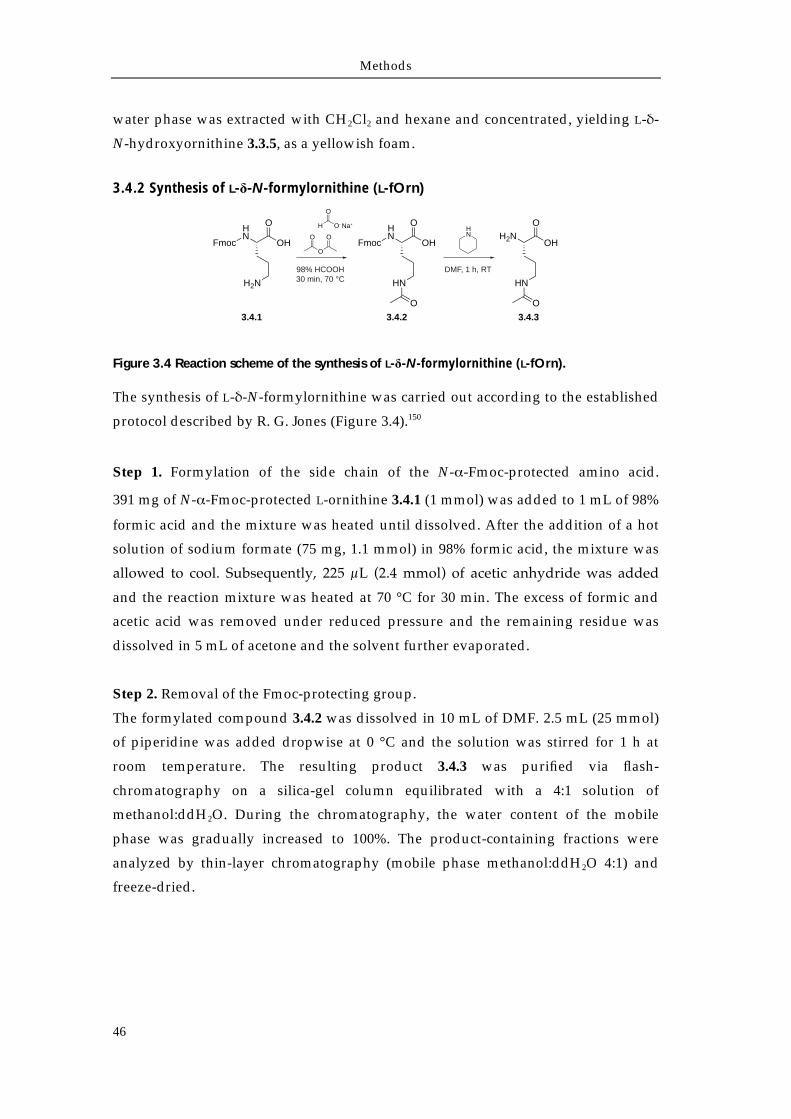
Methods
46
water phase was extracted with CH2Cl2 and hexane and concentrated, yielding L-δ-
N-hydroxyornithine 3.3.5, as a yellowish foam.
3.4.2 Synthesis of L-δ-N-formylornithine (L-fOrn)
Figure 3.4 Reaction scheme of the synthesis of L-δ-N-formylornithine (L-fOrn).
The synthesis of L-δ-N-formylornithine was carried out according to the established
protocol described by R. G. Jones (Figure 3.4).150
Step 1. Formylation of the side chain of the N-α-Fmoc-protected amino acid.
391 mg of N-α-Fmoc-protected L-ornithine 3.4.1 (1 mmol) was added to 1 mL of 98%
formic acid and the mixture was heated until dissolved. After the addition of a hot
solution of sodium formate (75 mg, 1.1 mmol) in 98% formic acid, the mixture was
allowed to cool. Subsequently, 225 μL (2.4 mmol) of acetic anhydride was added
and the reaction mixture was heated at 70 °C for 30 min. The excess of formic and
acetic acid was removed under reduced pressure and the remaining residue was
dissolved in 5 mL of acetone and the solvent further evaporated.
Step 2. Removal of the Fmoc-protecting group.
The formylated compound 3.4.2 was dissolved in 10 mL of DMF. 2.5 mL (25 mmol)
of piperidine was added dropwise at 0 °C and the solution was stirred for 1 h at
room temperature. The resulting product 3.4.3 was purified via flash-
chromatography on a silica-gel column equilibrated with a 4:1 solution of
methanol:ddH2O. During the chromatography, the water content of the mobile
phase was gradually increased to 100%. The product-containing fractions were
analyzed by thin-layer chromatography (mobile phase methanol:ddH2O 4:1) and
freeze-dried.
HN
Fmoc OH
O
H2N
HN
Fmoc OH
O
HN
O
H2NOH
O
HN
O
HN
DMF, 1 h, RT
O
O
O
H O-
O
98% HCOOH30 min, 70 °C
Na+
3.4.1 3.4.2 3.4.3
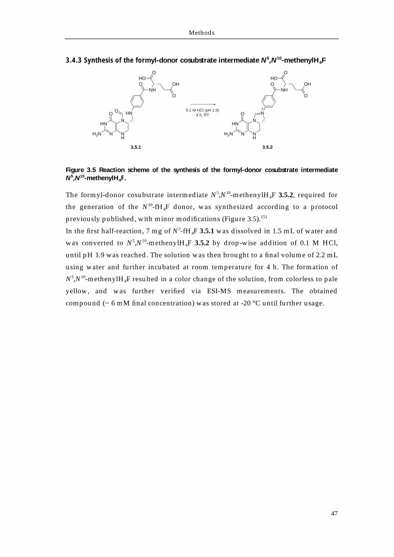
Methods
47
3.4.3 Synthesis of the formyl-donor cosubstrate intermediate N5,N10-methenylH4F
Figure 3.5 Reaction scheme of the synthesis of the formyl-donor cosubstrate intermediate N5,N10-methenylH4F.
The formyl-donor cosubstrate intermediate N5,N10-methenylH4F 3.5.2, required for
the generation of the N10-fH4F donor, was synthesized according to a protocol
previously published, with minor modifications (Figure 3.5).151
In the first half-reaction, 7 mg of N5-fH4F 3.5.1 was dissolved in 1.5 mL of water and
was converted to N5,N10-methenylH4F 3.5.2 by drop-wise addition of 0.1 M HCl,
until pH 1.9 was reached. The solution was then brought to a final volume of 2.2 mL
using water and further incubated at room temperature for 4 h. The formation of
N5,N10-methenylH4F resulted in a color change of the solution, from colorless to pale
yellow, and was further verified via ESI-MS measurements. The obtained
compound (~ 6 mM final concentration) was stored at -20 °C until further usage.
HN
N NH
N
H2N
O HN
NHO
OHO
O
OH
O
HN
N NH
N
H2N
O N
NHO
OHO
O
OH
0.1 M HCl (pH 1.9)4 h, RT
3.5.1 3.5.2
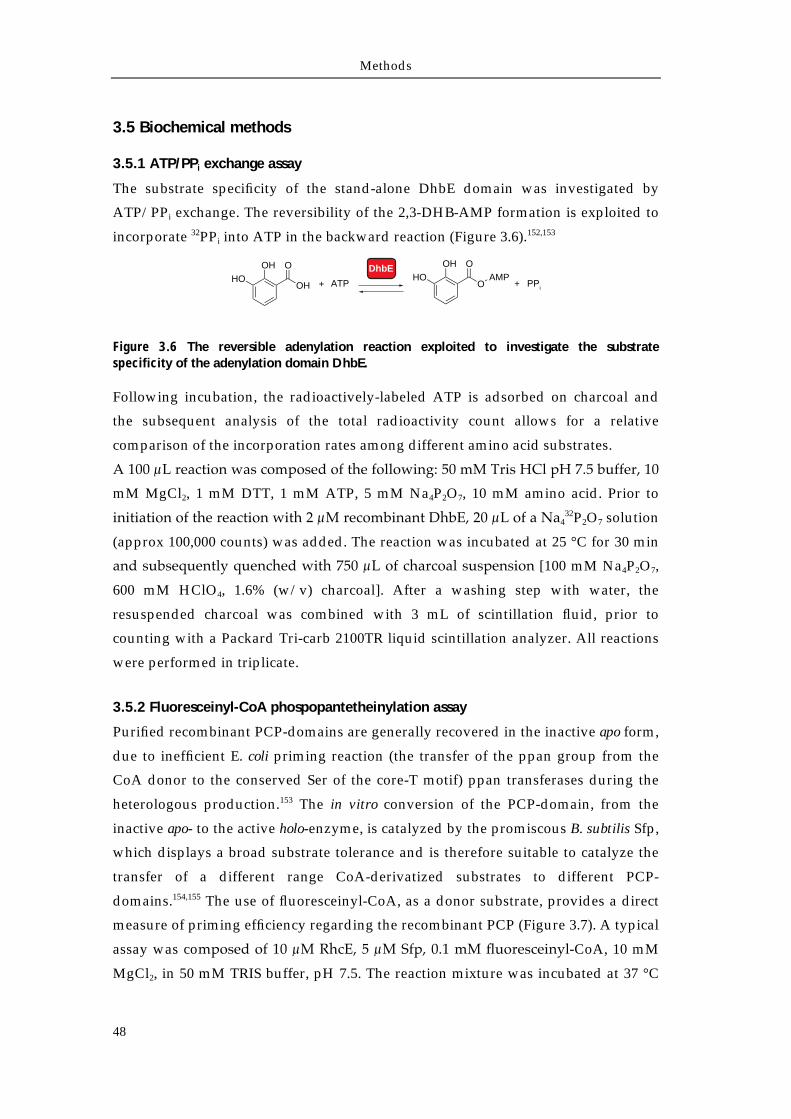
Methods
48
3.5 Biochemical methods
3.5.1 ATP/PPi exchange assay
The substrate specificity of the stand-alone DhbE domain was investigated by
ATP/PPi exchange. The reversibility of the 2,3-DHB-AMP formation is exploited to
incorporate 32PPi into ATP in the backward reaction (Figure 3.6).152,153
Figure 3.6 The reversible adenylation reaction exploited to investigate the substrate specificity of the adenylation domain DhbE.
Following incubation, the radioactively-labeled ATP is adsorbed on charcoal and
the subsequent analysis of the total radioactivity count allows for a relative
comparison of the incorporation rates among different amino acid substrates.
A 100 μL reaction was composed of the following: 50 mM Tris HCl pH 7.5 buffer, 10
mM MgCl2, 1 mM DTT, 1 mM ATP, 5 mM Na4P2O7, 10 mM amino acid. Prior to
initiation of the reaction with 2 μM recombinant DhbE, 20 μL of a Na432P2O7 solution
(approx 100,000 counts) was added. The reaction was incubated at 25 °C for 30 min
and subsequently quenched with 750 μL of charcoal suspension [100 mM Na4P2O7,
600 mM HClO4, 1.6% (w/v) charcoal]. After a washing step with water, the
resuspended charcoal was combined with 3 mL of scintillation fluid, prior to
counting with a Packard Tri-carb 2100TR liquid scintillation analyzer. All reactions
were performed in triplicate.
3.5.2 Fluoresceinyl-CoA phospopantetheinylation assay
Purified recombinant PCP-domains are generally recovered in the inactive apo form,
due to inefficient E. coli priming reaction (the transfer of the ppan group from the
CoA donor to the conserved Ser of the core-T motif) ppan transferases during the
heterologous production.153 The in vitro conversion of the PCP-domain, from the
inactive apo- to the active holo-enzyme, is catalyzed by the promiscous B. subtilis Sfp,
which displays a broad substrate tolerance and is therefore suitable to catalyze the
transfer of a different range CoA-derivatized substrates to different PCP-
domains.154,155 The use of fluoresceinyl-CoA, as a donor substrate, provides a direct
measure of priming efficiency regarding the recombinant PCP (Figure 3.7). A typical
assay was composed of 10 μM RhcE, 5 μM Sfp, 0.1 mM fluoresceinyl-CoA, 10 mM
MgCl2, in 50 mM TRIS buffer, pH 7.5. The reaction mixture was incubated at 37 °C
DhbEOHHO
OH
O OHHO
O
OAMP
ATP+ + PPi
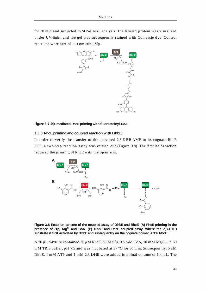
Methods
49
for 30 min and subjected to SDS-PAGE analysis. The labeled protein was visualized
under UV-light, and the gel was subsequently stained with Comassie dye. Control
reactions were carried out omitting Sfp.
Figure 3.7 Sfp-mediated RhcE priming with fluoresceinyl-CoA.
3.5.3 RhcE priming and coupled reaction with DhbE
In order to verify the transfer of the activated 2,3-DHB-AMP to its cognate RhcE
PCP, a two-step reaction assay was carried out (Figure 3.8). The first half-reaction
required the priming of RhcE with the ppan arm.
Figure 3.8 Reaction scheme of the coupled assay of DhbE and RhcE. (A) RhcE priming in the presence of Sfp, Mg2+ and CoA. (B) DhbE and RhcE coupled assay, where the 2,3-DHB substrate is first activated by DhbE and subsequently on the cognate primed ArCP RhcE.
A 50 μL mixture contained 50 μM RhcE, 5 μM Sfp, 0.5 mM CoA, 10 mM MgCl2, in 50
mM TRIS buffer, pH 7.5 and was incubated at 37 °C for 30 min. Subsequently, 5 μM
DhbE, 1 mM ATP and 1 mM 2,3-DHB were added to a final volume of 100 μL. The
OO OH
N OO
CoAS
COOH
RhcE
HO
+ RhcE
O OHO
NO O
S
HOOC
HNO
HNO
HO
OP
OO O-
Sfp
5'-3'-ADP
Mg2+
DhbE
RhcERhcE
A Sfp
SH
CoA 5'-3'-ADP
BOH
HOOH
O
Mg2+
ATP PPi
Mg2+
OHHO
O
OAMP +
RhcE
SH
RhcE
OS
HO
HO
+ AMP
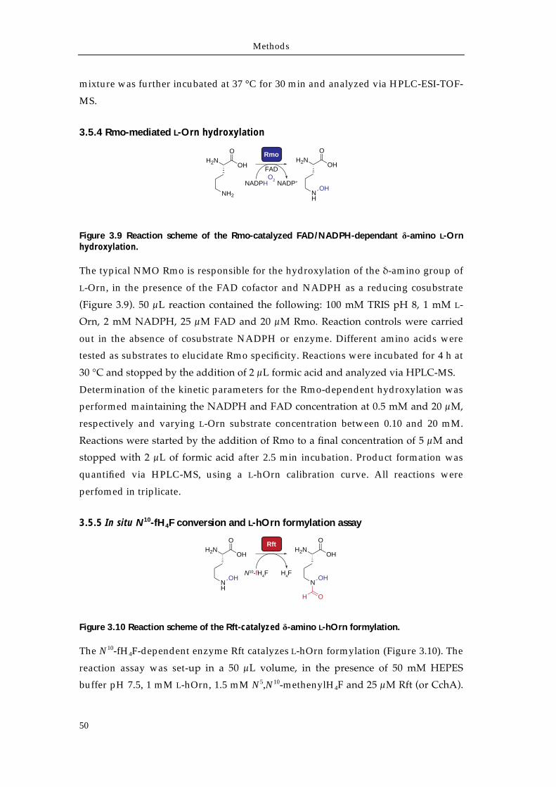
Methods
50
mixture was further incubated at 37 °C for 30 min and analyzed via HPLC-ESI-TOF-
MS.
3.5.4 Rmo-mediated L-Orn hydroxylation
Figure 3.9 Reaction scheme of the Rmo-catalyzed FAD/NADPH-dependant δ-amino L-Orn hydroxylation.
The typical NMO Rmo is responsible for the hydroxylation of the δ-amino group of
L-Orn, in the presence of the FAD cofactor and NADPH as a reducing cosubstrate
(Figure 3.9). 50 μL reaction contained the following: 100 mM TRIS pH 8, 1 mM L-
Orn, 2 mM NADPH, 25 μM FAD and 20 μM Rmo. Reaction controls were carried
out in the absence of cosubstrate NADPH or enzyme. Different amino acids were
tested as substrates to elucidate Rmo specificity. Reactions were incubated for 4 h at
30 °C and stopped by the addition of 2 μL formic acid and analyzed via HPLC-MS.
Determination of the kinetic parameters for the Rmo-dependent hydroxylation was
performed maintaining the NADPH and FAD concentration at 0.5 mM and 20 μM,
respectively and varying L-Orn substrate concentration between 0.10 and 20 mM.
Reactions were started by the addition of Rmo to a final concentration of 5 μM and
stopped with 2 μL of formic acid after 2.5 min incubation. Product formation was
quantified via HPLC-MS, using a L-hOrn calibration curve. All reactions were
perfomed in triplicate.
3.5.5 In situ N10-fH4F conversion and L-hOrn formylation assay
Figure 3.10 Reaction scheme of the Rft-catalyzed δ-amino L-hOrn formylation.
The N10-fH4F-dependent enzyme Rft catalyzes L-hOrn formylation (Figure 3.10). The
reaction assay was set-up in a 50 μL volume, in the presence of 50 mM HEPES
buffer pH 7.5, 1 mM L-hOrn, 1.5 mM N5,N10-methenylH4F and 25 μM Rft (or CchA).
Rmo
NADPH NADP+
FADO2
H2NOH
O
NH2
H2NOH
O
NH
OH
Rft
N10-fH4F
H2NOH
O
NH
OHH4F
H2NOH
O
NOH
OH
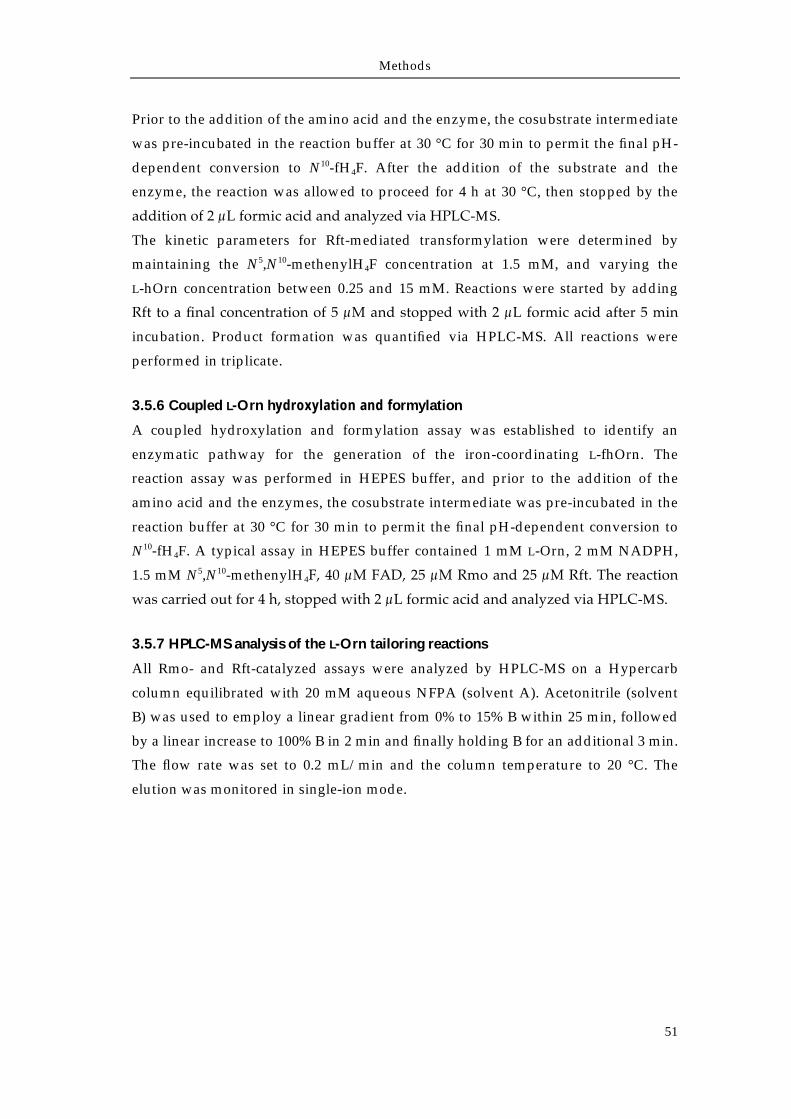
Methods
51
Prior to the addition of the amino acid and the enzyme, the cosubstrate intermediate
was pre-incubated in the reaction buffer at 30 °C for 30 min to permit the final pH-
dependent conversion to N10-fH4F. After the addition of the substrate and the
enzyme, the reaction was allowed to proceed for 4 h at 30 °C, then stopped by the
addition of 2 μL formic acid and analyzed via HPLC-MS.
The kinetic parameters for Rft-mediated transformylation were determined by
maintaining the N5,N10-methenylH4F concentration at 1.5 mM, and varying the
L-hOrn concentration between 0.25 and 15 mM. Reactions were started by adding
Rft to a final concentration of 5 μM and stopped with 2 μL formic acid after 5 min
incubation. Product formation was quantified via HPLC-MS. All reactions were
performed in triplicate.
3.5.6 Coupled L-Orn hydroxylation and formylation
A coupled hydroxylation and formylation assay was established to identify an
enzymatic pathway for the generation of the iron-coordinating L-fhOrn. The
reaction assay was performed in HEPES buffer, and prior to the addition of the
amino acid and the enzymes, the cosubstrate intermediate was pre-incubated in the
reaction buffer at 30 °C for 30 min to permit the final pH-dependent conversion to
N10-fH4F. A typical assay in HEPES buffer contained 1 mM L-Orn, 2 mM NADPH,
1.5 mM N5,N10-methenylH4F, 40 μM FAD, 25 μM Rmo and 25 μM Rft. The reaction
was carried out for 4 h, stopped with 2 μL formic acid and analyzed via HPLC-MS.
3.5.7 HPLC-MS analysis of the L-Orn tailoring reactions
All Rmo- and Rft-catalyzed assays were analyzed by HPLC-MS on a Hypercarb
column equilibrated with 20 mM aqueous NFPA (solvent A). Acetonitrile (solvent
B) was used to employ a linear gradient from 0% to 15% B within 25 min, followed
by a linear increase to 100% B in 2 min and finally holding B for an additional 3 min.
The flow rate was set to 0.2 mL/min and the column temperature to 20 °C. The
elution was monitored in single-ion mode.
Methods
52
3.6 Bioinformatic Methods
Web-browser-based and stand-alone programs were used to analyze nucleic acid
and protein sequences. Genome analysis and operon visualization was performed
with Artemis.156 Given a genome sequence, ANTIsmash allowed a rapid
identification of the major known secondary metabolites gene clusters.157 The
prediction of the adenylation domain substrate specificity was carried out with
NRPSpredictor and its updated version NRPSpredictor2.40,158 These tools analyze the
physico-chemical properties of the residues surrounding the A-domain substrate
binding pocket, extending the ten amino acid code defined by Stachelhaus et al., and
thus, improving the overall prediction.39 Sequence-homology searches were
executed using the BLAST algorithm, and multiple sequences alignments were
conducted with ClustalW, ClustalO or Muscle.159-162 Genome-BLAST comparisons
were carried out using Mauve.163 All sequences were retrieved from the NCBI
database (http://www.ncbi.nlm.nih.gov/).

Chapter 4
Results
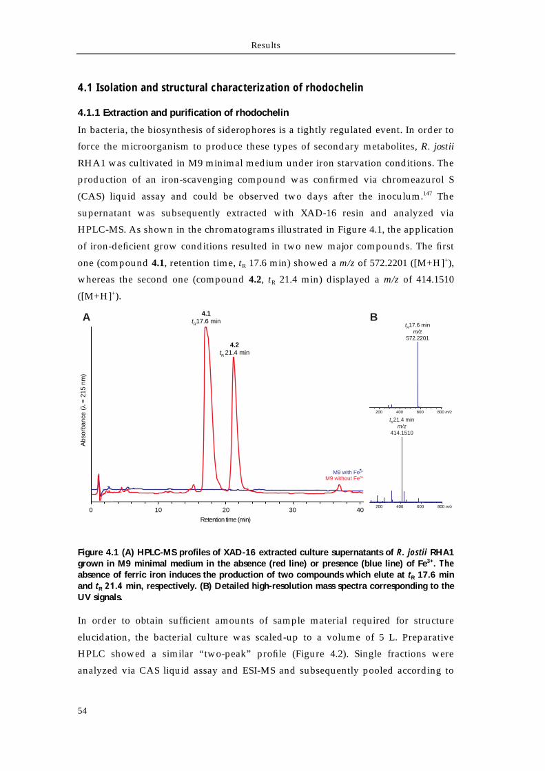
Results
54
4.1 Isolation and structural characterization of rhodochelin
4.1.1 Extraction and purification of rhodochelin
In bacteria, the biosynthesis of siderophores is a tightly regulated event. In order to
force the microorganism to produce these types of secondary metabolites, R. jostii
RHA1 was cultivated in M9 minimal medium under iron starvation conditions. The
production of an iron-scavenging compound was confirmed via chromeazurol S
(CAS) liquid assay and could be observed two days after the inoculum.147 The
supernatant was subsequently extracted with XAD-16 resin and analyzed via
HPLC-MS. As shown in the chromatograms illustrated in Figure 4.1, the application
of iron-deficient grow conditions resulted in two new major compounds. The first
one (compound 4.1, retention time, tR 17.6 min) showed a m/z of 572.2201 ([M+H]+),
whereas the second one (compound 4.2, tR 21.4 min) displayed a m/z of 414.1510
([M+H]+).
Figure 4.1 (A) HPLC-MS profiles of XAD-16 extracted culture supernatants of R. jostii RHA1 grown in M9 minimal medium in the absence (red line) or presence (blue line) of Fe3+. The absence of ferric iron induces the production of two compounds which elute at tR 17.6 min and tR 21.4 min, respectively. (B) Detailed high-resolution mass spectra corresponding to the UV signals.
In order to obtain sufficient amounts of sample material required for structure
elucidation, the bacterial culture was scaled-up to a volume of 5 L. Preparative
HPLC showed a similar “two-peak” profile (Figure 4.2). Single fractions were
analyzed via CAS liquid assay and ESI-MS and subsequently pooled according to
M9 without Fe3+M9 with Fe3+
Retention time (min)
Abs
orba
nce
(=
215
nm
)
0 10 20 30 40
200 400 600 800 m/z
tR17.6 minm/z
572.2201
200 400 600 800 m/z
tR21.4 minm/z
414.1510
4.1tR17.6 min
4.2tR 21.4 min
A B
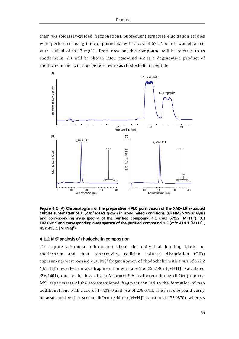
Results
55
their m/z (bioassay-guided fractionation). Subsequent structure elucidation studies
were performed using the compound 4.1 with a m/z of 572.2, which was obtained
with a yield of to 13 mg/L. From now on, this compound will be referred to as
rhodochelin. As will be shown later, comound 4.2 is a degradation product of
rhodochelin and will thus be referred to as rhodochelin tripeptide.
Figure 4.2 (A) Chromatogram of the preparative HPLC purification of the XAD-16 extracted culture supernatant of R. jostii RHA1 grown in iron-limited conditions. (B) HPLC-MS analysis and corresponding mass spectra of the purified compound 4.1 (m/z 572.2 [M+H]+). (C) HPLC-MS and corresponding mass spectra of the purified compound 4.2 (m/z 414.1 [M+H]+, m/z 436.1 [M+Na]+).
4.1.2 MSn analysis of rhodochelin composition
To acquire additional information about the individual building blocks of
rhodochelin and their connectivity, collision induced dissociation (CID)
experiments were carried out. MS2 fragmentation of rhodochelin with a m/z of 572.2
([M+H]+) revealed a major fragment ion with a m/z of 396.1402 ([M+H]+, calculated
396.1401), due to the loss of a δ-N-formyl-δ-N-hydroxyornithine (fhOrn) moiety.
MS3 experiments of the aforementioned fragment ion led to the formation of two
additional ions with a m/z of 177.0870 and m/z of 238.0711. The first one could easily
be associated with a second fhOrn residue ([M+H]+, calculated 177.0870), whereas
0 10 20 30 40
Abs
orba
nce
( =
215
nm
)
Retention time (min)
Retention time (min)0 10 20 30 40
550 600 m/z
572.2
tR16.6 min
SIC
[414
.1, 5
72.2
]
Retention time (min)
400 450 m/z
414.1
436.1
tR 20.3 min
A
B C
SIC
[414
.1, 5
72.2
]
4.1, rhodochelin
4.2, r. tripeptide
0 10 20 30 40
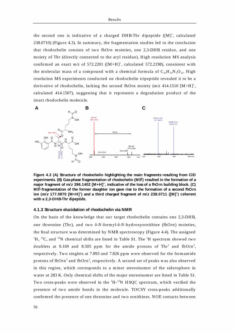
Results
56
the second one is indicative of a charged DHB-Thr dipeptide ([M]+, calculated
238.0710) (Figure 4.3). In summary, the fragmentation studies led to the conclusion
that rhodochelin consists of two fhOrn moieties, one 2,3-DHB residue, and one
moiety of Thr (directly connected to the aryl residue). High resolution MS analysis
confirmed an exact m/z of 572.2201 ([M+H]+, calculated 572.2198), consistent with
the molecular mass of a compound with a chemical formula of C23H33N5O12. High
resolution MS experiments conducted on rhodochelin tripeptide revealed it to be a
derivative of rhodochelin, lacking the second fhOrn moiety (m/z 414.1510 [M+H]+,
calculated 414.1507), suggesting that it represents a degradation product of the
intact rhodochelin molecule.
Figure 4.3 (A) Structure of rhodochelin highlighting the main fragments resulting from CID experiments. (B) Gas-phase fragmentation of rhodochelin (MS2) resulted in the formation of a major fragment of m/z 396.1402 [M+H]+, indicative of the loss of a fhOrn building block. (C) MS3-fragmentation of the former daughter ion gave rise to the formation of a second fhOrn ion (m/z 177.0870 [M+H]+) and a third charged fragment of m/z 238.0711 ([M]+) coherent with a 2,3-DHB-Thr dipeptide.
4.1.3 Structure elucidation of rhodochelin via NMR
On the basis of the knowledge that our target rhodochelin contains one 2,3-DHB,
one threonine (Thr), and two δ-N-formyl-δ-N-hydroxyornithine (fhOrn) moieties,
the final structure was determined by NMR spectroscopy (Figure 4.4). The assigned 1H, 13C, and 15N chemical shifts are listed in Table S1. The 1H spectrum showed two
doublets at 9.169 and 8.505 ppm for the amide protons of Thr1 and fhOrn2,
respectively. Two singlets at 7.893 and 7.826 ppm were observed for the formamide
protons of fhOrn2 and fhOrn3, respectively. A second set of peaks was also observed
in this region, which corresponds to a minor stereoisomer of the siderophore in
water at 283 K. Only chemical shifts of the major stereoisomer are listed in Table S1.
Two cross-peaks were observed in the 1H-15N HSQC spectrum, which verified the
presence of two amide bonds in the molecule. TOCSY cross-peaks additionally
confirmed the presence of one threonine and two ornithines. NOE contacts between
B
120
238.0711
m/z180 240 300
[fhOrn+H]+
177.0870
CA
OHHO
NH
O
H
OHN
OOH
O
NOH
OH
ONH2
NOH
OH
23
8.0
71
1
17
7.0
87
0
396.1402
396.1402
m/z380 440 500 560
573.2238554.2099
414.1514
-fhOrn
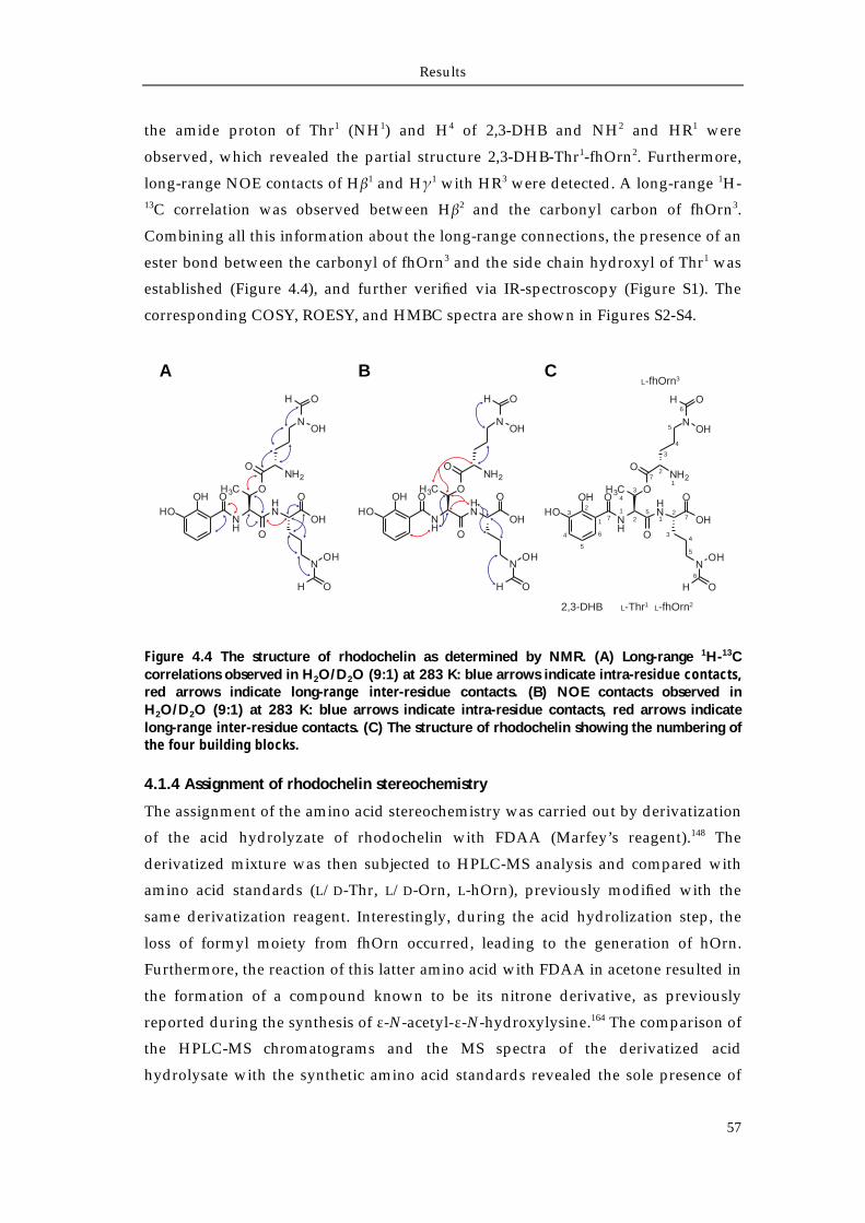
Results
57
the amide proton of Thr1 (NH1) and H4 of 2,3-DHB and NH2 and HR1 were
observed, which revealed the partial structure 2,3-DHB-Thr1-fhOrn2. Furthermore,
long-range NOE contacts of Hβ1 and Hγ1 with HR3 were detected. A long-range 1H-13C correlation was observed between Hβ2 and the carbonyl carbon of fhOrn3.
Combining all this information about the long-range connections, the presence of an
ester bond between the carbonyl of fhOrn3 and the side chain hydroxyl of Thr1 was
established (Figure 4.4), and further verified via IR-spectroscopy (Figure S1). The
corresponding COSY, ROESY, and HMBC spectra are shown in Figures S2-S4.
Figure 4.4 The structure of rhodochelin as determined by NMR. (A) Long-range 1H-13C correlations observed in H2O/D2O (9:1) at 283 K: blue arrows indicate intra-residue contacts, red arrows indicate long-range inter-residue contacts. (B) NOE contacts observed in H2O/D2O (9:1) at 283 K: blue arrows indicate intra-residue contacts, red arrows indicate long-range inter-residue contacts. (C) The structure of rhodochelin showing the numbering of the four building blocks.
4.1.4 Assignment of rhodochelin stereochemistry
The assignment of the amino acid stereochemistry was carried out by derivatization
of the acid hydrolyzate of rhodochelin with FDAA (Marfey’s reagent).148 The
derivatized mixture was then subjected to HPLC-MS analysis and compared with
amino acid standards (L/D-Thr, L/D-Orn, L-hOrn), previously modified with the
same derivatization reagent. Interestingly, during the acid hydrolization step, the
loss of formyl moiety from fhOrn occurred, leading to the generation of hOrn.
Furthermore, the reaction of this latter amino acid with FDAA in acetone resulted in
the formation of a compound known to be its nitrone derivative, as previously
reported during the synthesis of ε-N-acetyl-ε-N-hydroxylysine.164 The comparison of
the HPLC-MS chromatograms and the MS spectra of the derivatized acid
hydrolysate with the synthetic amino acid standards revealed the sole presence of
A B
1
6
5
4
2,3-DHB L-Thr1 L-fhOrn2
L-fhOrn3
7
4
3
3
2
25
12
7
34
5
6
72
1
3
4
5
6
1
C
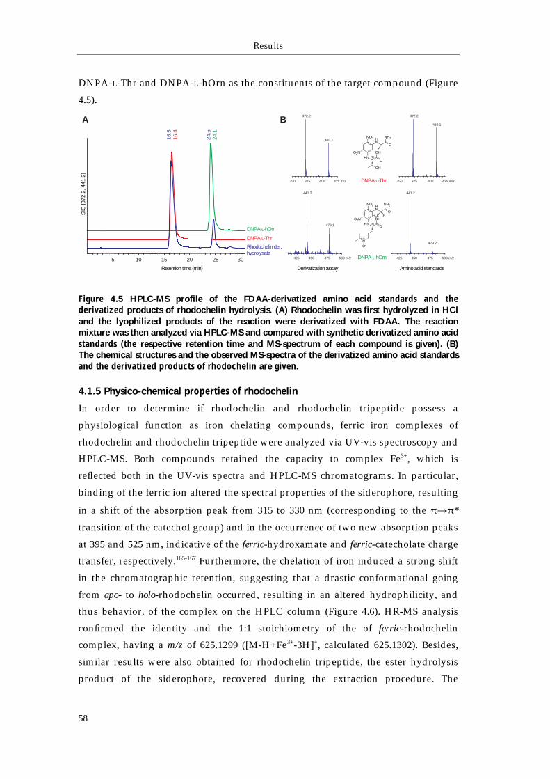
Results
58
DNPA-L-Thr and DNPA-L-hOrn as the constituents of the target compound (Figure
4.5).
Figure 4.5 HPLC-MS profile of the FDAA-derivatized amino acid standards and the derivatized products of rhodochelin hydrolysis. (A) Rhodochelin was first hydrolyzed in HCl and the lyophilized products of the reaction were derivatized with FDAA. The reaction mixture was then analyzed via HPLC-MS and compared with synthetic derivatized amino acid standards (the respective retention time and MS-spectrum of each compound is given). (B) The chemical structures and the observed MS-spectra of the derivatized amino acid standards and the derivatized products of rhodochelin are given.
4.1.5 Physico-chemical properties of rhodochelin
In order to determine if rhodochelin and rhodochelin tripeptide possess a
physiological function as iron chelating compounds, ferric iron complexes of
rhodochelin and rhodochelin tripeptide were analyzed via UV-vis spectroscopy and
HPLC-MS. Both compounds retained the capacity to complex Fe3+, which is
reflected both in the UV-vis spectra and HPLC-MS chromatograms. In particular,
binding of the ferric ion altered the spectral properties of the siderophore, resulting
in a shift of the absorption peak from 315 to 330 nm (corresponding to the π→π*
transition of the catechol group) and in the occurrence of two new absorption peaks
at 395 and 525 nm, indicative of the ferric-hydroxamate and ferric-catecholate charge
transfer, respectively.165-167 Furthermore, the chelation of iron induced a strong shift
in the chromatographic retention, suggesting that a drastic conformational going
from apo- to holo-rhodochelin occurred, resulting in an altered hydrophilicity, and
thus behavior, of the complex on the HPLC column (Figure 4.6). HR-MS analysis
confirmed the identity and the 1:1 stoichiometry of the of ferric-rhodochelin
complex, having a m/z of 625.1299 ([M-H+Fe3+-3H]+, calculated 625.1302). Besides,
similar results were also obtained for rhodochelin tripeptide, the ester hydrolysis
product of the siderophore, recovered during the extraction procedure. The
5 10 15 20 25 30
Retention time (min)
SIC
[372
.2, 4
41.2
]
Rhodochelin der. hydrolysate
DNPA-L-Thr
DNPA-L-hOrn
16.3
16.4
24.6
24.1
350 375 400 425 m/z
425 450 475 500 m/z 425 450 475 500 m/zDNPA-L-hOrn
350 375 400 425 m/zDNPA-L-Thr
NO2
O2NHN
HN
O
NH2
O
OH
OH
372.2
410.1
372.2
410.1
441.2 441.2
479.1
479.2
Derivatization assay Amino acid standards
NO2
O2NHN
HN
O
NH2
H3C H
O
OH
NO-
A B
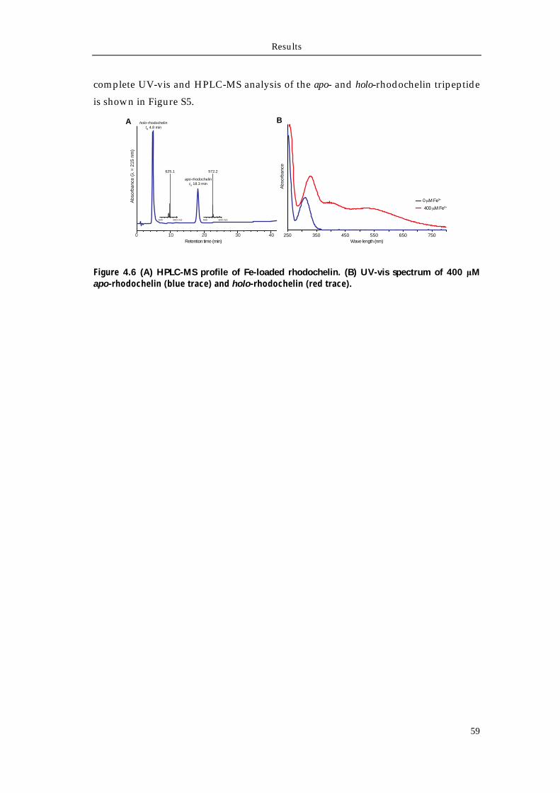
Results
59
complete UV-vis and HPLC-MS analysis of the apo- and holo-rhodochelin tripeptide
is shown in Figure S5.
Figure 4.6 (A) HPLC-MS profile of Fe-loaded rhodochelin. (B) UV-vis spectrum of 400 μM apo-rhodochelin (blue trace) and holo-rhodochelin (red trace).
0 10 20 30 40 250 350 450 550 650 750
600 650 m/z
Abs
orba
nce
( =
215
nm
)
Retention time (min)
550 600 m/z
625.1 572.2
holo-rhodochelintR 4.8 min
apo-rhodochelintR 18.3 min
Wave length (nm)
0 M Fe3+
400 M Fe3+
A B
Abs
orba
nce
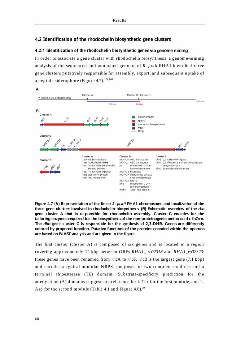
Results
60
4.2 Identification of the rhodochelin biosynthetic gene clusters
4.2.1 Identification of the rhodochelin biosynthetic genes via genome mining
In order to associate a gene cluster with rhodochelin biosynthesis, a genome-mining
analysis of the sequenced and annotated genome of R. jostii RHA1 identified three
gene clusters putatively responsible for assembly, export, and subsequent uptake of
a peptide siderophore (Figure 4.7).124,168
Figure 4.7 (A) Representation of the linear R. jostii RHA1 chromosome and localization of the three gene clusters involved in rhodochelin biosynthesis. (B) Schematic overview of the rhc gene cluster A that is responsible for rhodochelin assembly. Cluster C encodes for the tailoring enzymes required for the biosynthesis of the non-proteinogenic amino acid L-fhOrn. The dhb gene cluster C is responsible for the synthesis of 2,3-DHB. Genes are differently colored by proposed function. Putative functions of the proteins encoded within the operons are based on BLAST-analysis and are given in the figure.
The first cluster (cluster A) is composed of six genes and is located in a region
covering approximately 12 kbp between ORFs RHA1_ ro02318 and RHA1_ro02323:
these genes have been renamed from rhcA to rhcF. rhcB is the largest gene (7.1 kbp)
and encodes a typical modular NRPS, composed of two complete modules and a
terminal thioesterase (TE) domain. Substrate-specificity prediction for the
adenylation (A) domains suggests a preference for L-Thr for the first module, and L-
Asp for the second module (Table 4.1 and Figure 4.8).39
02.5 Mbp
7.8 Mbp
R. jostii RHA1 chromosomeCluster B Cluster CCluster A
Cluster A
rhcA
rhcB
rhcC
rhcD
rhcE
rhcF
dhbE
dhbA
dhbC
rft rmo
mbt
H
ro04
710
ro04
711
ro04
713
ro04
714
ro04
715
1kbp
export/import
NRPSprecursor biosynthesisMbtH
A
B
Cluster B
Cluster CCluster ArhcA isochorismataserhcB rhodochelin NRPSrhcC rhodochelin extracellular binding proteinrhcD rhodochelin exporterrhcE aryl carrier proteinrhcF ABC transporter
Cluster Bro04710 ABC transporterro04711 ABC transporterrft rhodochelin L-hOrn formyltransferasero04713 hydrolasero04714 diguanylate cyclase phosphodiesterasero04715 NRPSrmo rhodochelin L-Orn monooxygenasembtH MbtH-like protein
Cluster CdhbE 2,3-DHB AMP-ligasedhbA 2,3-dihydro-2,3-dihydroxybenzoate dehydrogenasedhbC isochorismate synthase
70 kbp
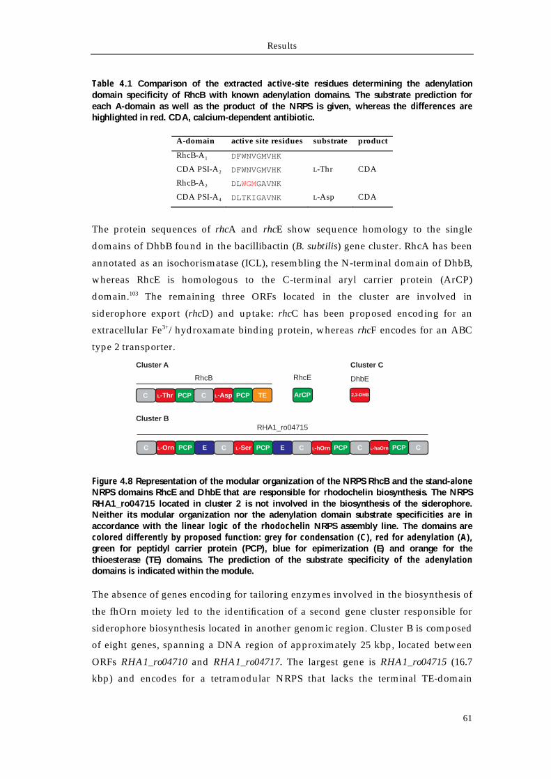
Results
61
Table 4.1 Comparison of the extracted active-site residues determining the adenylation domain specificity of RhcB with known adenylation domains. The substrate prediction for each A-domain as well as the product of the NRPS is given, whereas the differences are highlighted in red. CDA, calcium-dependent antibiotic.
A-domain active site residues substrate product
RhcB-A1 DFWNVGMVHK
CDA PSI-A2 DFWNVGMVHK L-Thr CDA
RhcB-A2 DLWGMGAVNK
CDA PSI-A4 DLTKIGAVNK L-Asp CDA
The protein sequences of rhcA and rhcE show sequence homology to the single
domains of DhbB found in the bacillibactin (B. subtilis) gene cluster. RhcA has been
annotated as an isochorismatase (ICL), resembling the N-terminal domain of DhbB,
whereas RhcE is homologous to the C-terminal aryl carrier protein (ArCP)
domain.103 The remaining three ORFs located in the cluster are involved in
siderophore export (rhcD) and uptake: rhcC has been proposed encoding for an
extracellular Fe3+/hydroxamate binding protein, whereas rhcF encodes for an ABC
type 2 transporter.
Figure 4.8 Representation of the modular organization of the NRPS RhcB and the stand-alone NRPS domains RhcE and DhbE that are responsible for rhodochelin biosynthesis. The NRPS RHA1_ro04715 located in cluster 2 is not involved in the biosynthesis of the siderophore. Neither its modular organization nor the adenylation domain substrate specificities are in accordance with the linear logic of the rhodochelin NRPS assembly line. The domains are colored differently by proposed function: grey for condensation (C), red for adenylation (A), green for peptidyl carrier protein (PCP), blue for epimerization (E) and orange for the thioesterase (TE) domains. The prediction of the substrate specificity of the adenylation domains is indicated within the module.
The absence of genes encoding for tailoring enzymes involved in the biosynthesis of
the fhOrn moiety led to the identification of a second gene cluster responsible for
siderophore biosynthesis located in another genomic region. Cluster B is composed
of eight genes, spanning a DNA region of approximately 25 kbp, located between
ORFs RHA1_ro04710 and RHA1_ro04717. The largest gene is RHA1_ro04715 (16.7
kbp) and encodes for a tetramodular NRPS that lacks the terminal TE-domain
ArCPC L-Asp PCP TEC L-Thr PCP
C L-Orn PCP E C L-Ser PCP E C L-hOrn PCP CC L-haOrn PCP
Cluster A
Cluster B
Cluster C
RhcB RhcE
RHA1_ro04715
2,3-DHB
DhbE
Results
62
(substituted by a C-domain). In addition, both the first two modules also contain an
epimerization domain. This large NRPS gene shows high sequence homology (51%
identity, 64% similarity) to EtcD, the NRPS responsible for erythrochelin
biosynthesis, a siderophore isolated from S. erythraea.14,169 The modular organization
of the NRPS exactly matches EtcD, and, in addition, the comparison of the
adenylation domain substrate specificity prediction (Table 5.2), together with the
presence of two tailoring enzymes annotated as a formyltransferase (ORF RHA1_
ro04712, renamed rft) and an ornithine monooxygenase (ORF RHA1_ro04716,
renamed rmo), respectively, led to the hypothesis that this cluster could be
responsible for the synthesis of a foroxymithine derivative (Figure 5.4).170 In
addition, the 3' end of rmo overlaps the 5' end of the following ORF (RHA1_ro04717)
by four nucleotides. Due to the high sequence identity of the latter ORF with MbtH-
like family proteins, it was renamed mbtH.171 Interestingly, this gene is present as a
single copy in the R. jostii RHA1 genome. ORFs RHA1_ro04710 and RHA1_ ro04711
are predicted to encode ABC-transporters, while RHA1_ro04713 and RHA1_ro04714
are thought not to be directly involved in rhodochelin biosynthesis and/or
transport, as they share homology with hydrolases and diguanilate
cyclase/phosphodiesterase. Taken together these results support the hypothesis
that while the rhc cluster is responsible for the synthesis of the catecholate-
hydroxamate siderophore rhodochelin and its export and uptake, the second cluster
contains tailoring enzymes required for the synthesis of the L-fhOrn building block.
In addition, cluster B contains the MbtH-like protein that was shown to be an
essential cofactor for the amino acid activation in some other systems, especially
under severe growth conditions, such as iron starvation.172,173
It is important to note that the incorporation of a 2,3-DHB building block into the
NRPS assembly line of bacillibactin requires its activation as an adenylate:174 this
reaction is carried out by the aryl acid-activating domain DhbE.35 Previous studies
reported that the presence of 2,3-DHB moieties within catecholate siderophores
requires additional genes necessary for its biosynthesis from the chorismate
precursor.175 In this study, no genes encoding enzymes involved in the chorismate
pathway have been annotated in the surroundings of both gene clusters, except for
the ICL RhcA. A BLAST search using DhbE as a query led to the identification of a
homologue (RHA1_ro04793) in another locus of the R. jostii RHA1 genome (cluster
C).159 This gene was renamed to dhbE. In addition, two more genes located upstream
of the adenylation domain, which where previously annotated as an isochorismate
synthase (dhbC) and a 2,3-dihydro-2,3-dihydroxybenzoate dehydrogenase (dhbA),
have been found, identifying all the enzymes necessary for the biosynthesis of the
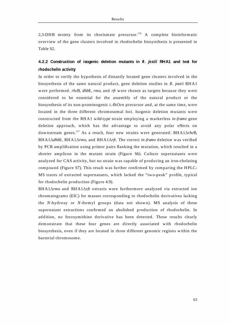
Results
63
2,3-DHB moiety from its chorismate precursor.176 A complete bioinformatic
overview of the gene clusters involved in rhodochelin biosynthesis is presented in
Table S2.
4.2.2 Construction of isogenic deletion mutants in R. jostii RHA1 and test for
rhodochelin activity
In order to verify the hypothesis of distantly located gene clusters involved in the
biosynthesis of the same natural product, gene deletion studies in R. jostii RHA1
were performed. rhcB, dhbE, rmo, and rft were chosen as targets because they were
considered to be essential for the assembly of the natural product or the
biosynthesis of its non-proteinogenic L-fhOrn precursor and, at the same time, were
located in the three different chromosomal loci. Isogenic deletion mutants were
constructed from the RHA1 wild-type strain employing a markerless in-frame gene
deletion approach, which has the advantage to avoid any polar effects on
downstream genes.177 As a result, four new strains were generated: RHA1ΔrhcB,
RHA1ΔdhbE, RHA1Δrmo, and RHA1Δrft. The correct in-frame deletion was verified
by PCR amplification using primer pairs flanking the mutation, which resulted in a
shorter amplicon in the mutant strain (Figure S6). Culture supernatants were
analyzed for CAS activity, but no strain was capable of producing an iron-chelating
compound (Figure S7). This result was further confirmed by comparing the HPLC-
MS traces of extracted supernatants, which lacked the “two-peak” profile, typical
for rhodochelin production (Figure 4.9).
RHA1Δrmo and RHA1Δrft extracts were furthermore analyzed via extracted ion
chromatograms (EIC) for masses corresponding to rhodochelin derivatives lacking
the N-hydroxy or N-formyl groups (data not shown). MS analysis of these
supernatant extractions confirmed an abolished production of rhodochelin. In
addition, no foroxymithine derivative has been detected. These results clearly
demonstrate that these four genes are directly associated with rhodochelin
biosynthesis, even if they are located in three different genomic regions within the
bacterial chromosome.
Results
64
Figure 4.9 (A) Comparative HPLC-MS profiles obtained from the extracts of the culture supernatants of R. jostii RHA1 wild-type strain and nonproducing rhodochelin mutant strains. The single deletion of one of any four genes (which are located in three different chromosomal loci) resulted in an abolished rhodochelin production, implying an unprecedented cross-talk mechanism. (B) Schematic overview of the genes (and their relative location) that have been deleted from the bacterial chromosome.
0 10 20 30 40 50
Abs
orba
nce
( =
215
nm
)
Retention time (min)
RHA1 wild-typerhcBdhbErmorft
rft rmo
1kbp
Genes involved in rhodochelin biosynthesis
Genes not involved in rhodochelin biosynthesis
rhcB
dhbE rhcB rhodochelin NRPS
dhbE 2,3-DHB adenylation domain rmo rhodochelin L-Orn monooxygenaserft rhodochelin L-hOrn formyltransferase
×× ×
×
A
B
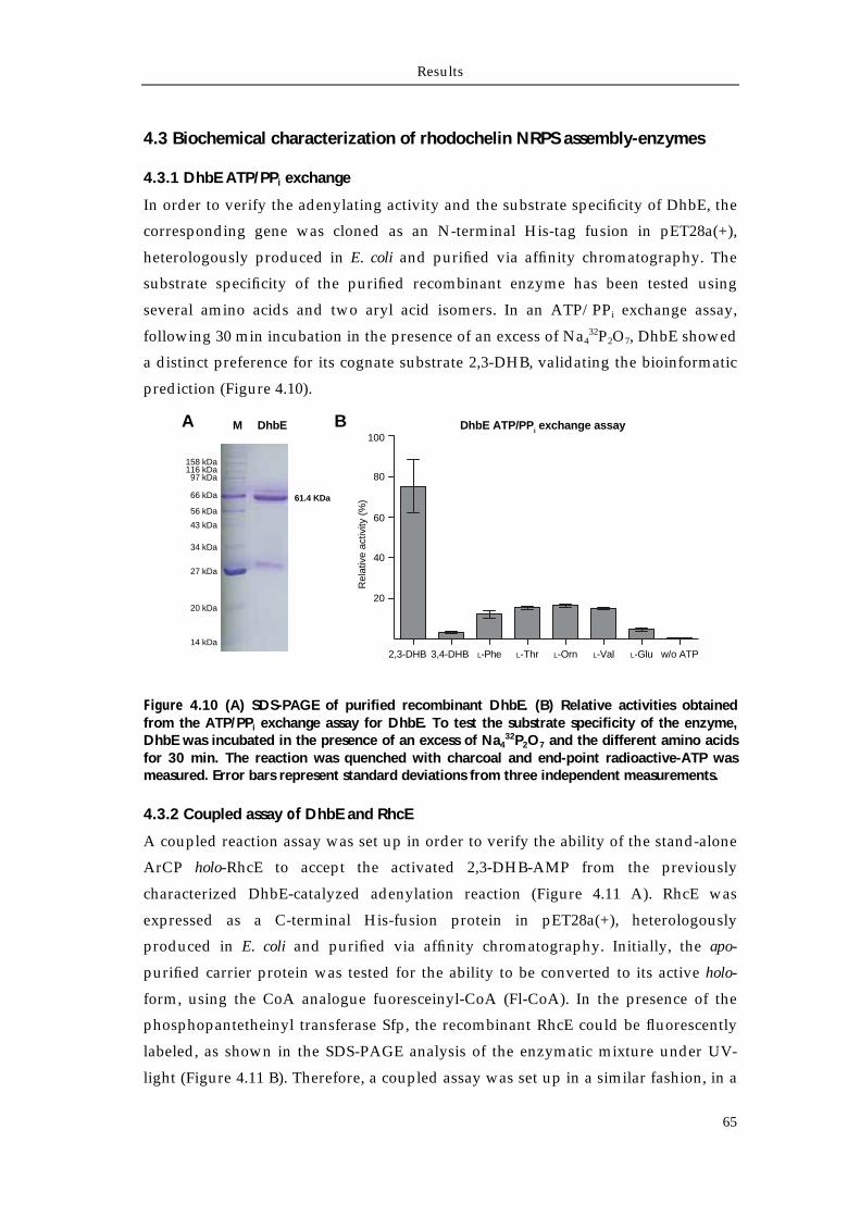
Results
65
4.3 Biochemical characterization of rhodochelin NRPS assembly-enzymes
4.3.1 DhbE ATP/PPi exchange
In order to verify the adenylating activity and the substrate specificity of DhbE, the
corresponding gene was cloned as an N-terminal His-tag fusion in pET28a(+),
heterologously produced in E. coli and purified via affinity chromatography. The
substrate specificity of the purified recombinant enzyme has been tested using
several amino acids and two aryl acid isomers. In an ATP/PPi exchange assay,
following 30 min incubation in the presence of an excess of Na432P2O7, DhbE showed
a distinct preference for its cognate substrate 2,3-DHB, validating the bioinformatic
prediction (Figure 4.10).
Figure 4.10 (A) SDS-PAGE of purified recombinant DhbE. (B) Relative activities obtained from the ATP/PPi exchange assay for DhbE. To test the substrate specificity of the enzyme, DhbE was incubated in the presence of an excess of Na4
32P2O7 and the different amino acids for 30 min. The reaction was quenched with charcoal and end-point radioactive-ATP was measured. Error bars represent standard deviations from three independent measurements.
4.3.2 Coupled assay of DhbE and RhcE
A coupled reaction assay was set up in order to verify the ability of the stand-alone
ArCP holo-RhcE to accept the activated 2,3-DHB-AMP from the previously
characterized DhbE-catalyzed adenylation reaction (Figure 4.11 A). RhcE was
expressed as a C-terminal His-fusion protein in pET28a(+), heterologously
produced in E. coli and purified via affinity chromatography. Initially, the apo-
purified carrier protein was tested for the ability to be converted to its active holo-
form, using the CoA analogue fuoresceinyl-CoA (Fl-CoA). In the presence of the
phosphopantetheinyl transferase Sfp, the recombinant RhcE could be fluorescently
labeled, as shown in the SDS-PAGE analysis of the enzymatic mixture under UV-
light (Figure 4.11 B). Therefore, a coupled assay was set up in a similar fashion, in a
DhbE ATP/PPi exchange assayDhbEM
66 kDa
27 kDa
56 kDa
43 kDa
34 kDa
97 kDa116 kDa158 kDa
61.4 KDa
20 kDa
14 kDa
A B
2,3-DHB 3,4-DHB L-Phe L-Thr L-Orn L-Val L-Glu w/o ATP
Rel
ativ
e ac
tivity
(%
)
20
40
60
80
100
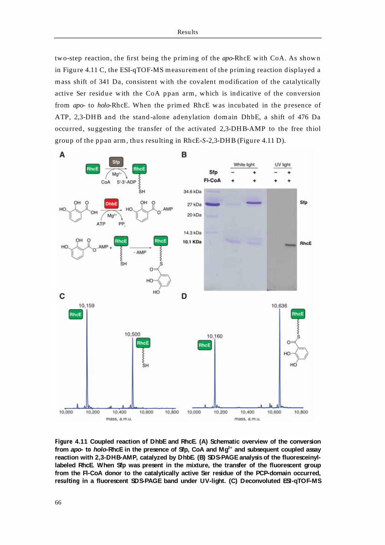
Results
66
two-step reaction, the first being the priming of the apo-RhcE with CoA. As shown
in Figure 4.11 C, the ESI-qTOF-MS measurement of the priming reaction displayed a
mass shift of 341 Da, consistent with the covalent modification of the catalytically
active Ser residue with the CoA ppan arm, which is indicative of the conversion
from apo- to holo-RhcE. When the primed RhcE was incubated in the presence of
ATP, 2,3-DHB and the stand-alone adenylation domain DhbE, a shift of 476 Da
occurred, suggesting the transfer of the activated 2,3-DHB-AMP to the free thiol
group of the ppan arm, thus resulting in RhcE-S-2,3-DHB (Figure 4.11 D).
Figure 4.11 Coupled reaction of DhbE and RhcE. (A) Schematic overview of the conversion from apo- to holo-RhcE in the presence of Sfp, CoA and Mg2+ and subsequent coupled assay reaction with 2,3-DHB-AMP, catalyzed by DhbE. (B) SDS-PAGE analysis of the fluoresceinyl-labeled RhcE. When Sfp was present in the mixture, the transfer of the fluorescent group from the Fl-CoA donor to the catalytically active Ser residue of the PCP-domain occurred, resulting in a fluorescent SDS-PAGE band under UV-light. (C) Deconvoluted ESI-qTOF-MS
Results
67
measurements of the Sfp-catalyzed RhcE priming reaction. The mass shift of 340 Da is indicative of the transfer of the ppan arm from the CoA donor to the catalytically active Ser residue of the PCP, resulting in the conversion from apo-enzyme to holo-enzyme. (D) When the primed RhcE was incubated in the presence of 2,3-DHB, ATP and DhbE, the coupled transfer of the DhbE-activated 2,3-DHB to the free thiol group of the ppan arm occurred, suggesting the biochemical cross-talk between the two proteins, that are encoded in two distantly-located genome regions.
Results
68
4.4 Biochemical characterization of L-Orn tailoring enzymes
4.4.1 Biochemical characterization of Rmo L-Orn Monooxygenase
4.4.1.1 Bioinformatic analysis of the NMO Rmo
In the previous section, it has been demonstrated that rmo encodes for a putative L-
Orn monooxygenase which is involved in the biosynthesis of rhodochelin, since its
deletion from the chromosome resulted in an abolished siderophore production.
A bioinformatic analysis showed that Rmo belongs to the class of NAD(P)H/FAD-
dependent monooxygenases and, when compared to already characterized
homologues (CchB, EtcB, PvdA, SidA, and IucD), it displays high similarity along
the overall sequence.88,94,178-181 As depicted in the multiple sequence alignment shown
in Figure 4.12, highly conserved motifs involved in substrate, NADPH, and FAD
binding could be easily identified. The FAD cofactor-binding consensus GxGxxN is
located at the N-terminus region of the protein in a conserved dinucleotide-binding
βαβ-motif. A similar α/β-nucleotide-binding architecture is responsible for the
binding of the NADPH cosubstrate, enclosed in a similar GxGQS motif. Lastly, the
residues involved in binding the L-Orn substrate are located in a small domain
located at the interface between the largest FAD and NADPH binding motifs (an
homology model is presented in Figure 5.5)15,182
4.4.1.2 Recombinant production and purification of active apo-Rmo
rmo was amplified from the R. jostii RHA1 chromosome and cloned into the
pET28a(+) expression vector. The recombinant protein was heterologously
produced in E. coli as an N-terminal His-tag fusion and purified via Ni-NTA affinity
chromatography. UV-vis spectroscopy analysis of the purified recombinant enzyme
indicates Rmo to be in the apo form, without bound FAD cofactor (Figure 4.13).
Analytical size exclusion chromatography estimated the molecular mass of Rmo in
solution to be equal to 220 kDa, suggesting the enzyme to adopt a tetrameric
quaternary structure, as previously reported for SidA and PvdA (Figure 4.14).178,179
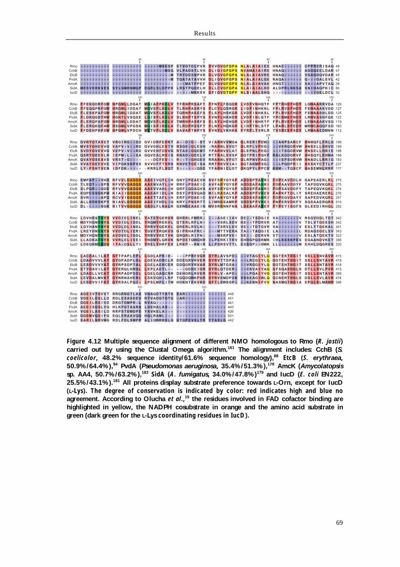
Results
69
Figure 4.12 Multiple sequence alignment of different NMO homologous to Rmo (R. jostii) carried out by using the Clustal Omega algorithm.161 The alignment includes: CchB (S. coelicolor, 48.2% sequence identity/61.6% sequence homology),88 EtcB (S. erythraea, 50.9%/64.4%),94 PvdA (Pseudomonas aeruginosa, 35.4%/51.3%),178 AmcK (Amycolatopsis sp. AA4, 50.7%/63.2%),183 SidA (A. fumigatus, 34.0%/47.8%)179 and IucD (E. coli EN222, 25.5%/43.1%).181 All proteins display substrate preference towards L-Orn, except for IucD (L-Lys). The degree of conservation is indicated by color: red indicates high and blue no agreement. According to Olucha et al.,15 the residues involved in FAD cofactor binding are highlighted in yellow, the NADPH cosubstrate in orange and the amino acid substrate in green (dark green for the L-Lys coordinating residues in IucD).
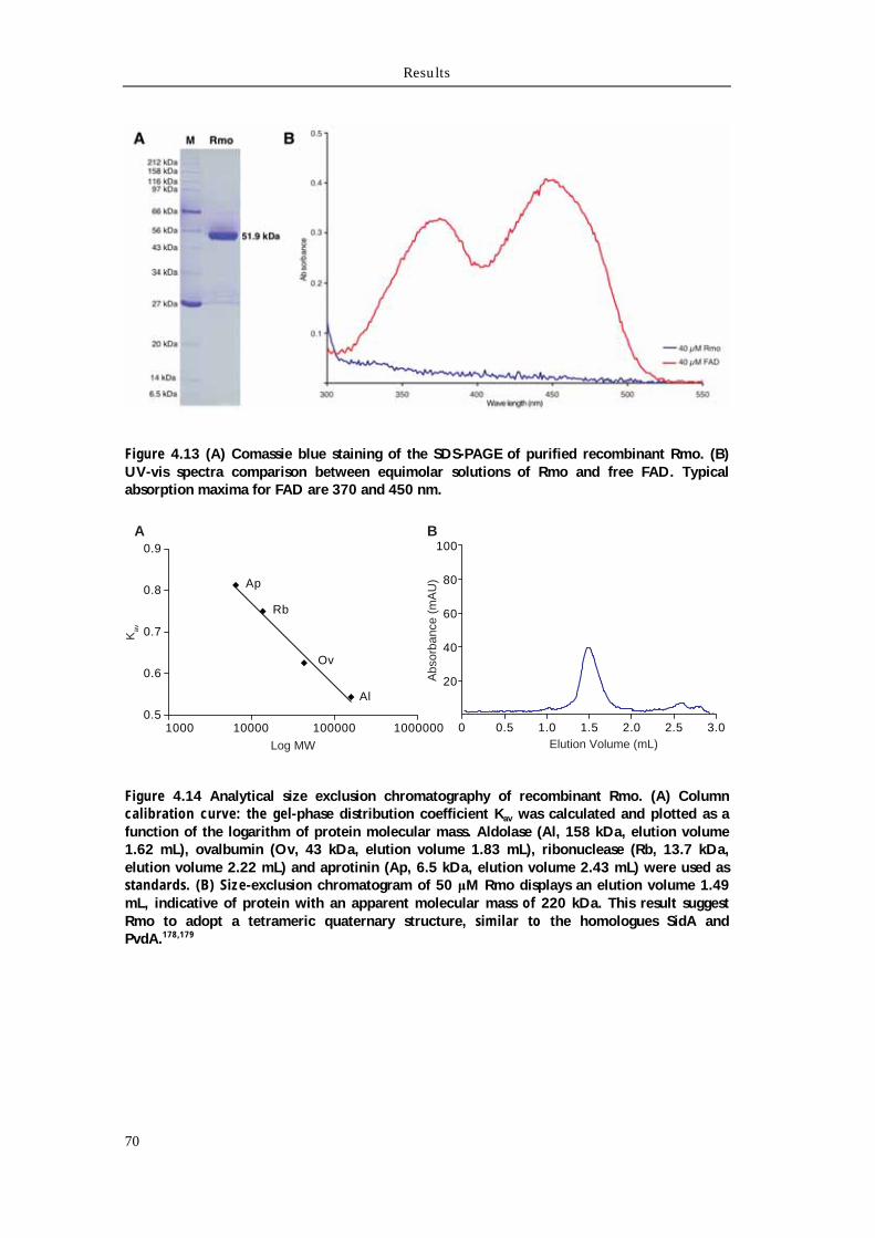
Results
70
Figure 4.13 (A) Comassie blue staining of the SDS-PAGE of purified recombinant Rmo. (B) UV-vis spectra comparison between equimolar solutions of Rmo and free FAD. Typical absorption maxima for FAD are 370 and 450 nm.
Figure 4.14 Analytical size exclusion chromatography of recombinant Rmo. (A) Column calibration curve: the gel-phase distribution coefficient Kav was calculated and plotted as a function of the logarithm of protein molecular mass. Aldolase (Al, 158 kDa, elution volume 1.62 mL), ovalbumin (Ov, 43 kDa, elution volume 1.83 mL), ribonuclease (Rb, 13.7 kDa, elution volume 2.22 mL) and aprotinin (Ap, 6.5 kDa, elution volume 2.43 mL) were used as standards. (B) Size-exclusion chromatogram of 50 μM Rmo displays an elution volume 1.49 mL, indicative of protein with an apparent molecular mass of 220 kDa. This result suggest Rmo to adopt a tetrameric quaternary structure, similar to the homologues SidA and PvdA.178,179
1000 10000 100000 10000000.5
0.6
0.7
0.8
0.9
Log MW
Kav
Al
Ov
Rb
Ap
0 0.5 1.0 1.5 2.0 2.5 3.0
20
40
60
80
100
Elution Volume (mL)
Abs
orba
nce
(mA
U)
A B
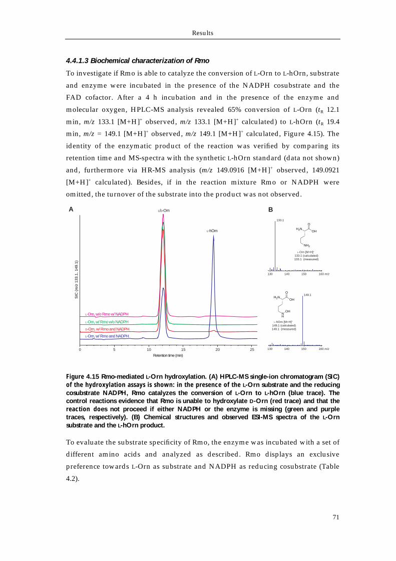
Results
71
4.4.1.3 Biochemical characterization of Rmo
To investigate if Rmo is able to catalyze the conversion of L-Orn to L-hOrn, substrate
and enzyme were incubated in the presence of the NADPH cosubstrate and the
FAD cofactor. After a 4 h incubation and in the presence of the enzyme and
molecular oxygen, HPLC-MS analysis revealed 65% conversion of L-Orn (tR 12.1
min, m/z 133.1 [M+H]+ observed, m/z 133.1 [M+H]+ calculated) to L-hOrn (tR 19.4
min, m/z = 149.1 [M+H]+ observed, m/z 149.1 [M+H]+ calculated, Figure 4.15). The
identity of the enzymatic product of the reaction was verified by comparing its
retention time and MS-spectra with the synthetic L-hOrn standard (data not shown)
and, furthermore via HR-MS analysis (m/z 149.0916 [M+H]+ observed, 149.0921
[M+H]+ calculated). Besides, if in the reaction mixture Rmo or NADPH were
omitted, the turnover of the substrate into the product was not observed.
Figure 4.15 Rmo-mediated L-Orn hydroxylation. (A) HPLC-MS single-ion chromatogram (SIC) of the hydroxylation assays is shown: in the presence of the L-Orn substrate and the reducing cosubstrate NADPH, Rmo catalyzes the conversion of L-Orn to L-hOrn (blue trace). The control reactions evidence that Rmo is unable to hydroxylate D-Orn (red trace) and that the reaction does not proceed if either NADPH or the enzyme is missing (green and purple traces, respectively). (B) Chemical structures and observed ESI-MS spectra of the L-Orn substrate and the L-hOrn product.
To evaluate the substrate specificity of Rmo, the enzyme was incubated with a set of
different amino acids and analyzed as described. Rmo displays an exclusive
preference towards L-Orn as substrate and NADPH as reducing cosubstrate (Table
4.2).
Retention time (min)
SIC
(m
/z 1
33.1
, 149
.1)
L-hOrn H2NOH
O
NH2
H2NOH
O
NH
OH
L-Orn [M+H]+ 133.1 (calculated)133.1 (measured)
L-hOrn [M+H]+ 149.1 (calculated)149.1 (measured)
A BL/D-Orn
0 5 10 15 20
D-Orn, w/ Rmo and NADPH
L-Orn, w/ Rmo and NADPH
L-Orn, w/ Rmo w/o NADPH
L-Orn, w/o Rmo w/ NADPH
25
133.1
130 140 150 160 m/z
149.1
130 140 150 160 m/z
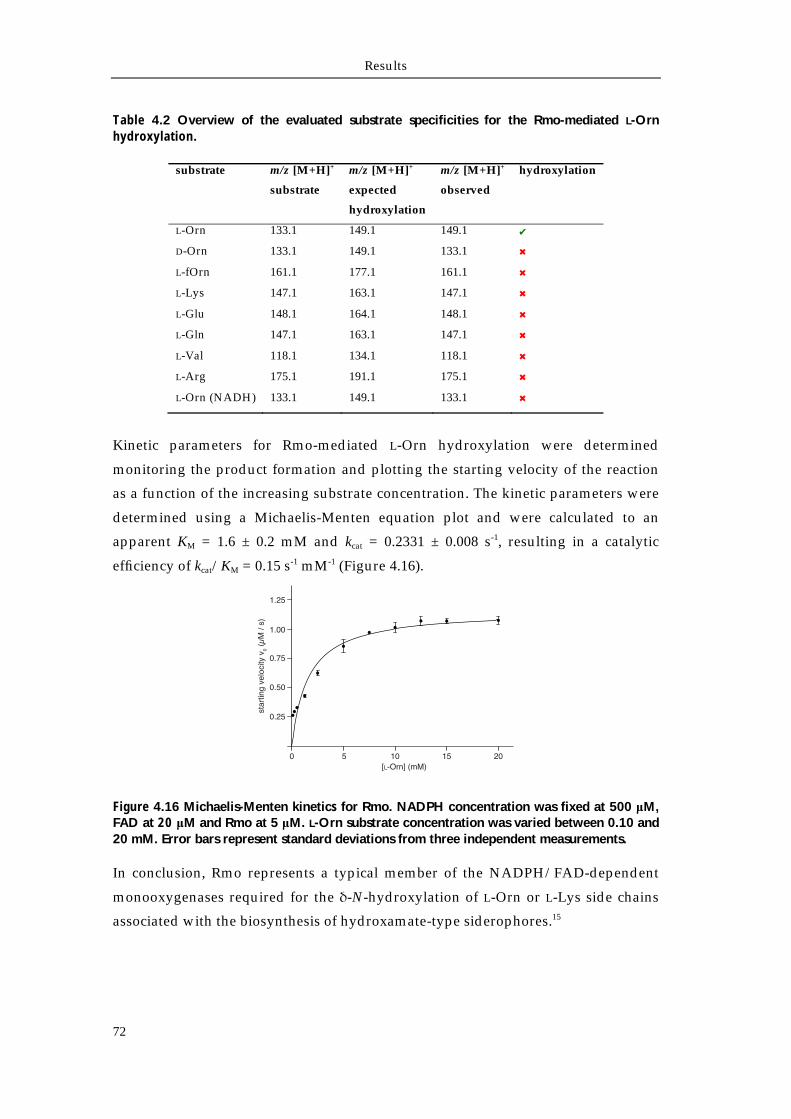
Results
72
Table 4.2 Overview of the evaluated substrate specificities for the Rmo-mediated L-Orn hydroxylation.
substrate m/z [M+H]+
substrate
m/z [M+H]+
expected
hydroxylation
m/z [M+H]+
observed
hydroxylation
L-Orn 133.1 149.1 149.1 ✔
D-Orn 133.1 149.1 133.1 ✖
L-fOrn 161.1 177.1 161.1 ✖
L-Lys 147.1 163.1 147.1 ✖
L-Glu 148.1 164.1 148.1 ✖
L-Gln 147.1 163.1 147.1 ✖
L-Val 118.1 134.1 118.1 ✖
L-Arg 175.1 191.1 175.1 ✖
L-Orn (NADH) 133.1 149.1 133.1 ✖
Kinetic parameters for Rmo-mediated L-Orn hydroxylation were determined
monitoring the product formation and plotting the starting velocity of the reaction
as a function of the increasing substrate concentration. The kinetic parameters were
determined using a Michaelis-Menten equation plot and were calculated to an
apparent KM = 1.6 ± 0.2 mM and kcat = 0.2331 ± 0.008 s-1, resulting in a catalytic
efficiency of kcat/KM = 0.15 s-1 mM-1 (Figure 4.16).
Figure 4.16 Michaelis-Menten kinetics for Rmo. NADPH concentration was fixed at 500 μM, FAD at 20 μM and Rmo at 5 μM. L-Orn substrate concentration was varied between 0.10 and 20 mM. Error bars represent standard deviations from three independent measurements.
In conclusion, Rmo represents a typical member of the NADPH/FAD-dependent
monooxygenases required for the δ-N-hydroxylation of L-Orn or L-Lys side chains
associated with the biosynthesis of hydroxamate-type siderophores.15
star
ting
velo
city
v0
(μM
/ s)
0 5 10 15 20[L-Orn] (mM)
0.25
0.50
0.75
1.00
1.25
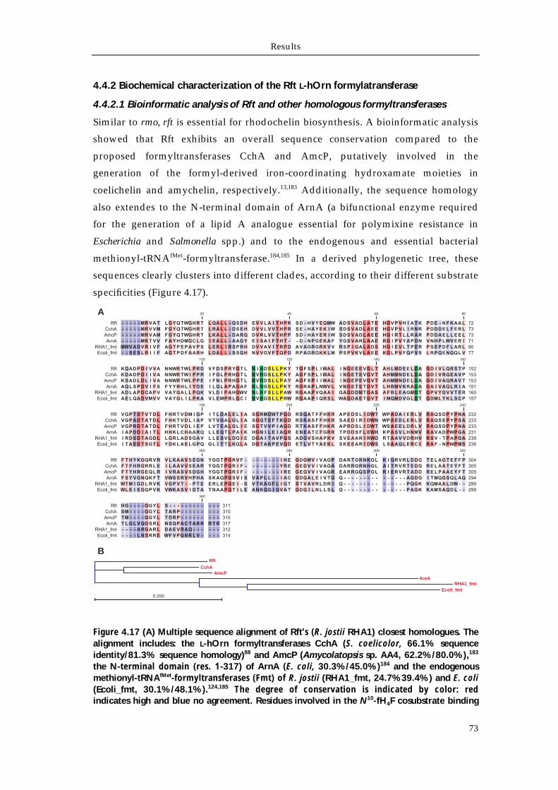
Results
73
4.4.2 Biochemical characterization of the Rft L-hOrn formylatransferase
4.4.2.1 Bioinformatic analysis of Rft and other homologous formyltransferases
Similar to rmo, rft is essential for rhodochelin biosynthesis. A bioinformatic analysis
showed that Rft exhibits an overall sequence conservation compared to the
proposed formyltransferases CchA and AmcP, putatively involved in the
generation of the formyl-derived iron-coordinating hydroxamate moieties in
coelichelin and amychelin, respectively.13,183 Additionally, the sequence homology
also extendes to the N-terminal domain of ArnA (a bifunctional enzyme required
for the generation of a lipid A analogue essential for polymixine resistance in
Escherichia and Salmonella spp.) and to the endogenous and essential bacterial
methionyl-tRNAfMet-formyltransferase.184,185 In a derived phylogenetic tree, these
sequences clearly clusters into different clades, according to their different substrate
specificities (Figure 4.17).
Figure 4.17 (A) Multiple sequence alignment of Rft’s (R. jostii RHA1) closest homologues. The alignment includes: the L-hOrn formyltransferases CchA (S. coelicolor, 66.1% sequence identity/81.3% sequence homology)88 and AmcP (Amycolatopsis sp. AA4, 62.2%/80.0%),183 the N-terminal domain (res. 1-317) of ArnA (E. coli, 30.3%/45.0%)184 and the endogenous methionyl-tRNAfMet-formyltransferases (Fmt) of R. jostii (RHA1_fmt, 24.7%39.4%) and E. coli (Ecoli_fmt, 30.1%/48.1%).124,185 The degree of conservation is indicated by color: red indicates high and blue no agreement. Residues involved in the N10-fH4F cosubstrate binding
RftCchA
AmcPArnA
RHA1_fmtEcoli_fmt
0.200
A
B
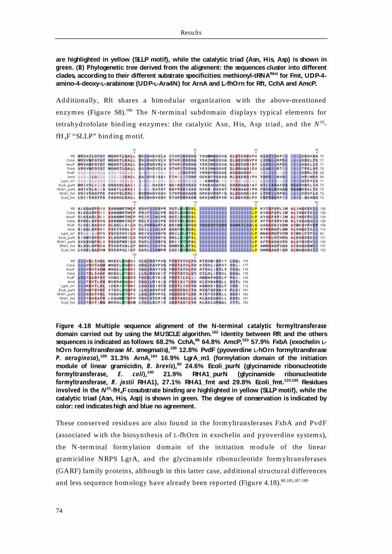
Results
74
are highlighted in yellow (SLLP motif), while the catalytic triad (Asn, His, Asp) is shown in green. (B) Phylogenetic tree derived from the alignment: the sequences cluster into different clades, according to their different substrate specificities: methionyl-tRNAfMet for Fmt, UDP-4-amino-4-deoxy-L-arabinose (UDP-L-Ara4N) for ArnA and L-fhOrn for Rft, CchA and AmcP.
Additionally, Rft shares a bimodular organization with the above-mentioned
enzymes (Figure S8).186 The N-terminal subdomain displays typical elements for
tetrahydrofolate binding enzymes: the catalytic Asn, His, Asp triad, and the N10-
fH4F “SLLP” binding motif.
Figure 4.18 Multiple sequence alignment of the N-terminal catalytic formyltransferase domain carried out by using the MUSCLE algorithm.162 Identity between Rft and the others sequences is indicated as follows: 68.2% CchA,88 64.8% AmcP,183 57.9% FxbA (exochelin L-hOrn formyltransferase M. smegmatis),188 12.8% PvdF (pyoverdine L-hOrn formyltransferase P. aeruginosa),189 31.3% ArnA,184 16.9% LgrA_m1 (formylation domain of the initiation module of linear gramicidin, B. brevis),80 24.6% Ecoli_purN (glycinamide ribonucleotide formyltransferase, E. coli),190 21.9% RHA1_purN (glycinamide ribonucleotide formyltransferase, R. jostii RHA1), 27.1% RHA1_fmt and 29.8% Ecoli_fmt.124,185 Residues involved in the N10-fH4F cosubstrate binding are highlighted in yellow (SLLP motif), while the catalytic triad (Asn, His, Asp) is shown in green. The degree of conservation is indicated by color: red indicates high and blue no agreement.
These conserved residues are also found in the formyltransferases FxbA and PvdF
(associated with the biosynthesis of L-fhOrn in exochelin and pyoverdine systems),
the N-terminal formylation domain of the initiation module of the linear
gramicidine NRPS LgrA, and the glycinamide ribonucleotide formyltransferases
(GARF) family proteins, although in this latter case, additional structural differences
and less sequence homology have already been reported (Figure 4.18).80,185,187-189
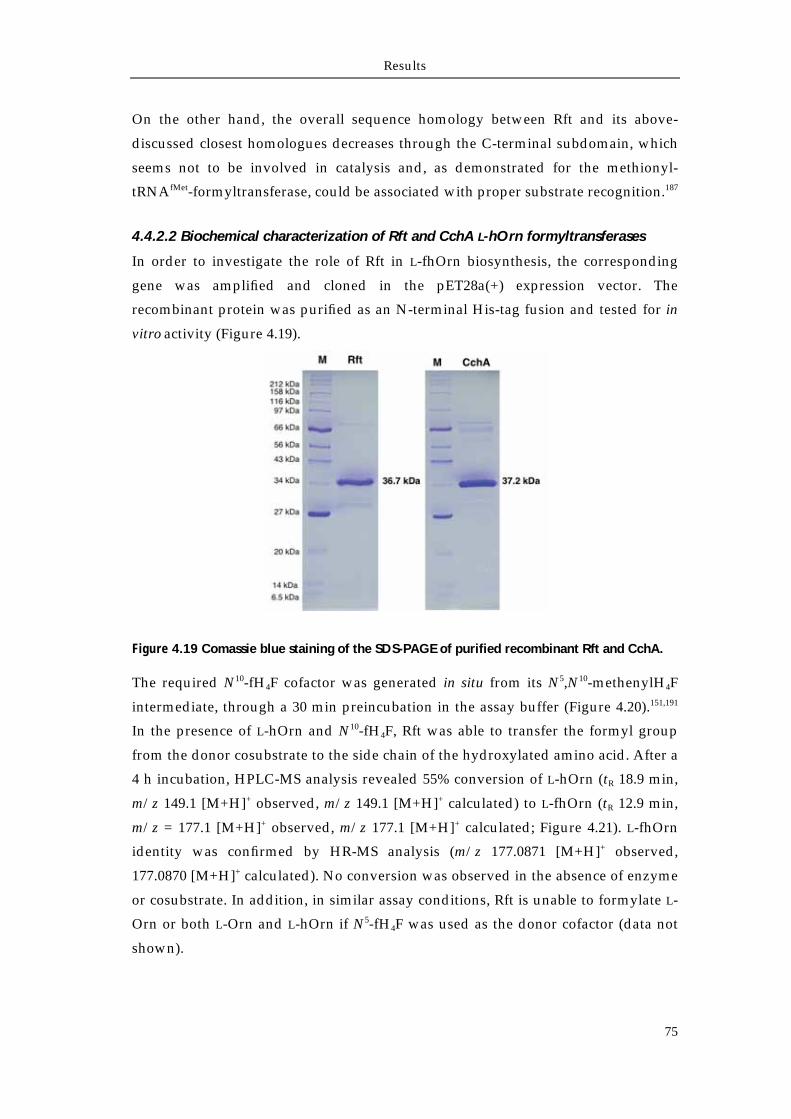
Results
75
On the other hand, the overall sequence homology between Rft and its above-
discussed closest homologues decreases through the C-terminal subdomain, which
seems not to be involved in catalysis and, as demonstrated for the methionyl-
tRNAfMet-formyltransferase, could be associated with proper substrate recognition.187
4.4.2.2 Biochemical characterization of Rft and CchA L-hOrn formyltransferases
In order to investigate the role of Rft in L-fhOrn biosynthesis, the corresponding
gene was amplified and cloned in the pET28a(+) expression vector. The
recombinant protein was purified as an N-terminal His-tag fusion and tested for in
vitro activity (Figure 4.19).
Figure 4.19 Comassie blue staining of the SDS-PAGE of purified recombinant Rft and CchA.
The required N10-fH4F cofactor was generated in situ from its N5,N10-methenylH4F
intermediate, through a 30 min preincubation in the assay buffer (Figure 4.20).151,191
In the presence of L-hOrn and N10-fH4F, Rft was able to transfer the formyl group
from the donor cosubstrate to the side chain of the hydroxylated amino acid. After a
4 h incubation, HPLC-MS analysis revealed 55% conversion of L-hOrn (tR 18.9 min,
m/z 149.1 [M+H]+ observed, m/z 149.1 [M+H]+ calculated) to L-fhOrn (tR 12.9 min,
m/z = 177.1 [M+H]+ observed, m/z 177.1 [M+H]+ calculated; Figure 4.21). L-fhOrn
identity was confirmed by HR-MS analysis (m/z 177.0871 [M+H]+ observed,
177.0870 [M+H]+ calculated). No conversion was observed in the absence of enzyme
or cosubstrate. In addition, in similar assay conditions, Rft is unable to formylate L-
Orn or both L-Orn and L-hOrn if N5-fH4F was used as the donor cofactor (data not
shown).
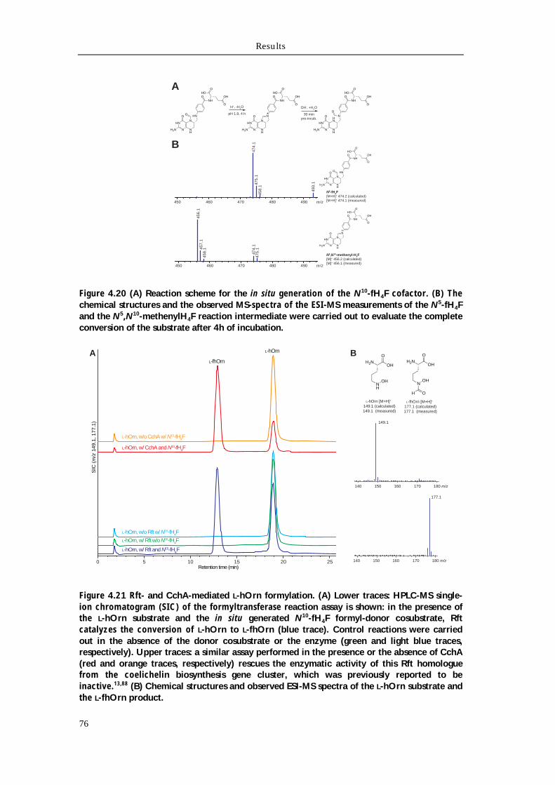
Results
76
Figure 4.20 (A) Reaction scheme for the in situ generation of the N10-fH4F cofactor. (B) The chemical structures and the observed MS-spectra of the ESI-MS measurements of the N5-fH4F and the N5,N10-methenylH4F reaction intermediate were carried out to evaluate the complete conversion of the substrate after 4h of incubation.
Figure 4.21 Rft- and CchA-mediated L-hOrn formylation. (A) Lower traces: HPLC-MS single-ion chromatogram (SIC) of the formyltransferase reaction assay is shown: in the presence of the L-hOrn substrate and the in situ generated N10-fH4F formyl-donor cosubstrate, Rft catalyzes the conversion of L-hOrn to L-fhOrn (blue trace). Control reactions were carried out in the absence of the donor cosubstrate or the enzyme (green and light blue traces, respectively). Upper traces: a similar assay performed in the presence or the absence of CchA (red and orange traces, respectively) rescues the enzymatic activity of this Rft homologue from the coelichelin biosynthesis gene cluster, which was previously reported to be inactive.13,88 (B) Chemical structures and observed ESI-MS spectra of the L-hOrn substrate and the L-fhOrn product.
m/z450 460 470 480 490
474
.1 4
75.1
493
.1
458
.1
m/z450 460 470 480 490
456
.1 4
57.1
474
.1 4
75.1
458
.1
N5-fH4F[M+H]+ 474.2 (calculated)[M+H]+ 474.1 (measured)
N5,N10-methenyl-H4F[M]+ 456.2 (calculated)[M]+ 456.1 (measured)
HN
N NH
N
H2N
O HN
NHO
OHO
O
OH
O
HN
N NH
N
H2N
O N
NHO
OHO
O
OH
HN
N NH
HN
H2N
O N
NHO
OHO
O
OH
OH+, -H2O
pH 1.9, 4 h
OH-, +H2O
30 minpre-incub.
A
B
HN
N NH
N
H2N
O HN
NHO
OHO
O
OH
O
HN
N NH
N
H2N
O N
NHO
OHO
O
OH
A
L-hOrn, w/ Rft and N10-fH4F
L-hOrn, w/ Rft w/o N10-fH4F
L-hOrn, w/o Rft w/ N10-fH4F
0 5 10 15 20 25Retention time (min)
L-hOrn, w/ CchA and N10-fH4F
L-hOrn, w/o CchA w/ N10-fH4F
B
SIC
(m
/z 1
49.1
, 177
.1)
L-hOrn
L-fhOrn H2NOH
O
NH
OH
L-hOrn [M+H]+ 149.1 (calculated)149.1 (measured)
H2NOH
O
NOH
OH
L-fhOrn [M+H]+ 177.1 (calculated)177.1 (measured)
149.1
177.1
160 180 m/z140 150 170
160 180 m/z140 150 170

Results
77
Previous attempts to characterize Rft using the N10-fH4F cosubstrate generated via a
chemoenzymatic synthesis approach (chemical conversion of H4F to N5,N10-
methyleneH4F in the presence of formaldehyde, followed by a FolD-catalyzed
regiospecific oxidation/cyclohydrolyzation) always resulted in no detectable
enzymatic activity (data not shown).192 Similar results were obtained during the
characterization of CchA, which left unclear whether the enzyme was purified in a
soluble but inactive form or was rendered inactive by the incubation in the reaction
assay.88 Therefore, on the basis of the obtained results for Rft, CchA was expressed,
purified (Figure 4.19), and assayed for enzymatic activity. As shown in Figure 4.21,
CchA was able to catalyze the conversion of L-hOrn to L-fhOrn (75% after 4 h
incubation).
Figure 4.22 (A) Sigmoidal plot of Rft kinetic characterization. N5,N10-methenylH4F concentration was fixed at 1.5 mM, while L-hOrn substrate concentration was varied between 0.25 and 15 mM. Error bars represent standard deviations from three independent measurements. (B) In order to elucidate the origin of this unusual cooperative behavior, analytical size exclusion chromatography of 50 μM Rft solution was carried out. Rft displays an elution volume 1.60 mL, indicative of protein with an apparent molecular mass of 146 kDa, suggesting the enzyme to adopt a tetrameric quaternary structure, likewise Rmo. The calibration curve for the analytical gel filtration is shared with the analytical gel filtration of the Rmo monooxygenase, which is illustrated in Figure 4.14.
The kinetic parameters of Rft tranformylation were determined monitoring the
conversion of substrate to product and plotting the starting velocities as a function
of the increasing substrate concentration. Surprisingly, the experimental data could
be fitted best using a sigmoidal curve, indicative of an allosteric kinetic mechanism,
rather than the typical rectangular hyperbola characteristic of classical Michaelis-
Menten kinetics (Figure 4.22 A).193 Normalized parameter values were calculated
through the nonlinear Hill-fit regression and were found to be equal to a Vmax =
0.078 ± 0.001 μmol L–1 s–1, a K0.5 = 1.2 ± 0.1 mM, and n = 2.7 ± 0.2. A value of the Hill
parameter n greater than 1 is indicative of a positive cooperative mechanism. To
0 0.5 1.0 1.5 2.0 2.5 3.0
20
40
60
80
100
Elution Volume (mL)
Abs
orba
nce
(mA
U)
A B0.08
0.06
0.04
0.02
star
ting
velo
city
v0
(μM
/ s)
0 5 10[L-hOrn] (mM)
15
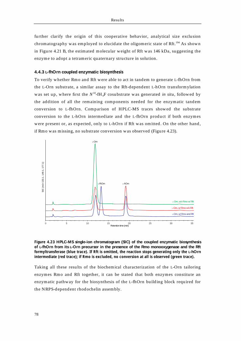
Results
78
further clarify the origin of this cooperative behavior, analytical size exclusion
chromatography was employed to elucidate the oligomeric state of Rft.194 As shown
in Figure 4.21 B, the estimated molecular weight of Rft was 146 kDa, suggesting the
enzyme to adopt a tetrameric quaternary structure in solution.
4.4.3 L-fhOrn coupled enzymatic biosynthesis
To verify whether Rmo and Rft were able to act in tandem to generate L-fhOrn from
the L-Orn substrate, a similar assay to the Rft-dependent L-hOrn transformylation
was set up, where first the N10-fH4F cosubstrate was generated in situ, followed by
the addition of all the remaining components needed for the enzymatic tandem
conversion to L-fhOrn. Comparison of HPLC-MS traces showed the substrate
conversion to the L-hOrn intermediate and the L-fhOrn product if both enzymes
were present or, as expected, only to L-hOrn if Rft was omitted. On the other hand,
if Rmo was missing, no substrate conversion was observed (Figure 4.23).
Figure 4.23 HPLC-MS single-ion chromatogram (SIC) of the coupled enzymatic biosynthesis of L-fhOrn from its L-Orn precursor in the presence of the Rmo monooxygenase and the Rft formyltransferase (blue trace). If Rft is omitted, the reaction stops generating only the L-hOrn intermediate (red trace); if Rmo is excluded, no conversion at all is observed (green trace).
Taking all these results of the biochemical characterization of the L-Orn tailoring
enzymes Rmo and Rft together, it can be stated that both enzymes constitute an
enzymatic pathway for the biosynthesis of the L-fhOrn building block required for
the NRPS-dependent rhodochelin assembly.
L-Orn, w/ Rmo w/o Rft
L-Orn, w/ Rmo and Rft
L-Orn, w/o Rmo w/ Rft
0 5 10 15 20 25
SIC
(m
/z 1
33.1
, 149
.1, 1
77.1
)
Retention time (min)
L-Orn
L-fhOrn L-hOrn
30 35
Chapter 5
Discussion
Discussion
80
5.1 Isolation and structural characterization of the siderophore rhodochelin
5.1.1 Rhodococcus spp. as a new source for secondary metabolites
In recent years, the increasing amount of sequenced microbial genomes has revealed
the presence of an impressive number of secondary metabolite gene clusters, most
of them considered “orphan” with respect to their natural product.44 Furthermore,
since the discovery of the capability of single strains to produce many natural
products, the concept “One Strain - MAny Compounds” (OSMAC) has been
introduced; thus, the interest to uncover new secondary metabolites in known
species is increasing.108 Despite the use of genome mining and its successful
application, the identification of new natural products still remains challenging. In
fact, without any experimental proof, it is difficult to define if an orphan cluster is
silent because it is not functional, the metabolite cannot be detected due to analytical
detection limits, or the laboratory cultivation methods are inappropriate for its
biosynthesis.43
The continuous interest in the Rhodococcus genus as a bioremediation and
bioconversion tool has shifted the focus to regard these species as natural product
producing strains.123 As streptomycetes, rhodococci belong to the actinomycetal
order and thus are predisposed to a putative and underestimated secondary
metabolism. In addition, they also offer experimental advantages that could ease the
comprehension of the physiology of closely related species like Mycobacterium and
Streptomyces, that are difficult to cultivate. Therefore, the complete genome sequence
of R. jostii RHA1 provides an excellent opportunity that facilitate both the
exploitation of the catabolic versatility of these industrially-important
microorganisms and natural product discovery.124 So far, the only secondary
metabolites isolated from Rhodococcus spp. were the siderophores heterobactin A,
rhodobactin and the ribosomally-derived lasso peptide lariatin.125,126,195 In addition,
with the exception of lariatin, the two siderophores still remain orphan with respect
to their biosynthetic gene clusters.196
Genome analysis of the R. jostii RHA1 chromosome identified 23 secondary
metabolite gene clusters, suggesting the possibility of an extensive secondary
metabolism (Table 5.1).124,157 Most of them encode for peptides of non-ribosomal
origin, though PKS and hybrid PKS-NRPS systems are present as well. Moreover,
six of the NRPS genes extent more than 25 kbp in length.168
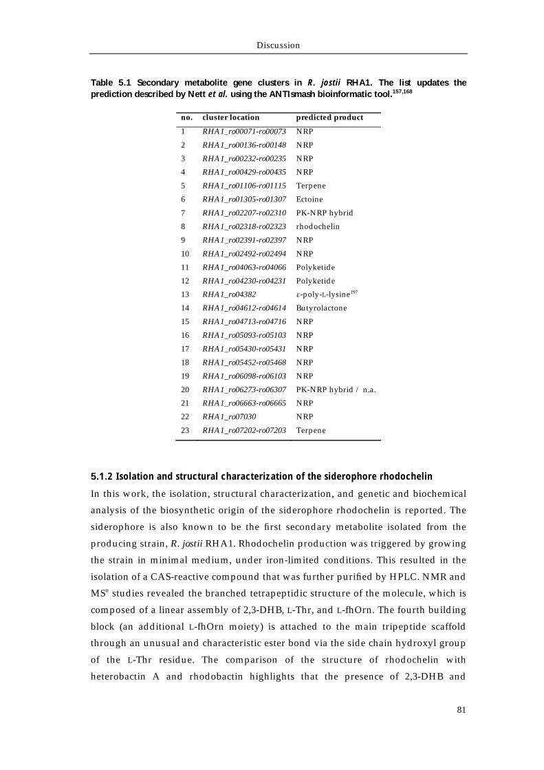
Discussion
81
Table 5.1 Secondary metabolite gene clusters in R. jostii RHA1. The list updates the prediction described by Nett et al. using the ANTIsmash bioinformatic tool.157,168
no. cluster location predicted product
1 RHA1_ro00071-ro00073 NRP
2 RHA1_ro00136-ro00148 NRP
3 RHA1_ro00232-ro00235 NRP
4 RHA1_ro00429-ro00435 NRP
5 RHA1_ro01106-ro01115 Terpene
6 RHA1_ro01305-ro01307 Ectoine
7 RHA1_ro02207-ro02310 PK-NRP hybrid
8 RHA1_ro02318-ro02323 rhodochelin
9 RHA1_ro02391-ro02397 NRP
10 RHA1_ro02492-ro02494 NRP
11 RHA1_ro04063-ro04066 Polyketide
12 RHA1_ro04230-ro04231 Polyketide
13 RHA1_ro04382 ε-poly-L-lysine197
14 RHA1_ro04612-ro04614 Butyrolactone
15 RHA1_ro04713-ro04716 NRP
16 RHA1_ro05093-ro05103 NRP
17 RHA1_ro05430-ro05431 NRP
18 RHA1_ro05452-ro05468 NRP
19 RHA1_ro06098-ro06103 NRP
20 RHA1_ro06273-ro06307 PK-NRP hybrid / n.a.
21 RHA1_ro06663-ro06665 NRP
22 RHA1_ro07030 NRP
23 RHA1_ro07202-ro07203 Terpene
5.1.2 Isolation and structural characterization of the siderophore rhodochelin
In this work, the isolation, structural characterization, and genetic and biochemical
analysis of the biosynthetic origin of the siderophore rhodochelin is reported. The
siderophore is also known to be the first secondary metabolite isolated from the
producing strain, R. jostii RHA1. Rhodochelin production was triggered by growing
the strain in minimal medium, under iron-limited conditions. This resulted in the
isolation of a CAS-reactive compound that was further purified by HPLC. NMR and
MSn studies revealed the branched tetrapeptidic structure of the molecule, which is
composed of a linear assembly of 2,3-DHB, L-Thr, and L-fhOrn. The fourth building
block (an additional L-fhOrn moiety) is attached to the main tripeptide scaffold
through an unusual and characteristic ester bond via the side chain hydroxyl group
of the L-Thr residue. The comparison of the structure of rhodochelin with
heterobactin A and rhodobactin highlights that the presence of 2,3-DHB and
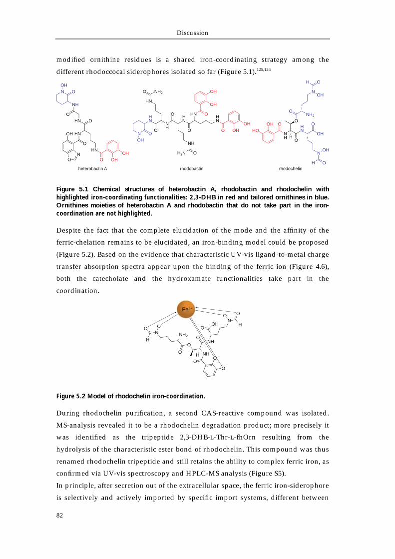
Discussion
82
modified ornithine residues is a shared iron-coordinating strategy among the
different rhodoccocal siderophores isolated so far (Figure 5.1).125,126
Figure 5.1 Chemical structures of heterobactin A, rhodobactin and rhodochelin with highlighted iron-coordinating functionalities: 2,3-DHB in red and tailored ornithines in blue. Ornithines moieties of heterobactin A and rhodobactin that do not take part in the iron-coordination are not highlighted.
Despite the fact that the complete elucidation of the mode and the affinity of the
ferric-chelation remains to be elucidated, an iron-binding model could be proposed
(Figure 5.2). Based on the evidence that characteristic UV-vis ligand-to-metal charge
transfer absorption spectra appear upon the binding of the ferric ion (Figure 4.6),
both the catecholate and the hydroxamate functionalities take part in the
coordination.
Figure 5.2 Model of rhodochelin iron-coordination.
During rhodochelin purification, a second CAS-reactive compound was isolated.
MS-analysis revealed it to be a rhodochelin degradation product; more precisely it
was identified as the tripeptide 2,3-DHB-L-Thr-L-fhOrn resulting from the
hydrolysis of the characteristic ester bond of rhodochelin. This compound was thus
renamed rhodochelin tripeptide and still retains the ability to complex ferric iron, as
confirmed via UV-vis spectroscopy and HPLC-MS analysis (Figure S5).
In principle, after secretion out of the extracellular space, the ferric iron-siderophore
is selectively and actively imported by specific import systems, different between
O
NH
HN
H
OOH
HO
OOH
O
NOH
OH
ONH2
NOH
H O
rhodochelin
N
OH
O
HN
O
NH
HN
HN
O NH2
O
NH
OH2N
O
HN O
OH
OH
HN
O OH
OH
rhodobactin
N O
OH
NH
OHN O
HN
HN
O OHOH
O
OH
ON
heterobactin A
O
OO
NHH
O
O
NH2NOO
H NHO
OHO
NO O
H
Fe3+
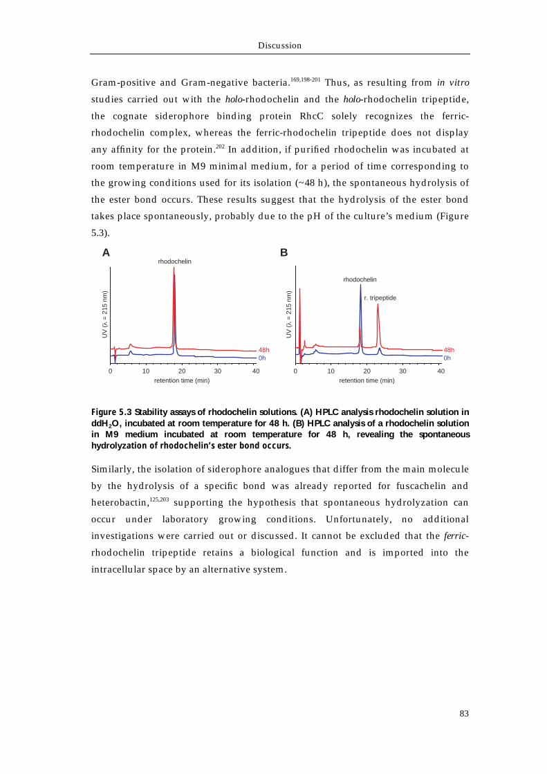
Discussion
83
Gram-positive and Gram-negative bacteria.169,198-201 Thus, as resulting from in vitro
studies carried out with the holo-rhodochelin and the holo-rhodochelin tripeptide,
the cognate siderophore binding protein RhcC solely recognizes the ferric-
rhodochelin complex, whereas the ferric-rhodochelin tripeptide does not display
any affinity for the protein.202 In addition, if purified rhodochelin was incubated at
room temperature in M9 minimal medium, for a period of time corresponding to
the growing conditions used for its isolation (~48 h), the spontaneous hydrolysis of
the ester bond occurs. These results suggest that the hydrolysis of the ester bond
takes place spontaneously, probably due to the pH of the culture’s medium (Figure
5.3).
Figure 5.3 Stability assays of rhodochelin solutions. (A) HPLC analysis rhodochelin solution in ddH2O, incubated at room temperature for 48 h. (B) HPLC analysis of a rhodochelin solution in M9 medium incubated at room temperature for 48 h, revealing the spontaneous hydrolyzation of rhodochelin’s ester bond occurs.
Similarly, the isolation of siderophore analogues that differ from the main molecule
by the hydrolysis of a specific bond was already reported for fuscachelin and
heterobactin,125,203 supporting the hypothesis that spontaneous hydrolyzation can
occur under laboratory growing conditions. Unfortunately, no additional
investigations were carried out or discussed. It cannot be excluded that the ferric-
rhodochelin tripeptide retains a biological function and is imported into the
intracellular space by an alternative system.
0 10 20 30 40
retention time (min)
UV
( =
215
nm
)
0h48h
rhodochelin
0 10 20 30 40
retention time (min)
0h48h
rhodochelin
r. tripeptide
A B
UV
( =
215
nm
)
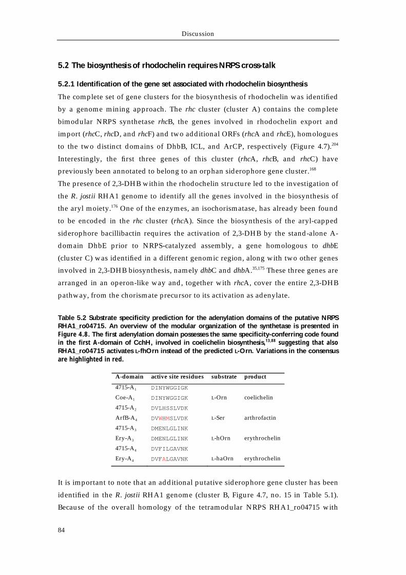
Discussion
84
5.2 The biosynthesis of rhodochelin requires NRPS cross-talk
5.2.1 Identification of the gene set associated with rhodochelin biosynthesis
The complete set of gene clusters for the biosynthesis of rhodochelin was identified
by a genome mining approach. The rhc cluster (cluster A) contains the complete
bimodular NRPS synthetase rhcB, the genes involved in rhodochelin export and
import (rhcC, rhcD, and rhcF) and two additional ORFs (rhcA and rhcE), homologues
to the two distinct domains of DhbB, ICL, and ArCP, respectively (Figure 4.7).204
Interestingly, the first three genes of this cluster (rhcA, rhcB, and rhcC) have
previously been annotated to belong to an orphan siderophore gene cluster.168
The presence of 2,3-DHB within the rhodochelin structure led to the investigation of
the R. jostii RHA1 genome to identify all the genes involved in the biosynthesis of
the aryl moiety.176 One of the enzymes, an isochorismatase, has already been found
to be encoded in the rhc cluster (rhcA). Since the biosynthesis of the aryl-capped
siderophore bacillibactin requires the activation of 2,3-DHB by the stand-alone A-
domain DhbE prior to NRPS-catalyzed assembly, a gene homologous to dhbE
(cluster C) was identified in a different genomic region, along with two other genes
involved in 2,3-DHB biosynthesis, namely dhbC and dhbA.35,175 These three genes are
arranged in an operon-like way and, together with rhcA, cover the entire 2,3-DHB
pathway, from the chorismate precursor to its activation as adenylate.
Table 5.2 Substrate specificity prediction for the adenylation domains of the putative NRPS RHA1_ro04715. An overview of the modular organization of the synthetase is presented in Figure 4.8. The first adenylation domain possesses the same specificity-conferring code found in the first A-domain of CchH, involved in coelichelin biosynthesis,13,88 suggesting that also RHA1_ro04715 activates L-fhOrn instead of the predicted L-Orn. Variations in the consensus are highlighted in red.
A-domain active site residues substrate product
4715-A1 DINYWGGIGK
Coe-A1 DINYWGGIGK L-Orn coelichelin
4715-A2 DVLHSSLVDK
ArfB-A4 DVWHMSLVDK L-Ser arthrofactin
4715-A3 DMENLGLINK
Ery-A3 DMENLGLINK L-hOrn erythrochelin
4715-A4 DVFILGAVNK
Ery-A4 DVFALGAVNK L-haOrn erythrochelin
It is important to note that an additional putative siderophore gene cluster has been
identified in the R. jostii RHA1 genome (cluster B, Figure 4.7, no. 15 in Table 5.1).
Because of the overall homology of the tetramodular NRPS RHA1_ro04715 with
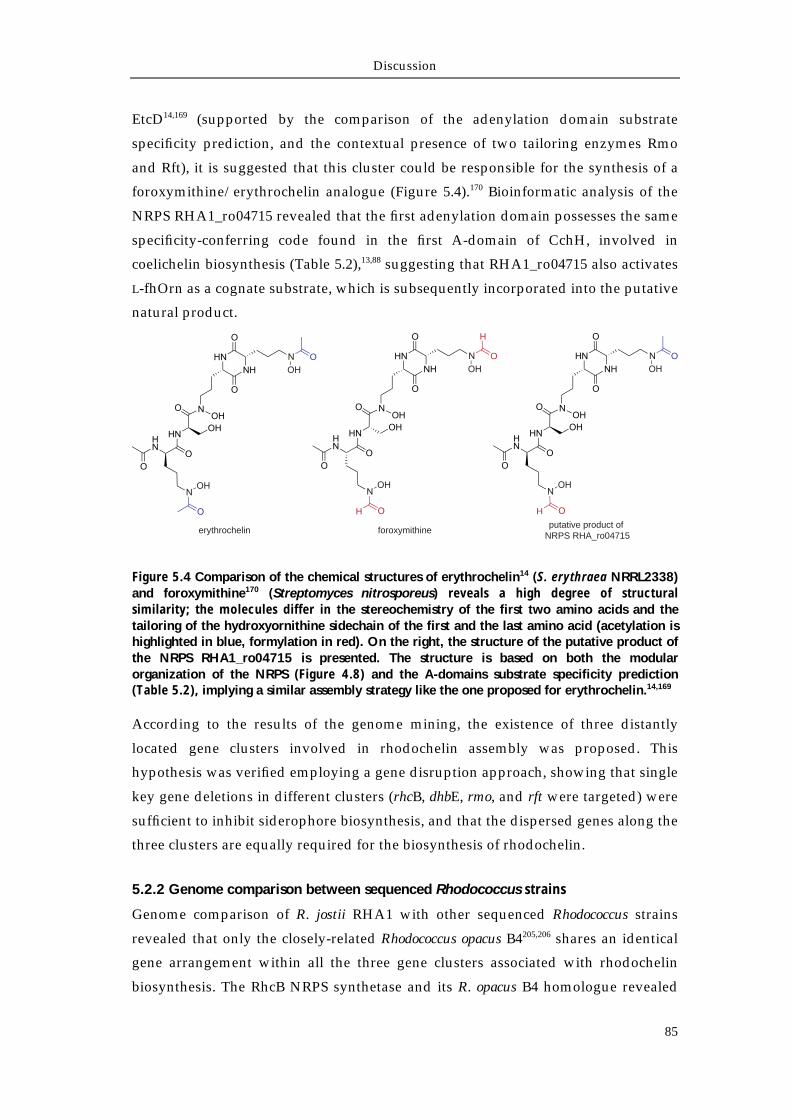
Discussion
85
EtcD14,169 (supported by the comparison of the adenylation domain substrate
specificity prediction, and the contextual presence of two tailoring enzymes Rmo
and Rft), it is suggested that this cluster could be responsible for the synthesis of a
foroxymithine/erythrochelin analogue (Figure 5.4).170 Bioinformatic analysis of the
NRPS RHA1_ro04715 revealed that the first adenylation domain possesses the same
specificity-conferring code found in the first A-domain of CchH, involved in
coelichelin biosynthesis (Table 5.2),13,88 suggesting that RHA1_ro04715 also activates
L-fhOrn as a cognate substrate, which is subsequently incorporated into the putative
natural product.
Figure 5.4 Comparison of the chemical structures of erythrochelin14 (S. erythraea NRRL2338) and foroxymithine170 (Streptomyces nitrosporeus) reveals a high degree of structural similarity; the molecules differ in the stereochemistry of the first two amino acids and the tailoring of the hydroxyornithine sidechain of the first and the last amino acid (acetylation is highlighted in blue, formylation in red). On the right, the structure of the putative product of the NRPS RHA1_ro04715 is presented. The structure is based on both the modular organization of the NRPS (Figure 4.8) and the A-domains substrate specificity prediction (Table 5.2), implying a similar assembly strategy like the one proposed for erythrochelin.14,169
According to the results of the genome mining, the existence of three distantly
located gene clusters involved in rhodochelin assembly was proposed. This
hypothesis was verified employing a gene disruption approach, showing that single
key gene deletions in different clusters (rhcB, dhbE, rmo, and rft were targeted) were
sufficient to inhibit siderophore biosynthesis, and that the dispersed genes along the
three clusters are equally required for the biosynthesis of rhodochelin.
5.2.2 Genome comparison between sequenced Rhodococcus strains
Genome comparison of R. jostii RHA1 with other sequenced Rhodococcus strains
revealed that only the closely-related Rhodococcus opacus B4205,206 shares an identical
gene arrangement within all the three gene clusters associated with rhodochelin
biosynthesis. The RhcB NRPS synthetase and its R. opacus B4 homologue revealed
erythrochelin foroxymithineputative product of
NRPS RHA_ro04715
O
H
O
H
O
H
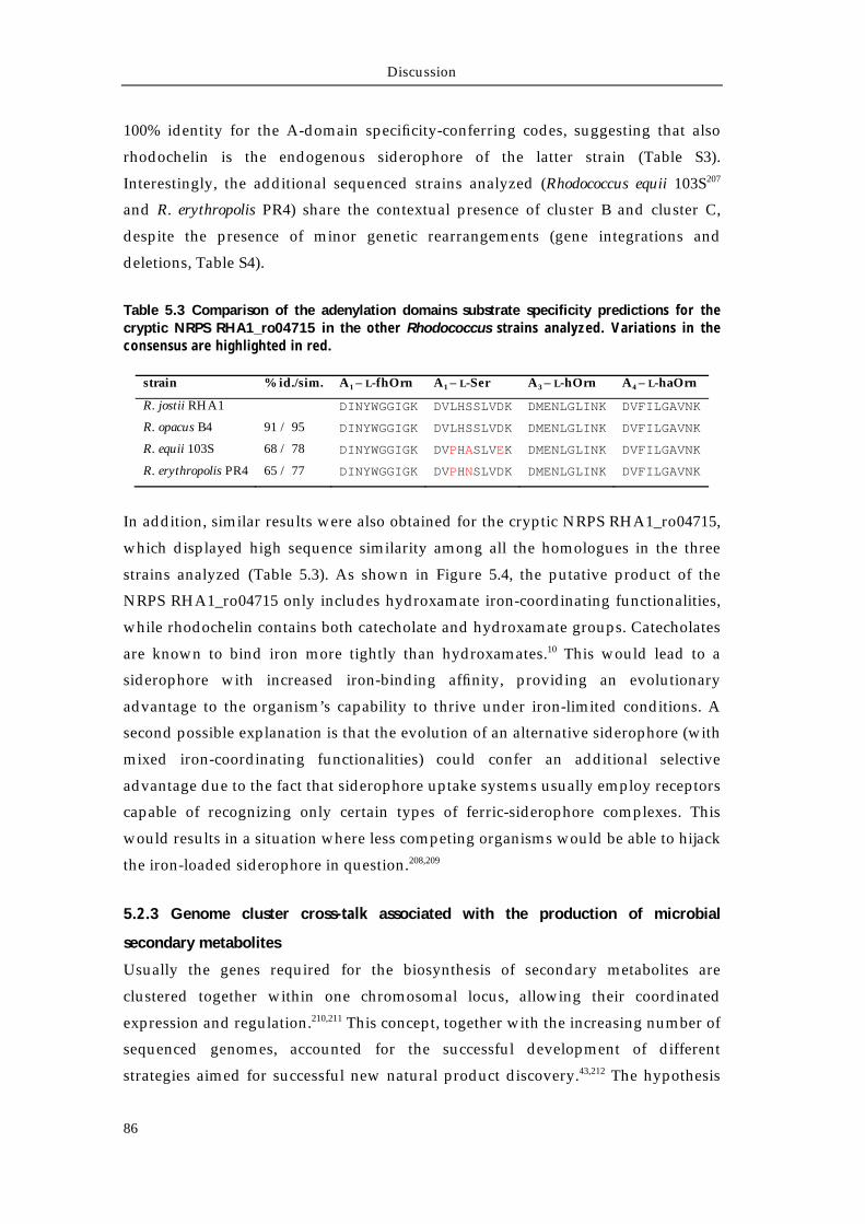
Discussion
86
100% identity for the A-domain specificity-conferring codes, suggesting that also
rhodochelin is the endogenous siderophore of the latter strain (Table S3).
Interestingly, the additional sequenced strains analyzed (Rhodococcus equii 103S207
and R. erythropolis PR4) share the contextual presence of cluster B and cluster C,
despite the presence of minor genetic rearrangements (gene integrations and
deletions, Table S4).
Table 5.3 Comparison of the adenylation domains substrate specificity predictions for the cryptic NRPS RHA1_ro04715 in the other Rhodococcus strains analyzed. Variations in the consensus are highlighted in red.
strain % id./sim. A1 – L-fhOrn A1 – L-Ser A3 – L-hOrn A4 – L-haOrn
R. jostii RHA1 DINYWGGIGK DVLHSSLVDK DMENLGLINK DVFILGAVNK
R. opacus B4 91 / 95 DINYWGGIGK DVLHSSLVDK DMENLGLINK DVFILGAVNK
R. equii 103S 68 / 78 DINYWGGIGK DVPHASLVEK DMENLGLINK DVFILGAVNK
R. erythropolis PR4 65 / 77 DINYWGGIGK DVPHNSLVDK DMENLGLINK DVFILGAVNK
In addition, similar results were also obtained for the cryptic NRPS RHA1_ro04715,
which displayed high sequence similarity among all the homologues in the three
strains analyzed (Table 5.3). As shown in Figure 5.4, the putative product of the
NRPS RHA1_ro04715 only includes hydroxamate iron-coordinating functionalities,
while rhodochelin contains both catecholate and hydroxamate groups. Catecholates
are known to bind iron more tightly than hydroxamates.10 This would lead to a
siderophore with increased iron-binding affinity, providing an evolutionary
advantage to the organism’s capability to thrive under iron-limited conditions. A
second possible explanation is that the evolution of an alternative siderophore (with
mixed iron-coordinating functionalities) could confer an additional selective
advantage due to the fact that siderophore uptake systems usually employ receptors
capable of recognizing only certain types of ferric-siderophore complexes. This
would results in a situation where less competing organisms would be able to hijack
the iron-loaded siderophore in question.208,209
5.2.3 Genome cluster cross-talk associated with the production of microbial
secondary metabolites
Usually the genes required for the biosynthesis of secondary metabolites are
clustered together within one chromosomal locus, allowing their coordinated
expression and regulation.210,211 This concept, together with the increasing number of
sequenced genomes, accounted for the successful development of different
strategies aimed for successful new natural product discovery.43,212 The hypothesis
Discussion
87
that natural product gene clusters are among the most diverse and rapidly-evolving
genetic elements of a bacterium’s chromosome has been recently proposed,
suggesting them to be the ideal candidates for the evolutionary study of bacterial
collectives.213 In this context, the discovery of different examples of cross-talk
mechanisms between the secondary metabolite gene clusters can be representative
of the bacterium’s genome plasticity towards the natural selective pressure,
continuously remodeling its elements (and thus its metabolome), fueling the fitness
for the environment.
Similarly to rhodochelin, the catecholate siderophore vibriobactin (Vibrio cholerae),
harbors its biosynthetic genes within two different genome regions. The genes for
the siderophore transport and utilization, viuA and viuB, and the biosynthetic NRPS
vibF214-216 are separated from those associated with the biosynthesis and activation of
2,3-DHB and for the periplasmic binding and ABC-dependent intracellular import
of the iron-siderophore complex.217-219 On the other hand, to our knowledge,
rhodochelin is the first example where the genes associated with the biosynthesis of
the aryl moiety are not clustered together.175 Another siderophore, erythrochelin, is
synthetized by genes dispersed between two diverse gene clusters:14,169 the erc/etc
cluster encodes the enzymes responsible for precursor biosynthesis and siderophore
assembly and export, whereas the δ-N-L-acetyltransferase mcd (also essential for the
biosynthesis of erythromycin and the red pigment flaviolin220) is located in the
nonfunctional nrps1 cluster.221 Moreover, PPTases, the enzymes responsible for the
apo- to holo- conversion of carrier protein domains of FAS, PKS and NRPS, are an
additional example of enzymes being shared by different biosynthetic pathway,
sometimes displaying a high degree of substrate tolerance towards the carrier
protein, like B. subtilis Sfp.154,155
Recently, a few additional examples of functional secondary metabolism cross-talk
mechanisms have been reported, demonstrating that a more complex biosynthetic
network exists. For example, either one of two essential malonyl-CoA
acyltransferases of the endogenous fatty acid synthase complex are required for the
biosynthesis of the anticancer depsipeptide FK288 in Chromobacterium violaceum 968,
since the two modules DepB and DepC of the PKS lacks the essential AT function
which is replaced by an AT-docking domain. Additionally, in the same species, the
activation of the FK288 PKS ACP domains requires a broad-substrate-range Sfp-
type PPTase which is encoded in a different locus.222 Similar to the last discussed
example, the initiation reaction of the assembly of the quinoxaline antibiotics
requires the transfer of the activated chromophore quinoxaline-2-carboxylic acid
from the cognate stand-alone A-domain TrsI to the first elongation NRPS TrsII. This
Discussion
88
step is carried out by an ACP from the primary FAS II, to form an initiation module,
showing a functional interaction between the primary and the secondary
metabolism.49
Cross-talk between different secondary metabolites gene clusters can also occur on
the regulatory level. For example, in P. fluorescens Pf-5, phloroglucinol act either as a
precursor, an inducer and a repressor of the biosynthesis of the antibiotics
pyoluteorin and 2,4-diacetylphloroglucinol. At nanomolar concentration, the
presence of the 2,4-diacetylphloroglucinol precursor phloroglucinol is required to
stimulate the biosynthesis of pyoluteorin, which is inhibited at higher
concentrations.223 Another similar cross-talk mechanism has also been observed in
fungi. In Aspergillus nidulans, the overexpression of a putative pathway-specific
NRPS-regulatory gene scpR induces the activation and, thus, the formation of a
cryptic NRP, which further downstream upregulates a second transcription factor,
afoA. This latter one activates the biosynthesis of the polyketide asperfuranone
through its specific biosynthetic pathway. In addition, these results represent the
first report of regulatory cross-talk between two biosynthetic gene clusters located
on different fungal chromosomes.224
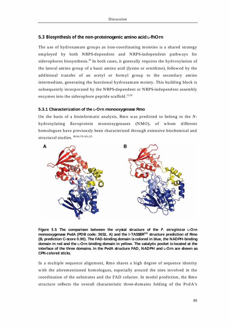
Discussion
89
5.3 Biosynthesis of the non-proteinogenic amino acid L-fhOrn
The use of hydroxamate groups as iron-coordinating moieties is a shared strategy
employed by both NRPS-dependent and NRPS-independent pathways for
siderophores biosynthesis.20 In both cases, it generally requires the hydroxylation of
the lateral amino group of a basic amino acid (lysine or ornithine), followed by the
additional transfer of an acetyl or formyl group to the secondary amine
intermediate, generating the functional hydroxamate moiety. This building block is
subsequently incorporated by the NRPS-dependent or NRPS-independent assembly
enzymes into the siderophore peptide scaffold.15,26
5.3.1 Characterization of the L-Orn monooxygenase Rmo
On the basis of a bioinformatic analysis, Rmo was predicted to belong to the N-
hydroxylating flavoprotein monooxygenases (NMO), of whom different
homologues have previously been characterized through extensive biochemical and
structural studies. 88,94,178-181,225
Figure 5.5 The comparison between the crystal structure of the P. aeruginosa L-Orn monooxygenase PvdA (PDB code: 36S1, A) and the I-TASSER226 structure prediction of Rmo (B, prediction C-score 0.90). The FAD-binding domain is colored in blue, the NADPH-binding domain in red and the L-Orn binding domain in yellow. The catalytic pocket is located at the interface of the three domains. In the PvdA structure FAD, NADPH and L-Orn are shown as CPK-colored sticks.
In a multiple sequence alignment, Rmo shares a high degree of sequence identity
with the aforementioned homologues, especially around the sites involved in the
coordination of the substrates and the FAD cofactor. In model prediction, the Rmo
structure reflects the overall characteristic three-domains folding of the PvdA’s
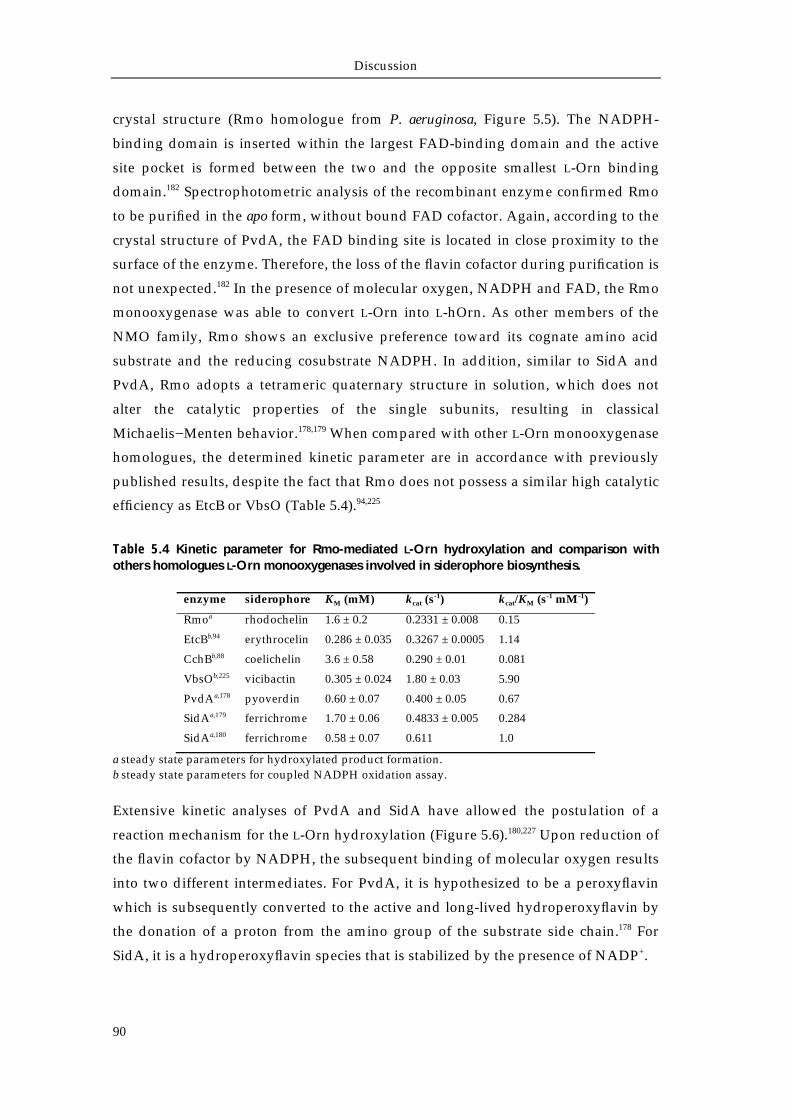
Discussion
90
crystal structure (Rmo homologue from P. aeruginosa, Figure 5.5). The NADPH-
binding domain is inserted within the largest FAD-binding domain and the active
site pocket is formed between the two and the opposite smallest L-Orn binding
domain.182 Spectrophotometric analysis of the recombinant enzyme confirmed Rmo
to be purified in the apo form, without bound FAD cofactor. Again, according to the
crystal structure of PvdA, the FAD binding site is located in close proximity to the
surface of the enzyme. Therefore, the loss of the flavin cofactor during purification is
not unexpected.182 In the presence of molecular oxygen, NADPH and FAD, the Rmo
monooxygenase was able to convert L-Orn into L-hOrn. As other members of the
NMO family, Rmo shows an exclusive preference toward its cognate amino acid
substrate and the reducing cosubstrate NADPH. In addition, similar to SidA and
PvdA, Rmo adopts a tetrameric quaternary structure in solution, which does not
alter the catalytic properties of the single subunits, resulting in classical
Michaelis−Menten behavior.178,179 When compared with other L-Orn monooxygenase
homologues, the determined kinetic parameter are in accordance with previously
published results, despite the fact that Rmo does not possess a similar high catalytic
efficiency as EtcB or VbsO (Table 5.4).94,225
Table 5.4 Kinetic parameter for Rmo-mediated L-Orn hydroxylation and comparison with others homologues L-Orn monooxygenases involved in siderophore biosynthesis.
enzyme siderophore KM (mM) kcat (s-1) kcat/KM (s-1 mM-1)
Rmoa rhodochelin 1.6 ± 0.2 0.2331 ± 0.008 0.15
EtcBb,94 erythrocelin 0.286 ± 0.035 0.3267 ± 0.0005 1.14
CchBb,88 coelichelin 3.6 ± 0.58 0.290 ± 0.01 0.081
VbsOb,225 vicibactin 0.305 ± 0.024 1.80 ± 0.03 5.90
PvdAa,178 pyoverdin 0.60 ± 0.07 0.400 ± 0.05 0.67
SidAa,179 ferrichrome 1.70 ± 0.06 0.4833 ± 0.005 0.284
SidAa,180 ferrichrome 0.58 ± 0.07 0.611 1.0
a steady state parameters for hydroxylated product formation. b steady state parameters for coupled NADPH oxidation assay.
Extensive kinetic analyses of PvdA and SidA have allowed the postulation of a
reaction mechanism for the L-Orn hydroxylation (Figure 5.6).180,227 Upon reduction of
the flavin cofactor by NADPH, the subsequent binding of molecular oxygen results
into two different intermediates. For PvdA, it is hypothesized to be a peroxyflavin
which is subsequently converted to the active and long-lived hydroperoxyflavin by
the donation of a proton from the amino group of the substrate side chain.178 For
SidA, it is a hydroperoxyflavin species that is stabilized by the presence of NADP+.
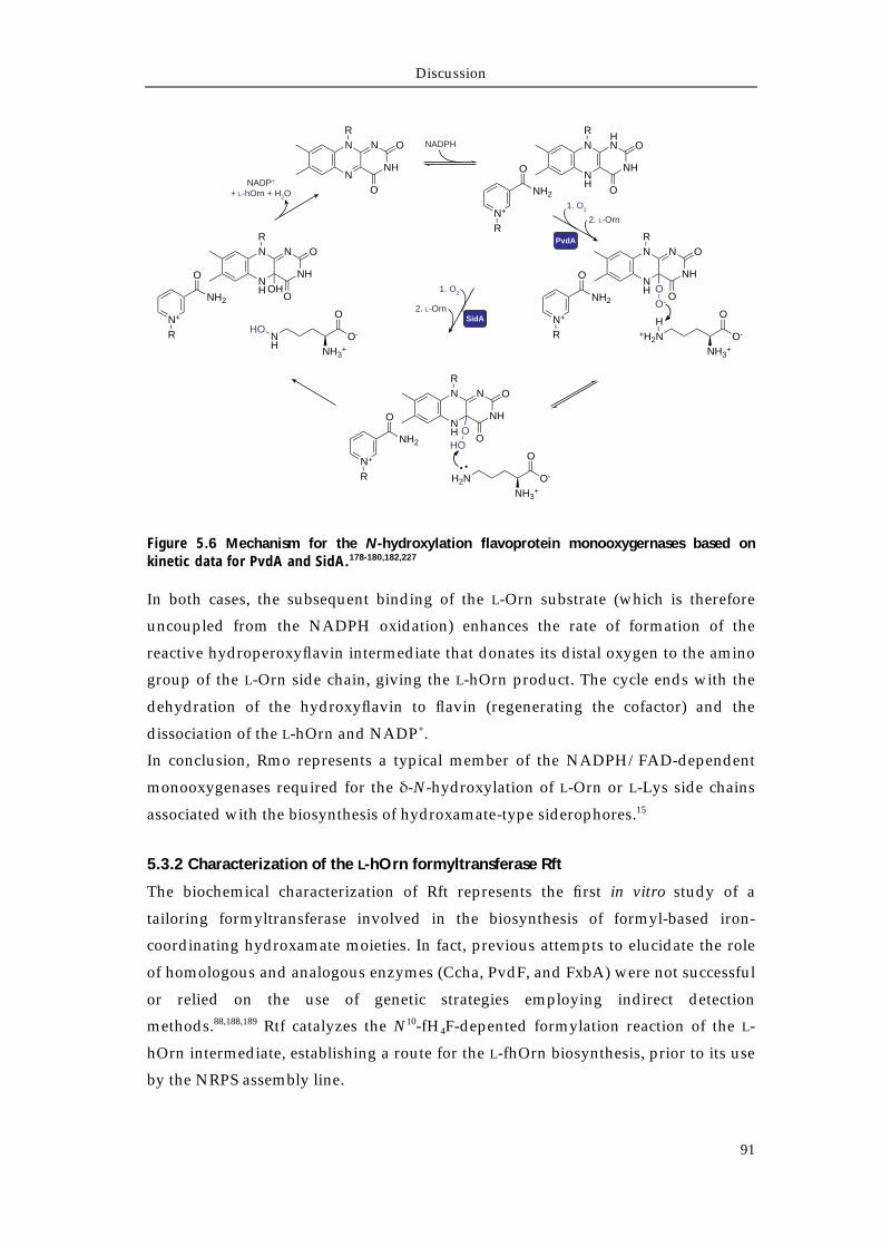
Discussion
91
Figure 5.6 Mechanism for the N-hydroxylation flavoprotein monooxygernases based on kinetic data for PvdA and SidA.178-180,182,227
In both cases, the subsequent binding of the L-Orn substrate (which is therefore
uncoupled from the NADPH oxidation) enhances the rate of formation of the
reactive hydroperoxyflavin intermediate that donates its distal oxygen to the amino
group of the L-Orn side chain, giving the L-hOrn product. The cycle ends with the
dehydration of the hydroxyflavin to flavin (regenerating the cofactor) and the
dissociation of the L-hOrn and NADP+.
In conclusion, Rmo represents a typical member of the NADPH/FAD-dependent
monooxygenases required for the δ-N-hydroxylation of L-Orn or L-Lys side chains
associated with the biosynthesis of hydroxamate-type siderophores.15
5.3.2 Characterization of the L-hOrn formyltransferase Rft
The biochemical characterization of Rft represents the first in vitro study of a
tailoring formyltransferase involved in the biosynthesis of formyl-based iron-
coordinating hydroxamate moieties. In fact, previous attempts to elucidate the role
of homologous and analogous enzymes (Ccha, PvdF, and FxbA) were not successful
or relied on the use of genetic strategies employing indirect detection
methods.88,188,189 Rtf catalyzes the N10-fH4F-depented formylation reaction of the L-
hOrn intermediate, establishing a route for the L-fhOrn biosynthesis, prior to its use
by the NRPS assembly line.
SidA
PvdA
N
N
NH
NR
O
ONH
N
NH
HN
R
O
O
N+
R
O
NH2
NH
N
NH
NR
O
O
N+
R
O
NH2O
HO
H2NNH3
+
O-
O
NH
N
NH
NR
O
O
N+
R
O
NH2OO-
+H2NH
NH3+
O-
O
NH
N
NH
NR
O
O
N+
R
O
NH2OH
NH
NH3+
O-
OHO
1. O2
NADP+
+ L-hOrn + H2O
2. L-Orn
2. L-Orn
NADPH
1. O2
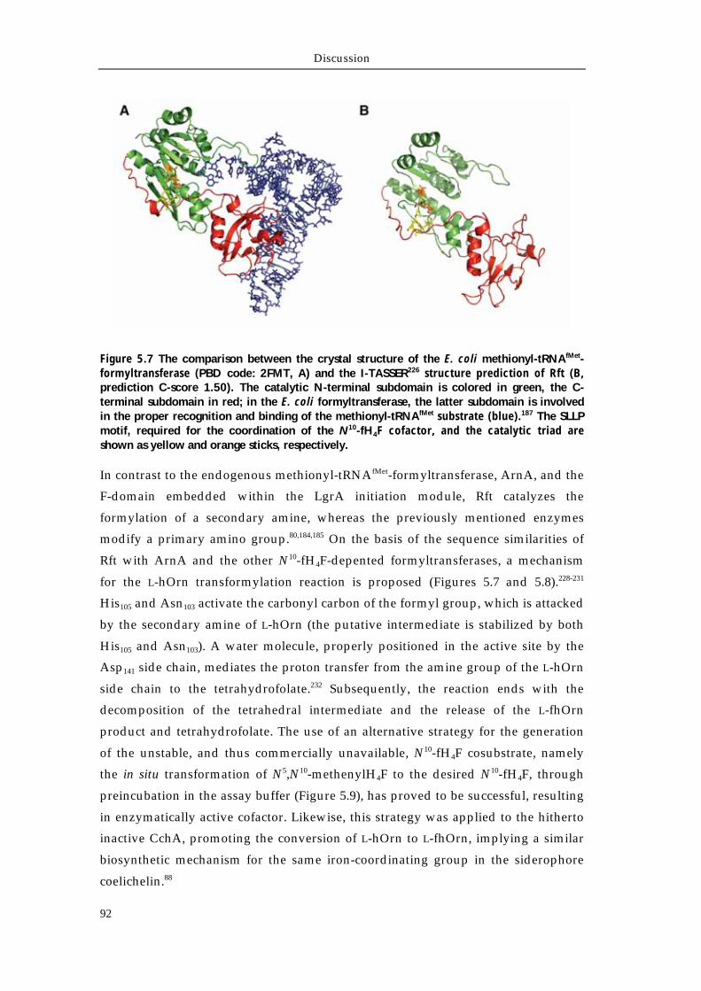
Discussion
92
Figure 5.7 The comparison between the crystal structure of the E. coli methionyl-tRNAfMet-formyltransferase (PBD code: 2FMT, A) and the I-TASSER226 structure prediction of Rft (B, prediction C-score 1.50). The catalytic N-terminal subdomain is colored in green, the C-terminal subdomain in red; in the E. coli formyltransferase, the latter subdomain is involved in the proper recognition and binding of the methionyl-tRNAfMet substrate (blue).187 The SLLP motif, required for the coordination of the N10-fH4F cofactor, and the catalytic triad are shown as yellow and orange sticks, respectively.
In contrast to the endogenous methionyl-tRNAfMet-formyltransferase, ArnA, and the
F-domain embedded within the LgrA initiation module, Rft catalyzes the
formylation of a secondary amine, whereas the previously mentioned enzymes
modify a primary amino group.80,184,185 On the basis of the sequence similarities of
Rft with ArnA and the other N10-fH4F-depented formyltransferases, a mechanism
for the L-hOrn transformylation reaction is proposed (Figures 5.7 and 5.8).228-231
His105 and Asn103 activate the carbonyl carbon of the formyl group, which is attacked
by the secondary amine of L-hOrn (the putative intermediate is stabilized by both
His105 and Asn103). A water molecule, properly positioned in the active site by the
Asp141 side chain, mediates the proton transfer from the amine group of the L-hOrn
side chain to the tetrahydrofolate.232 Subsequently, the reaction ends with the
decomposition of the tetrahedral intermediate and the release of the L-fhOrn
product and tetrahydrofolate. The use of an alternative strategy for the generation
of the unstable, and thus commercially unavailable, N10-fH4F cosubstrate, namely
the in situ transformation of N5,N10-methenylH4F to the desired N10-fH4F, through
preincubation in the assay buffer (Figure 5.9), has proved to be successful, resulting
in enzymatically active cofactor. Likewise, this strategy was applied to the hitherto
inactive CchA, promoting the conversion of L-hOrn to L-fhOrn, implying a similar
biosynthetic mechanism for the same iron-coordinating group in the siderophore
coelichelin.88
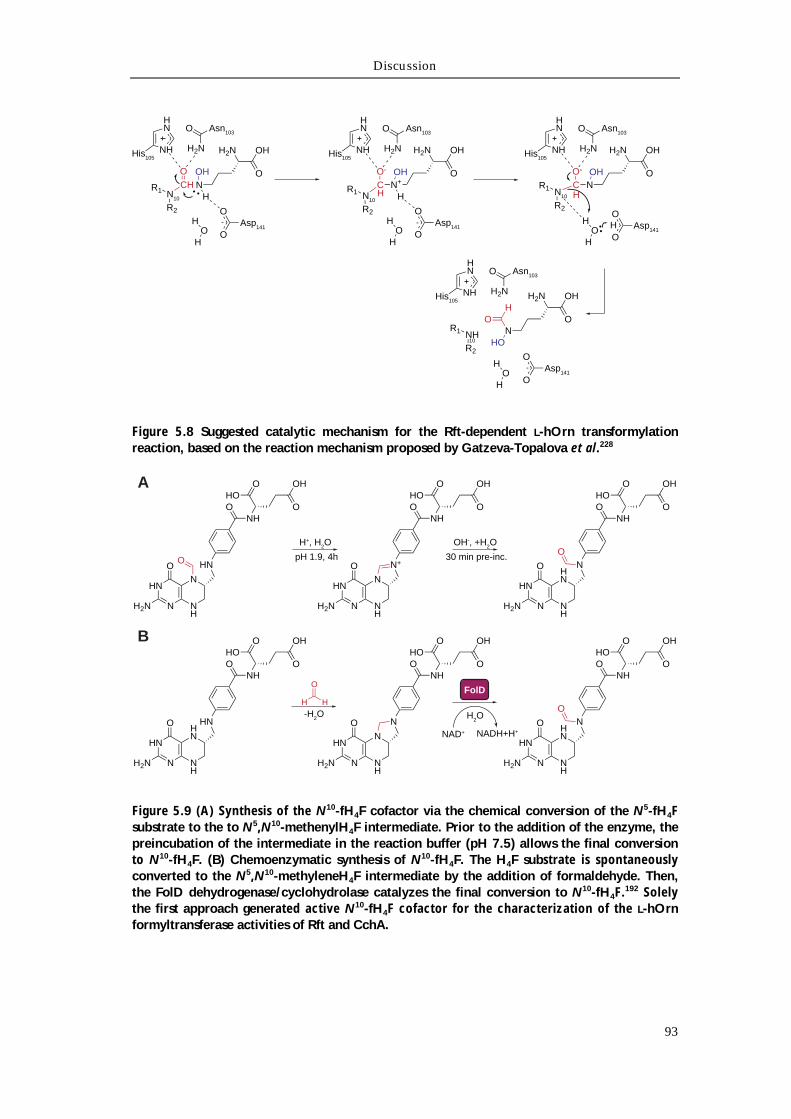
Discussion
93
Figure 5.8 Suggested catalytic mechanism for the Rft-dependent L-hOrn transformylation reaction, based on the reaction mechanism proposed by Gatzeva-Topalova et al.228
Figure 5.9 (A) Synthesis of the N10-fH4F cofactor via the chemical conversion of the N5-fH4F substrate to the to N5,N10-methenylH4F intermediate. Prior to the addition of the enzyme, the preincubation of the intermediate in the reaction buffer (pH 7.5) allows the final conversion to N10-fH4F. (B) Chemoenzymatic synthesis of N10-fH4F. The H4F substrate is spontaneously converted to the N5,N10-methyleneH4F intermediate by the addition of formaldehyde. Then, the FolD dehydrogenase/cyclohydrolase catalyzes the final conversion to N10-fH4F.192 Solely the first approach generated active N10-fH4F cofactor for the characterization of the L-hOrn formyltransferase activities of Rft and CchA.
Asp141
O-
O
HO
H
NH
HN
His105
O-
Asn103O
H2N
CHN10
N+OH
HR2
R1
H2N
O
OH
Asp141
O-
O
HO
H
NH
HN
His105
O
Asn103O
H2N
CHN10
NOH
HR2
R1
H2N
O
OH
Asp141
O
O
HO
H
NH
HN
His105
O-
Asn103O
H2N
CHN10
NOH
R2
R1
H2N
O
OH
H
Asp141
O
O
HO
H
NH
HN
His105
Asn103O
H2N
NHR2
R110
O
OHH2N
NHO
OH
-
HN
N NH
N
H2N
O HN
NHO
OH
O
OHO
O
HN
N NH
N
H2N
O N+
NHO
OH
O
OHO
HN
N NH
HN
H2N
O N
NHO
OH
O
OHO
O
HN
N NH
HN
H2N
O HN
NHO
OH
O
OHO
HN
N NH
N
H2N
O N
NHO
OH
O
OHO
HN
N NH
HN
H2N
O N
NHO
OH
O
OHO
O
pH 1.9, 4h
FolD
NAD+ NADH+H+
H2O
H+, H2O
30 min pre-inc.
OH-, +H2O
-H2O
O
HH
A
B
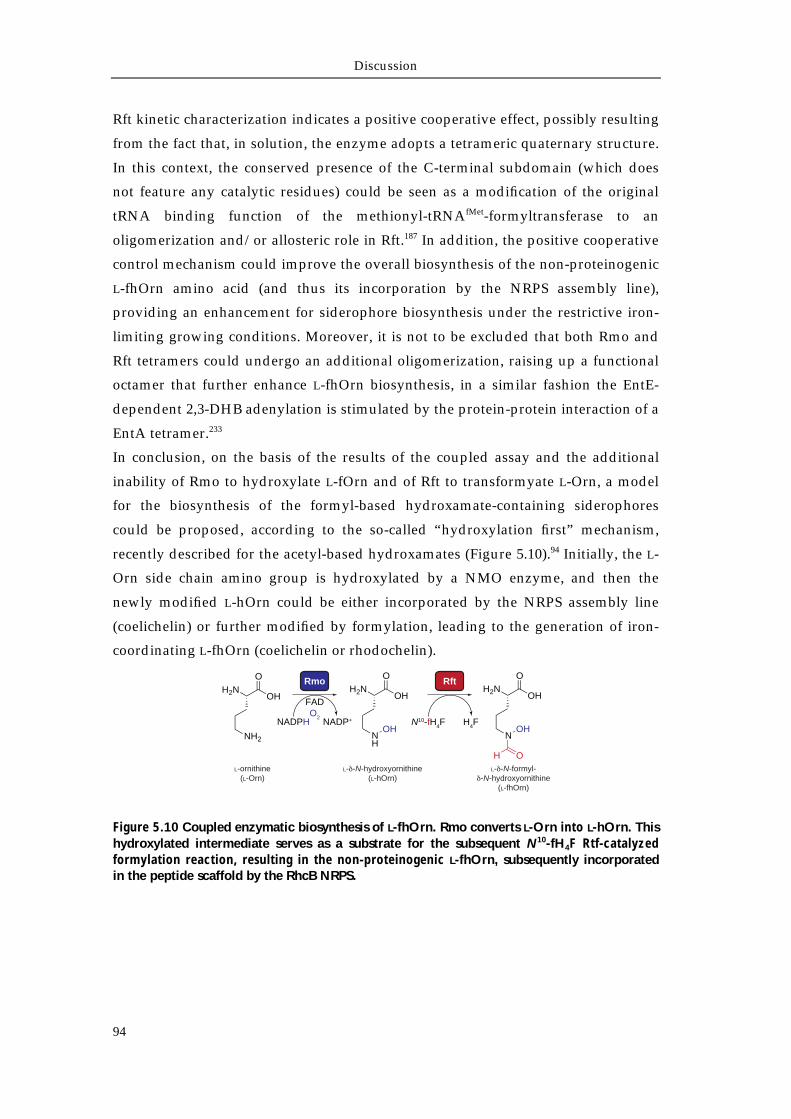
Discussion
94
Rft kinetic characterization indicates a positive cooperative effect, possibly resulting
from the fact that, in solution, the enzyme adopts a tetrameric quaternary structure.
In this context, the conserved presence of the C-terminal subdomain (which does
not feature any catalytic residues) could be seen as a modification of the original
tRNA binding function of the methionyl-tRNAfMet-formyltransferase to an
oligomerization and/or allosteric role in Rft.187 In addition, the positive cooperative
control mechanism could improve the overall biosynthesis of the non-proteinogenic
L-fhOrn amino acid (and thus its incorporation by the NRPS assembly line),
providing an enhancement for siderophore biosynthesis under the restrictive iron-
limiting growing conditions. Moreover, it is not to be excluded that both Rmo and
Rft tetramers could undergo an additional oligomerization, raising up a functional
octamer that further enhance L-fhOrn biosynthesis, in a similar fashion the EntE-
dependent 2,3-DHB adenylation is stimulated by the protein-protein interaction of a
EntA tetramer.233
In conclusion, on the basis of the results of the coupled assay and the additional
inability of Rmo to hydroxylate L-fOrn and of Rft to transformyate L-Orn, a model
for the biosynthesis of the formyl-based hydroxamate-containing siderophores
could be proposed, according to the so-called “hydroxylation first” mechanism,
recently described for the acetyl-based hydroxamates (Figure 5.10).94 Initially, the L-
Orn side chain amino group is hydroxylated by a NMO enzyme, and then the
newly modified L-hOrn could be either incorporated by the NRPS assembly line
(coelichelin) or further modified by formylation, leading to the generation of iron-
coordinating L-fhOrn (coelichelin or rhodochelin).
Figure 5.10 Coupled enzymatic biosynthesis of L-fhOrn. Rmo converts L-Orn into L-hOrn. This hydroxylated intermediate serves as a substrate for the subsequent N10-fH4F Rtf-catalyzed formylation reaction, resulting in the non-proteinogenic L-fhOrn, subsequently incorporated in the peptide scaffold by the RhcB NRPS.
Rft
N10-fH4F H4F
H2NOH
O
NOH
OH
Rmo
NADPH NADP+
FADO2
H2NOH
O
NH2
H2NOH
O
NH
OH
L-ornithine(L-Orn)
L- -N-hydroxyornithine(L-hOrn)
L- -N-formyl--N-hydroxyornithine
(L-fhOrn)
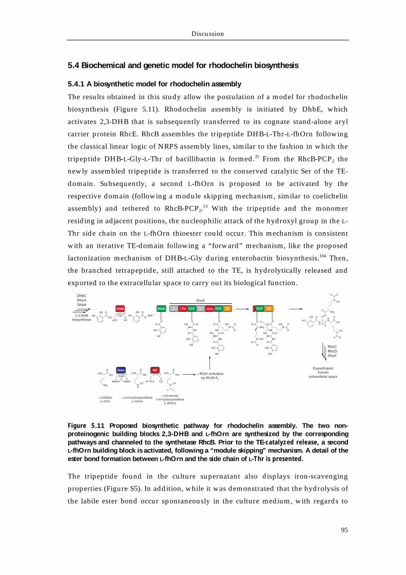
Discussion
95
5.4 Biochemical and genetic model for rhodochelin biosynthesis
5.4.1 A biosynthetic model for rhodochelin assembly
The results obtained in this study allow the postulation of a model for rhodochelin
biosynthesis (Figure 5.11). Rhodochelin assembly is initiated by DhbE, which
activates 2,3-DHB that is subsequently transferred to its cognate stand-alone aryl
carrier protein RhcE. RhcB assembles the tripeptide DHB-L-Thr-L-fhOrn following
the classical linear logic of NRPS assembly lines, similar to the fashion in which the
tripeptide DHB-L-Gly-L-Thr of bacillibactin is formed.35 From the RhcB-PCP2 the
newly assembled tripeptide is transferred to the conserved catalytic Ser of the TE-
domain. Subsequently, a second L-fhOrn is proposed to be activated by the
respective domain (following a module skipping mechanism, similar to coelichelin
assembly) and tethered to RhcB-PCP2.13 With the tripeptide and the monomer
residing in adjacent positions, the nucleophilic attack of the hydroxyl group in the L-
Thr side chain on the L-fhOrn thioester could occur. This mechanism is consistent
with an iterative TE-domain following a “forward” mechanism, like the proposed
lactonization mechanism of DHB-L-Gly during enterobactin biosynthesis.104 Then,
the branched tetrapeptide, still attached to the TE, is hydrolytically released and
exported to the extracellular space to carry out its biological function.
Figure 5.11 Proposed biosynthetic pathway for rhodochelin assembly. The two non-proteinogenic building blocks 2,3-DHB and L-fhOrn are synthesized by the corresponding pathways and channeled to the synthetase RhcB. Prior to the TE-catalyzed release, a second L-fhOrn building block is activated, following a “module skipping” mechanism. A detail of the ester bond formation between L-fhOrn and the side chain of L-Thr is presented.
The tripeptide found in the culture supernatant also displays iron-scavenging
properties (Figure S5). In addition, while it was demonstrated that the hydrolysis of
the labile ester bond occur spontaneously in the culture medium, with regards to
C L-fhOrn PCP TEC L-Thr PCP
RhcB
DhbEOH
HOOH
O
ATP PPi
Mg2+
OHHO
O
OAMP
RhcE
OS
HO
HO
PCP TE
O
NH
HN
H
OOH
HO
OOH
O
NOH
OH
ONH2
NOH
H O
Export/Importfrom/to
extracellular space
DhbCRhcADhbA
2,3-DHBbiosynthesis
RhcCRhcDRhcF
Rft
N10-fH4F
H2NOH
O
NH
OHH4F
H2NOH
O
NOH
OH
Rmo
NADPH NADP+
FADO2
H2NOH
O
NH2
L-fhOrn activationby RhcB-A2
L-ornithine(L-Orn)
L- -N-hydroxyornithine(L-hOrn)
L- -N-formyl--N-hydroxyornithine
(L-fhOrn)
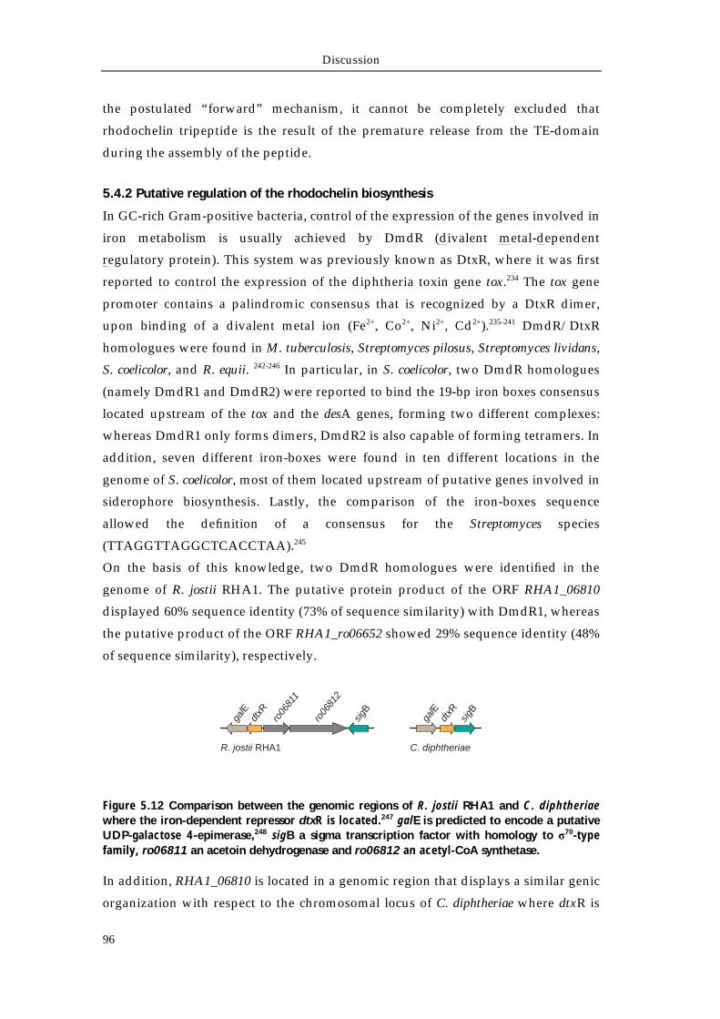
Discussion
96
the postulated “forward” mechanism, it cannot be completely excluded that
rhodochelin tripeptide is the result of the premature release from the TE-domain
during the assembly of the peptide.
5.4.2 Putative regulation of the rhodochelin biosynthesis
In GC-rich Gram-positive bacteria, control of the expression of the genes involved in
iron metabolism is usually achieved by DmdR (divalent metal-dependent
regulatory protein). This system was previously known as DtxR, where it was first
reported to control the expression of the diphtheria toxin gene tox.234 The tox gene
promoter contains a palindromic consensus that is recognized by a DtxR dimer,
upon binding of a divalent metal ion (Fe2+, Co2+, Ni2+, Cd2+).235-241 DmdR/DtxR
homologues were found in M. tuberculosis, Streptomyces pilosus, Streptomyces lividans,
S. coelicolor, and R. equii. 242-246 In particular, in S. coelicolor, two DmdR homologues
(namely DmdR1 and DmdR2) were reported to bind the 19-bp iron boxes consensus
located upstream of the tox and the desA genes, forming two different complexes:
whereas DmdR1 only forms dimers, DmdR2 is also capable of forming tetramers. In
addition, seven different iron-boxes were found in ten different locations in the
genome of S. coelicolor, most of them located upstream of putative genes involved in
siderophore biosynthesis. Lastly, the comparison of the iron-boxes sequence
allowed the definition of a consensus for the Streptomyces species
(TTAGGTTAGGCTCACCTAA).245
On the basis of this knowledge, two DmdR homologues were identified in the
genome of R. jostii RHA1. The putative protein product of the ORF RHA1_06810
displayed 60% sequence identity (73% of sequence similarity) with DmdR1, whereas
the putative product of the ORF RHA1_ro06652 showed 29% sequence identity (48%
of sequence similarity), respectively.
Figure 5.12 Comparison between the genomic regions of R. jostii RHA1 and C. diphtheriae where the iron-dependent repressor dtxR is located.247 galE is predicted to encode a putative UDP-galactose 4-epimerase,248 sigB a sigma transcription factor with homology to σ70-type family, ro06811 an acetoin dehydrogenase and ro06812 an acetyl-CoA synthetase.
In addition, RHA1_06810 is located in a genomic region that displays a similar genic
organization with respect to the chromosomal locus of C. diphtheriae where dtxR is
galE
dtxR
ro06
811
ro06
812
sigB
galE
dtxR
sigB
R. jostii RHA1 C. diphtheriae
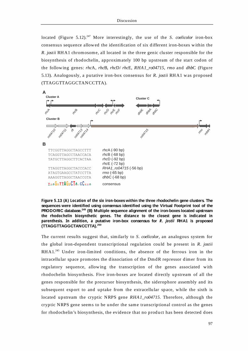
Discussion
97
located (Figure 5.12).247 More interestingly, the use of the S. coelicolor iron-box
consensus sequence allowed the identification of six different iron-boxes within the
R. jostii RHA1 chromosome, all located in the three genic cluster responsible for the
biosynthesis of rhodochelin, approximately 100 bp upstream of the start codon of
the following genes: rhcA, rhcB, rhcD/rhcE, RHA1_ro04715, rmo and dhbC (Figure
5.13). Analogously, a putative iron-box consensus for R. jostii RHA1 was proposed
(TTAGGTTAGGCTANCCTTA).
Figure 5.13 (A) Location of the six iron-boxes within the three rhodochelin gene clusters. The sequences were identified using consensus identified using the Virtual Footprint tool of the PRODORIC database.249 (B) Multiple sequence alignment of the iron-boxes located upstream the rhodochelin biosynthetic genes. The distance to the closest gene is indicated in parenthesis. In addition, a putative iron-box consensus for R. jostii RHA1 is proposed (TTAGGTTAGGCTANCCTTA).250
The current results suggest that, similarly to S. coelicolor, an analogous system for
the global iron-dependent transcriptional regulation could be present in R. jostii
RHA1.245 Under iron-limited conditions, the absence of the ferrous iron in the
intracellular space promotes the dissociation of the DmdR repressor dimer from its
regulatory sequence, allowing the transcription of the genes associated with
rhodochelin biosynthesis. Five iron-boxes are located directly upstream of all the
genes responsible for the precursor biosynthesis, the siderophore assembly and its
subsequent export to and uptake from the extracellular space, while the sixth is
located upstream the cryptic NRPS gene RHA1_ro04715. Therefore, although the
cryptic NRPS gene seems to be under the same transcriptional control as the genes
for rhodochelin’s biosynthesis, the evidence that no product has been detected does
Cluster A
rhcA
rhcB
rhcC
rhcD
rhcE
rhcF
dhbE
dhbA
dhbC
rft
rmo
mbt
H
ro04
710
ro04
711
ro04
713
ro04
714
ro04
715
Cluster B
Cluster C
TTCGGTTAGGCTAGCCTTT rhcA (-90 bp)TCAGGTTAGCCTAACCACA rhcB (-68 bp)TATGCTTAGGCTTCACTAA rhcD (-92 bp) rhcE (-72 bp)TTAGGTTAGGCTACCCACC RHA1_ro04715 (-56 bp)ATAGTGAAGCCTATCCTTA rmo (-65 bp)AAAGGTTAGGCTAACCGTA dhbC (-68 bp)
consensus
A
B
Discussion
98
not exclude the synthetase to be functional, to be silent (other unknown repressors
or activators could play a role at a different hierarchical transcriptional level) or the
growing conditions applied are inadequate to allow detection. On the other hand,
the absence on the iron-box consensus upstream of the rft gene still does not exclude
a different transcriptional control, or, that the biosynthesis of the nonproteinogenic
L-fhOrn amino acid is simply enhanced by the positive cooperative mechanism of
the Rft tetramer in vivo.
Discussion
99
5.5 Perspective and outlook
Microorganisms produces a multitude of small molecules that are not used in the
primary metabolism but take part in important physiological and ecological roles,
mediating the interactions among other species of the community and/or with
multicellular organisms.213 The discovery that the genes responsible for the
biosynthesis of natural products are often clustered together in a genome facilitated
the important genetic and the biochemical findings during the last two decades.
Conversely, the presented discovery of cross-talk mechanisms between three gene
clusters for the biosynthesis of a single secondary metabolite in R. jostii RHA1,251
supports an unexpected level of complexity, especially for a molecule with a relative
simple (but still important) function, like a siderophore. In this context, the
hypothesis that natural product gene clusters are among the most diverse and
rapidly evolving genetic elements of a chromosome reflects the genome plasticity
towards the natural selective pressure, continuously remodeling its elements to best
fit the environment. In addition, understanding the molecules and the mechanisms
that microorganism use to interact with each other can lead to a deeper
understanding of their physiology, ecology and evolution and, lately, to a new and
better drug discovery for human therapy.252
The existence of approximately 20 secondary metabolite gene clusters in R. jostii
RHA1 that remains still orphan with respect to their biosynthesized molecule put
the strain into account for the isolation and the structural characterization of new
natural products. Several strategies have already been reported being successful
and can be employed to elicit cryptic pathways in other species; for example,
triggering the global nutritional regulators of primary metabolism under phosphate
or nitrogen starvation253, using of small chemical as elicitors,254 or with the co-
cultivation in the presence of other species of the same ecosystem, in order to
attempt to simulate the natural habitat and prompt the inter-species
communication.255 Apart from culture-dependent techniques, the availability of
different genetic tools specifically designed for Rhodoccocus spp. allows the genetic
engineering of the strain and thus the identification of new natural products
through comparative metabolic profiling.44 In particular, the use of plasmid systems
specifically engineered for heterologous gene expression in Rhodoccocus spp.256 can
be employed to activate cryptic biosynthetic gene clusters through the inducible
and/or constitutive manipulation of pathway-specific activators, as recently
demonstrated for Streptomyces ambofaciens and Burkholderia spp.257-259

References
101
References
1. Pierre, J. L., Fontecave, M. & Crichton, R. R. Chemistry for an essential biological process: the reduction of ferric iron. Biometals 15, 341–346 (2002).
2. Miethke, M. & Marahiel, M. A. Siderophore-based iron acquisition and pathogen control. Microbiol Mol Biol Rev 71, 413–451 (2007).
3. Andrews, S. C., Robinson, A. K. & Rodríguez-Quiñones, F. Bacterial iron homeostasis. FEMS Microbiol Rev 27, 215–237 (2003).
4. Wandersman, C. & Delepelaire, P. Bacterial iron sources: from siderophores to hemophores. Annu Rev Microbiol 58, 611–647 (2004).
5. Schaible, U. E. & Kaufmann, S. H. E. Iron and microbial infection. Nat Rev Microbiol 2, 946–953 (2004).
6. Winkelmann, G. & Drechsel, H. Microbial Siderophores. Biotechnology 8, 199–246 (2008). 7. Miethke, M. et al. Ferri-bacillibactin uptake and hydrolysis in Bacillus subtilis. Mol Microbiol
61, 1413–1427 (2006). 8. Peuckert, F., Miethke, M., Albrecht, A. G., Essen, L.-O. & Marahiel, M. A. Structural basis and
stereochemistry of triscatecholate siderophore binding by FeuA. Angew Chem Int Ed Engl 48, 7924–7927 (2009).
9. Sandy, M. & Butler, A. Microbial iron acquisition: marine and terrestrial siderophores. Chem Rev 109, 4580–4595 (2009).
10. Crumbliss, A. L. & Harrington, J. M. Iron sequestration by small molecules: Thermodynamic and kinetic studies of natural siderophores and synthetic model compounds. Advances in Inorganic Chemistry Volume 61, 179–250 T2 – (2009).
11. Raymond, K. N., Dertz, E. A. & Kim, S. S. Enterobactin: an archetype for microbial iron transport. Proc Natl Acad Sci USA 100, 3584–3588 (2003).
12. May, J. J., Wendrich, T. M. & Marahiel, M. A. The dhb operon of Bacillus subtilis encodes the biosynthetic template for the catecholic siderophore 2,3-dihydroxybenzoate-glycine-threonine trimeric ester bacillibactin. J Biol Chem 276, 7209–7217 (2001).
13. Lautru, S., Deeth, R. J., Bailey, L. M. & Challis, G. L. Discovery of a new peptide natural product by Streptomyces coelicolor genome mining. Nat Chem Biol 1, 265–269 (2005).
14. Robbel, L., Knappe, T. A., Linne, U., Xie, X. & Marahiel, M. A. Erythrochelin--a hydroxamate-type siderophore predicted from the genome of Saccharopolyspora erythraea. FEBS J 277, 663–676 (2010).
15. Olucha, J. & Lamb, A. L. Mechanistic and structural studies of the N-hydroxylating flavoprotein monooxygenases. Bioorg. Chem. 39, 171–177 (2011).
16. Martinez, J. S. et al. Structure and membrane affinity of a suite of amphiphilic siderophores produced by a marine bacterium. Proc Natl Acad Sci USA 100, 3754–3759 (2003).
17. Martinez, J. S. et al. Self-assembling amphiphilic siderophores from marine bacteria. Science 287, 1245–1247 (2000).
18. Meiwes, J. et al. Isolation and characterization of staphyloferrin A, a compound with siderophore activity from Staphylococcus hyicus DSM 20459. FEMS Microbiol Rev 55, 201–205 (1990).
19. Oves-Costales, D., Kadi, N. & Challis, G. L. The long-overlooked enzymology of a nonribosomal peptide synthetase-independent pathway for virulence-conferring siderophore biosynthesis. Chem. Commun. VL - 6530–6541 (2009).doi:10.1039/b913092f
20. Barry, S. M. & Challis, G. L. Recent advances in siderophore biosynthesis. Current Opinion in Chemical Biology 13, 205–215 (2009).
21. de Lorenzo, V., Bindereif, A., Paw, B. H. & Neilands, J. B. Aerobactin biosynthesis and transport genes of plasmid ColV-K30 in Escherichia coli K-12. J Bacteriol 165, 570–578 (1986).
22. Thariath, A., Socha, D., Valvano, M. A. & Viswanatha, T. Construction and biochemical characterization of recombinant cytoplasmic forms of the IucD protein (lysine:N6-hydroxylase) encoded by the pColV-K30 aerobactin gene cluster. J Bacteriol 175, 589–596 (1993).
23. Coy, M., Paw, B. H., Bindereif, A. & Neilands, J. B. Isolation and properties of N epsilon-hydroxylysine:acetyl coenzyme A N epsilon-transacetylase from Escherichia coli pABN11. Biochemistry 25, 2485–2489 (1986).
24. Kadi, N., Oves-Costales, D., Barona-Gómez, F. & Challis, G. L. A new family of ATP-dependent oligomerization-macrocyclization biocatalysts. Nat Chem Biol 3, 652–656 (2007).
25. Kadi, N., Arbache, S., Song, L., Oves-Costales, D. & Challis, G. L. Identification of a gene cluster that directs putrebactin biosynthesis in Shewanella species: PubC catalyzes cyclodimerization of N-hydroxy-N-succinylputrescine. J Am Chem Soc 130, 10458–10459 (2008).
26. Challis, G. L. A widely distributed bacterial pathway for siderophore biosynthesis independent of nonribosomal peptide synthetases. Chembiochem 6, 601–611 (2005).
27. Schwarzer, D. & Marahiel, M. A. Multimodular biocatalysts for natural product assembly. Naturwissenschaften 88, 93–101 (2001).
References
102
28. Nolan, E. M. & Walsh, C. T. How nature morphs peptide scaffolds into antibiotics. Chembiochem 10, 34–53 (2009).
29. Finking, R. & Marahiel, M. A. Biosynthesis of nonribosomal peptides. Annu Rev Microbiol 58, 453–488 (2004).
30. Newman, D. J., Cragg, G. M. & Snader, K. M. The influence of natural products upon drug discovery. Nat Prod Rep 17, 215–234 (2000).
31. Hopwood, D. A. Streptomyces in Nature And Medicine. 250 (Oxford University Press, USA: 2007).
32. Walsh, C. T. & Fischbach, M. A. Natural products version 2.0: connecting genes to molecules. J Am Chem Soc 132, 2469–2493 (2010).
33. Tanovic, A., Samel, S. A., Essen, L.-O. & Marahiel, M. A. Crystal structure of the termination module of a nonribosomal peptide synthetase. Science 321, 659–663 (2008).
34. Conti, E., Stachelhaus, T., Marahiel, M. A. & Brick, P. Structural basis for the activation of phenylalanine in the non-ribosomal biosynthesis of gramicidin S. EMBO J 16, 4174–4183 (1997).
35. May, J. J., Kessler, N., Marahiel, M. A. & Stubbs, M. T. Crystal structure of DhbE, an archetype for aryl acid activating domains of modular nonribosomal peptide synthetases. Proc Natl Acad Sci USA 99, 12120–12125 (2002).
36. Yonus, H. et al. Crystal structure of DltA. Implications for the reaction mechanism of non-ribosomal peptide synthetase adenylation domains. Journal of Biological Chemistry 283, 32484–32491 (2008).
37. Hegg, E. L. Unraveling the structure and mechanism of acetyl-coenzyme A synthase. Acc Chem Res 37, 775–783 (2004).
38. Conti, E., Lloyd, L. F., Akins, J., Franks, N. P. & Brick, P. Crystallization and preliminary diffraction studies of firefly luciferase from Photinus pyralis. Acta Crystallogr. D Biol. Crystallogr. 52, 876–878 (1996).
39. Stachelhaus, T., Mootz, H. D. & Marahiel, M. A. The specificity-conferring code of adenylation domains in nonribosomal peptide synthetases. Chem Biol 6, 493–505 (1999).
40. Rausch, C., Weber, T., Kohlbacher, O., Wohlleben, W. & Huson, D. H. Specificity prediction of adenylation domains in nonribosomal peptide synthetases (NRPS) using transductive support vector machines (TSVMs). Nucleic acids research 33, 5799–5808 (2005).
41. Bachmann, B. O. & Ravel, J. Methods for in silico prediction of microbial polyketide and nonribosomal peptide biosynthetic pathways from DNA sequence data. Meth Enzymol 458, 181–217 (2009).
42. Weber, T. et al. CLUSEAN: a computer-based framework for the automated analysis of bacterial secondary metabolite biosynthetic gene clusters. Journal of Biotechnology 140, 13–17 (2009).
43. Challis, G. L. Mining microbial genomes for new natural products and biosynthetic pathways. Microbiology 154, 1555–1569 (2008).
44. Zerikly, M. & Challis, G. L. Strategies for the discovery of new natural products by genome mining. Chembiochem 10, 625–633 (2009).
45. Francklyn, C. S. DNA polymerases and aminoacyl-tRNA synthetases: shared mechanisms for ensuring the fidelity of gene expression. Biochemistry 47, 11695–11703 (2008).
46. Stachelhaus, T., Hüser, A. & Marahiel, M. A. Biochemical characterization of peptidyl carrier protein (PCP), the thiolation domain of multifunctional peptide synthetases. Chem Biol 3, 913–921 (1996).
47. Lambalot, R. H. et al. A new enzyme superfamily - the phosphopantetheinyl transferases. Chem Biol 3, 923–936 (1996).
48. Mootz, H. D., Finking, R. & Marahiel, M. A. 4'-phosphopantetheine transfer in primary and secondary metabolism of Bacillus subtilis. Journal of Biological Chemistry 276, 37289–37298 (2001).
49. Schmoock, G. et al. Functional cross-talk between fatty acid synthesis and nonribosomal peptide synthesis in quinoxaline antibiotic-producing streptomycetes. Journal of Biological Chemistry 280, 4339–4349 (2005).
50. Koglin, A. et al. Conformational switches modulate protein interactions in peptide antibiotic synthetases. Science 312, 273–276 (2006).
51. Belshaw, P. J., Walsh, C. T. & Stachelhaus, T. Aminoacyl-CoAs as probes of condensation domain selectivity in nonribosomal peptide synthesis. Science 284, 486–489 (1999).
52. Stachelhaus, T., Mootz, H. D., Bergendahl, V. & Marahiel, M. A. Peptide bond formation in nonribosomal peptide biosynthesis. Catalytic role of the condensation domain. Journal of Biological Chemistry 273, 22773–22781 (1998).
53. Linne, U. & Marahiel, M. A. Control of directionality in nonribosomal peptide synthesis: role of the condensation domain in preventing misinitiation and timing of epimerization. Biochemistry 39, 10439–10447 (2000).
54. Luo, L. et al. Timing of epimerization and condensation reactions in nonribosomal peptide assembly lines: kinetic analysis of phenylalanine activating elongation modules of tyrocidine synthetase B. Biochemistry 41, 9184–9196 (2002).
References
103
55. Keating, T. A., Marshall, C. G., Walsh, C. T. & Keating, A. E. The structure of VibH represents nonribosomal peptide synthetase condensation, cyclization and epimerization domains. Nat. Struct. Biol. 9, 522–526 (2002).
56. Samel, S. A., Schoenafinger, G., Knappe, T. A., Marahiel, M. A. & Essen, L.-O. Structural and functional insights into a peptide bond-forming bidomain from a nonribosomal peptide synthetase. Structure 15, 781–792 (2007).
57. Leslie, A. G. Refined crystal structure of type III chloramphenicol acetyltransferase at 1.75 A resolution. J Mol Biol 213, 167–186 (1990).
58. Kopp, F. & Marahiel, M. A. Where chemistry meets biology: the chemoenzymatic synthesis of nonribosomal peptides and polyketides. Curr. Opin. Biotechnol. 18, 513–520 (2007).
59. Kopp, F. & Marahiel, M. A. Macrocyclization strategies in polyketide and nonribosomal peptide biosynthesis. Nat Prod Rep 24, 735–749 (2007).
60. Bruner, S. D. et al. Structural basis for the cyclization of the lipopeptide antibiotic surfactin by the thioesterase domain SrfTE. Structure 10, 301–310 (2002).
61. Samel, S. A., Wagner, B., Marahiel, M. A. & Essen, L.-O. The thioesterase domain of the fengycin biosynthesis cluster: a structural base for the macrocyclization of a non-ribosomal lipopeptide. J Mol Biol 359, 876–889 (2006).
62. Pazirandeh, M., Chirala, S. S., Huang, W. Y. & Wakil, S. J. Characterization of recombinant thioesterase and acyl carrier protein domains of chicken fatty acid synthase expressed in Escherichia coli. Journal of Biological Chemistry 264, 18195–18201 (1989).
63. Tseng, C. C. et al. Characterization of the surfactin synthetase C-terminal thioesterase domain as a cyclic depsipeptide synthase. Biochemistry 41, 13350–13359 (2002).
64. Hubbard, B. K. & Walsh, C. T. Vancomycin assembly: nature's way. Angew Chem Int Ed Engl 42, 730–765 (2003).
65. Chiu, H. T. et al. Molecular cloning and sequence analysis of the complestatin biosynthetic gene cluster. Proc Natl Acad Sci USA 98, 8548–8553 (2001).
66. Miller, D. A., Luo, L., Hillson, N., Keating, T. A. & Walsh, C. T. Yersiniabactin synthetase: a four-protein assembly line producing the nonribosomal peptide/polyketide hybrid siderophore of Yersinia pestis. Chem Biol 9, 333–344 (2002).
67. Kessler, N., Schuhmann, H., Morneweg, S., Linne, U. & Marahiel, M. A. The linear pentadecapeptide gramicidin is assembled by four multimodular nonribosomal peptide synthetases that comprise 16 modules with 56 catalytic domains. Journal of Biological Chemistry 279, 7413–7419 (2004).
68. Kopp, F., Mahlert, C., Grünewald, J. & Marahiel, M. A. Peptide macrocyclization: the reductase of the nostocyclopeptide synthetase triggers the self-assembly of a macrocyclic imine. J Am Chem Soc 128, 16478–16479 (2006).
69. Magarvey, N. A., Haltli, B., He, M., Greenstein, M. & Hucul, J. A. Biosynthetic pathway for mannopeptimycins, lipoglycopeptide antibiotics active against drug-resistant gram-positive pathogens. Antimicrobial Agents and Chemotherapy 50, 2167–2177 (2006).
70. Debono, M. et al. Enzymatic and chemical modifications of lipopeptide antibiotic A21978C: the synthesis and evaluation of daptomycin (LY146032). J Antibiot 41, 1093–1105 (1988).
71. Stein, D. B., Linne, U., Hahn, M. & Marahiel, M. A. Impact of epimerization domains on the intermodular transfer of enzyme-bound intermediates in nonribosomal peptide synthesis. Chembiochem 7, 1807–1814 (2006).
72. Konz, D. & Marahiel, M. A. How do peptide synthetases generate structural diversity? Chem Biol 6, R39–48 (1999).
73. Schneider, T. L., Shen, B. & Walsh, C. T. Oxidase domains in epothilone and bleomycin biosynthesis: thiazoline to thiazole oxidation during chain elongation. Biochemistry 42, 9722–9730 (2003).
74. Gehring, A. M., Mori, I., Perry, R. D. & Walsh, C. T. The nonribosomal peptide synthetase HMWP2 forms a thiazoline ring during biogenesis of yersiniabactin, an iron-chelating virulence factor of Yersinia pestis. Biochemistry 37, 11637–11650 (1998).
75. Shen, B. et al. Cloning and Characterization of the Bleomycin Biosynthetic Gene Cluster from StreptomycesverticillusATCC15003 1. J Nat Prod 65, 422–431 (2002).
76. Konz, D., Klens, A., Schörgendorfer, K. & Marahiel, M. A. The bacitracin biosynthesis operon of Bacillus licheniformis ATCC 10716: molecular characterization of three multi-modular peptide synthetases. Chem Biol 4, 927–937 (1997).
77. Du, L., Chen, M., Zhang, Y. & Shen, B. BlmIII and BlmIV nonribosomal peptide synthetase-catalyzed biosynthesis of the bleomycin bithiazole moiety involving both in cis and in trans aminoacylation. Biochemistry 42, 9731–9740 (2003).
78. Reimmann, C. et al. Essential PchG-Dependent Reduction in Pyochelin Biosynthesis of Pseudomonas aeruginosa. (2001).
79. Ansari, M. Z., Sharma, J., Gokhale, R. S. & Mohanty, D. In silico analysis of methyltransferase domains involved in biosynthesis of secondary metabolites. BMC Bioinformatics 9, 454 (2008).
80. Schoenafinger, G., Schracke, N., Linne, U. & Marahiel, M. A. Formylation Domain: An Essential Modifying Enzyme for the Nonribosomal Biosynthesis of Linear Gramicidin. J Am Chem Soc 128, 7406–7407 (2006).
References
104
81. Schubert, H. L., Blumenthal, R. M. & Cheng, X. Many paths to methyltransfer: a chronicle of convergence. Trends in Biochemical Sciences 28, 329–335 (2003).
82. Hojati, Z. et al. Structure, biosynthetic origin, and engineered biosynthesis of calcium-dependent antibiotics from Streptomyces coelicolor. Chem Biol 9, 1175–1187 (2002).
83. Mahlert, C., Kopp, F., Thirlway, J., Micklefield, J. & Marahiel, M. A. Stereospecific enzymatic transformation of alpha-ketoglutarate to (2S,3R)-3-methyl glutamate during acidic lipopeptide biosynthesis. J Am Chem Soc 129, 12011–12018 (2007).
84. Huang, Y.-T. et al. In vitro characterization of enzymes involved in the synthesis of nonproteinogenic residue (2S,3S)-beta-methylphenylalanine in glycopeptide antibiotic mannopeptimycin. Chembiochem 10, 2480–2487 (2009).
85. He, H. et al. Mannopeptimycins, novel antibacterial glycopeptides from Streptomyces hygroscopicus, LL-AC98. J Am Chem Soc 124, 9729–9736 (2002).
86. van Berkel, W. J. H., Kamerbeek, N. M. & Fraaije, M. W. Flavoprotein monooxygenases, a diverse class of oxidative biocatalysts. Journal of Biotechnology 124, 670–689 (2006).
87. Kopp, F., Linne, U., Oberthür, M. & Marahiel, M. A. Harnessing the chemical activation inherent to carrier protein-bound thioesters for the characterization of lipopeptide fatty acid tailoring enzymes. J Am Chem Soc 130, 2656–2666 (2008).
88. Pohlmann, V. & Marahiel, M. A. Delta-amino group hydroxylation of L-ornithine during coelichelin biosynthesis. Org Biomol Chem 6, 1843–1848 (2008).
89. Strieker, M., Kopp, F., Mahlert, C., Essen, L.-O. & Marahiel, M. A. Mechanistic and Structural Basis of Stereospecific Cβ-Hydroxylation in Calcium-Dependent Antibiotic, a Daptomycin-Type Lipopeptide. ACS Chem Biol 2, 187–196 (2007).
90. Helmetag, V., Samel, S. A., Thomas, M. G., Marahiel, M. A. & Essen, L.-O. Structural basis for the erythro-stereospecificity of the L-arginine oxygenase VioC in viomycin biosynthesis. FEBS J 276, 3669–3682 (2009).
91. Strieker, M., Nolan, E. M., Walsh, C. T. & Marahiel, M. A. Stereospecific Synthesis of threo- and erythro-β-Hydroxyglutamic Acid During Kutzneride Biosynthesis. J Am Chem Soc 131, 13523–13530 (2009).
92. Bruijnincx, P. C. A., van Koten, G. & Klein Gebbink, R. J. M. Mononuclear non-heme iron enzymes with the 2-His-1-carboxylate facial triad: recent developments in enzymology and modeling studies. Chem Soc Rev 37, 2716–2744 (2008).
93. Costas, M., Mehn, M. P., Jensen, M. P. & Que, L. Dioxygen activation at mononuclear nonheme iron active sites: enzymes, models, and intermediates. Chem Rev 104, 939–986 (2004).
94. Robbel, L., Helmetag, V., Knappe, T. A. & Marahiel, M. A. Consecutive enzymatic modification of ornithine generates the hydroxamate moieties of the siderophore erythrochelin. Biochemistry 50, 6073–6080 (2011).
95. Linne, U., Schwarzer, D., Schroeder, G. N. & Marahiel, M. A. Mutational analysis of a type II thioesterase associated with nonribosomal peptide synthesis. Eur J Biochem 271, 1536–1545 (2004).
96. Schwarzer, D., Mootz, H. D., Linne, U. & Marahiel, M. A. Regeneration of misprimed nonribosomal peptide synthetases by type II thioesterases. Proc Natl Acad Sci USA 99, 14083–14088 (2002).
97. Yeh, E., Kohli, R. M., Bruner, S. D. & Walsh, C. T. Type II thioesterase restores activity of a NRPS module stalled with an aminoacyl-S-enzyme that cannot be elongated. Chembiochem 5, 1290–1293 (2004).
98. Schneider, A. & Marahiel, M. A. Genetic evidence for a role of thioesterase domains, integrated in or associated with peptide synthetases, in non-ribosomal peptide biosynthesis in Bacillus subtilis. Arch Microbiol 169, 404–410 (1998).
99. Mootz, H. D. & Marahiel, M. A. The tyrocidine biosynthesis operon of Bacillus brevis: complete nucleotide sequence and biochemical characterization of functional internal adenylation domains. J Bacteriol 179, 6843–6850 (1997).
100. Cosmina, P. et al. Sequence and analysis of the genetic locus responsible for surfactin synthesis in Bacillus subtilis. Mol Microbiol 8, 821–831 (1993).
101. Miao, V. et al. Daptomycin biosynthesis in Streptomyces roseosporus: cloning and analysis of the gene cluster and revision of peptide stereochemistry. Microbiology 151, 1507–1523 (2005).
102. Smith, D. J. et al. Beta-lactam antibiotic biosynthetic genes have been conserved in clusters in prokaryotes and eukaryotes. EMBO J 9, 741–747 (1990).
103. Gehring, A. M., Mori, I. & Walsh, C. T. Reconstitution and characterization of the Escherichia coli enterobactin synthetase from EntB, EntE, and EntF. Biochemistry 37, 2648–2659 (1998).
104. Shaw-Reid, C. A. et al. Assembly line enzymology by multimodular nonribosomal peptide synthetases: the thioesterase domain of E. coli EntF catalyzes both elongation and cyclolactonization. Chem Biol 6, 385–400 (1999).
105. Juguet, M. et al. An iterative nonribosomal peptide synthetase assembles the pyrrole-amide antibiotic congocidine in Streptomyces ambofaciens. Chem Biol 16, 421–431 (2009).
106. Wenzel, S. C. et al. Structure and biosynthesis of myxochromides S1-3 in Stigmatella aurantiaca: evidence for an iterative bacterial type I polyketide synthase and for module skipping in nonribosomal peptide biosynthesis. Chembiochem 6, 375–385 (2005).
References
105
107. Bosello, M., Mielcarek, A., Giessen, T. W. & Marahiel, M. A. An Enzymatic Pathway for the Biosynthesis of the Formylhydroxyornithine Required for Rhodochelin Iron Coordination. Biochemistry 51, 3059–3066 (2012).
108. Bode, H., Bethe, B., Höfs, R. & Zeeck, A. Big effects from small changes: possible ways to explore nature's chemical diversity. Chembiochem (2002).
109. Margulies, M. et al. Genome sequencing in microfabricated high-density picolitre reactors. Nature 437, 376–380 (2005).
110. Donadio, S., Monciardini, P. & Sosio, M. Polyketide synthases and nonribosomal peptide synthetases: the emerging view from bacterial genomics. Nat Prod Rep 24, 1073–1109 (2007).
111. Ansari, M. Z., Yadav, G., Gokhale, R. S. & Mohanty, D. NRPS-PKS: a knowledge-based resource for analysis of NRPS/PKS megasynthases. Nucleic acids research 32, W405–13 (2004).
112. Bentley, S. D. et al. Complete genome sequence of the model actinomycete Streptomyces coelicolor A3(2). Nature 417, 141–147 (2002).
113. Gust, B., Challis, G. L., Fowler, K., Kieser, T. & Chater, K. F. PCR-targeted Streptomyces gene replacement identifies a protein domain needed for biosynthesis of the sesquiterpene soil odor geosmin. Proc Natl Acad Sci USA 100, 1541–1546 (2003).
114. Song, L. et al. Type III polyketide synthase beta-ketoacyl-ACP starter unit and ethylmalonyl-CoA extender unit selectivity discovered by Streptomyces coelicolor genome mining. J Am Chem Soc 128, 14754–14755 (2006).
115. Corre, C., Song, L., O'Rourke, S., Chater, K. F. & Challis, G. L. 2-Alkyl-4-hydroxymethylfuran-3-carboxylic acids, antibiotic production inducers discovered by Streptomyces coelicolor genome mining. Proc Natl Acad Sci USA 105, 17510–17515 (2008).
116. Udwary, D. W. et al. Genome sequencing reveals complex secondary metabolome in the marine actinomycete Salinispora tropica. Proc Natl Acad Sci USA 104, 10376–10381 (2007).
117. McClerren, A. L. et al. Discovery and in vitro biosynthesis of haloduracin, a two-component lantibiotic. Proc Natl Acad Sci USA 103, 17243–17248 (2006).
118. Fortin, P. D., Walsh, C. T. & Magarvey, N. A. A transglutaminase homologue as a condensation catalyst in antibiotic assembly lines. Nature 448, 824–827 (2007).
119. Watanabe, K. et al. Escherichia coli allows efficient modular incorporation of newly isolated quinomycin biosynthetic enzyme into echinomycin biosynthetic pathway for rational design and synthesis of potent antibiotic unnatural natural product. J Am Chem Soc 131, 9347–9353 (2009).
120. Watanabe, K., Oguri, H. & Oikawa, H. Diversification of echinomycin molecular structure by way of chemoenzymatic synthesis and heterologous expression of the engineered echinomycin biosynthetic pathway. Current Opinion in Chemical Biology 13, 189–196 (2009).
121. Gross, H. et al. The genomisotopic approach: a systematic method to isolate products of orphan biosynthetic gene clusters. Chem Biol 14, 53–63 (2007).
122. A, B., R, S. & U, B. The nitrile-degrading enzymes: current status and future prospects. Applied microbiology … 60, 33–44 (2002).
123. van der Geize, R. & Dijkhuizen, L. Harnessing the catabolic diversity of rhodococci for environmental and biotechnological applications. Curr Opin Microbiol 7, 255–261 (2004).
124. McLeod, M. P. et al. The complete genome of Rhodococcus sp. RHA1 provides insights into a catabolic powerhouse. Proc Natl Acad Sci USA 103, 15582–15587 (2006).
125. Carrano, C. J., Jordan, M., Drechsel, H., Schmid, D. G. & Winkelmann, G. Heterobactins: A new class of siderophores from Rhodococcus erythropolis IGTS8 containing both hydroxamate and catecholate donor groups. Biometals 14, 119–125 (2001).
126. Dhungana, S. et al. Purification and characterization of rhodobactin: a mixed ligand siderophore from Rhodococcus rhodochrous strain OFS. Biometals 20, 853–867 (2007).
127. Seto, M. et al. A Novel Transformation of Polychlorinated Biphenyls by Rhodococcus sp. Strain RHA1. Applied and … 61, 3353–3358 (1995).
128. Helmetag, V. Biochemische und strukturelle Untersuchungen der Biosynthese unnatürlicher Aminosäuren als Bausteine nicht-ribosomaler Peptide. (Philipps-Universität Marburg: Marburg an der Lahn, 2009).
129. Schäfer, A. et al. Small mobilizable multi-purpose cloning vectors derived from the Escherichia coli plasmids pK18 and pK19: selection of defined deletions in the chromosome of Corynebacterium glutamicum. Gene 145, 69–73 (1994).
130. Pridmore, R. D. New and versatile cloning vectors with kanamycin-resistance marker. Gene 56, 309–312 (1987).
131. Datta, N., Hedges, R., Shaw, E. J., Sykes, R. & Richmond, M. Properties of an R factor from Pseudomonas aeruginosa. J Bacteriol 108, 1244–1249 (1971).
132. Trieu-Cuot, P., Carlier, C., Martin, P. & Courvalin, P. Plasmid transfer by conjugation from Escherichia coli to Gram-positive bacteria. FEMS Microbiol Rev 48, 289–294 (1987).
133. Mazodier, P., Petter, R. & Thompson, C. Intergeneric conjugation between Escherichia coli and Streptomyces species. J Bacteriol 171, 3583–3585 (1989).
134. Schäfer, A., Kalinowski, J., Simon, R., Seep-Feldhaus, A. H. & Pühler, A. High-frequency conjugal plasmid transfer from gram-negative Escherichia coli to various gram-positive coryneform bacteria. J Bacteriol 172, 1663–1666 (1990).
References
106
135. Gay, P., Le Coq, D., Steinmetz, M., Berkelman, T. & Kado, C. I. Positive selection procedure for entrapment of insertion sequence elements in gram-negative bacteria. J Bacteriol 164, 918–921 (1985).
136. Jäger, W., Schäfer, A., Pühler, A., Labes, G. & Wohlleben, W. Expression of the Bacillus subtilis sacB gene leads to sucrose sensitivity in the gram-positive bacterium Corynebacterium glutamicum but not in Streptomyces lividans. J Bacteriol 174, 5462–5465 (1992).
137. Steinmetz, M., Le Coq, D., Djemia, H. B. & Gay, P. Genetic analysis of sacB, the structural gene of a secreted enzyme, levansucrase of Bacillus subtilis Marburg. Mol. Gen. Genet. 191, 138–144 (1983).
138. Studier, F. W. & Moffatt, B. A. Use of bacteriophage T7 RNA polymerase to direct selective high-level expression of cloned genes. J Mol Biol 189, 113–130 (1986).
139. Simon, R., Priefer, U. & Pühler, A. A broad host range mobilization system for in vivo genetic engineering: transposon mutagenesis in gram negative bacteria. Nature Biotechnology (1983).
140. Bauchop, T. & Elsden, S. R. The growth of micro-organisms in relation to their energy supply. J Gen Microbiol 23, 457–469 (1960).
141. Sambrook, J. & Russell, D. W. Molecular Cloning: A Laboratory Manual. (Cold Spring Harbor Laboratory Press: 2001).
142. Horton, R. M., Cai, Z. L., Ho, S. N. & Pease, L. R. Gene splicing by overlap extension: tailor-made genes using the polymerase chain reaction. BioTechniques 8, 528–535 (1990).
143. van der Geize, R., Hessels, G., van Gerwen, R., van der Meijden, P. & Dijkhuizen, L. Unmarked gene deletion mutagenesis of kstD, encoding 3-ketosteroid Delta1-dehydrogenase, in Rhodococcus erythropolis SQ1 using sacB as counter-selectable marker. FEMS Microbiol Rev 205, 197–202 (2001).
144. Bradford, M. M. A rapid and sensitive method for the quantitation of microgram quantities of protein utilizing the principle of protein-dye binding. Anal Biochem 72, 248–254 (1976).
145. Perkins, D. N., Pappin, D. J., Creasy, D. M. & Cottrell, J. S. Probability-based protein identification by searching sequence databases using mass spectrometry data. Electrophoresis 20, 3551–3567 (1999).
146. Koenig, T. et al. Robust prediction of the MASCOT score for an improved quality assessment in mass spectrometric proteomics. J Proteome Res 7, 3708–3717 (2008).
147. Schwyn, B. & Neilands, J. B. Universal chemical assay for the detection and determination of siderophores. Anal Biochem 160, 47–56 (1987).
148. Bhushan, R. & Brückner, H. Marfey's reagent for chiral amino acid analysis: a review. Amino Acids 27, 231–247 (2004).
149. Lin, Y. & Miller, M. Practical Synthesis of Hydroxamate-Derived Siderophore Components by an Indirect Oxidation Method and Syntheses of a DIG− Siderophore Conjugate and a Biotin …. J. Org. Chem (1999).
150. Jones, R. G. Studies on imidazoles. II. The Synthesis of 5-imidazolecarboxylates from glycine and substituted glycine esters. J Am Chem Soc 71, 644–647 (1949).
151. Blanquet, S., Dessen, P. & Kahn, D. Properties and specificity of methionyl-tRNAfMet formyltransferase from Escherichia coli. Meth Enzymol 106, 141–152 (1984).
152. Santi, D. V., Webster, R. W. & Cleland, W. W. Kinetics of aminoacyl-tRNA synthetases catalyzed ATP-PPi exchange. Meth Enzymol 29, 620–627 (1974).
153. Linne, U. & Marahiel, M. A. Reactions catalyzed by mature and recombinant nonribosomal peptide synthetases. Meth Enzymol 388, 293–315 (2004).
154. Reuter, K., Mofid, M. R., Marahiel, M. A. & Ficner, R. Crystal structure of the surfactin synthetase-activating enzyme sfp: a prototype of the 4'-phosphopantetheinyl transferase superfamily. EMBO J 18, 6823–6831 (1999).
155. La Clair, J. J., Foley, T. L., Schegg, T. R., Regan, C. M. & Burkart, M. D. Manipulation of carrier proteins in antibiotic biosynthesis. Chem Biol 11, 195–201 (2004).
156. Rutherford, K. et al. Artemis: sequence visualization and annotation. Bioinformatics 16, 944–945 (2000).
157. Medema, M. H. et al. antiSMASH: rapid identification, annotation and analysis of secondary metabolite biosynthesis gene clusters in bacterial and fungal genome sequences. Nucleic acids research (2011).doi:10.1093/nar/gkr466
158. Röttig, M. et al. NRPSpredictor2--a web server for predicting NRPS adenylation domain specificity. Nucleic acids research 39, W362–7 (2011).
159. Altschul, S. F., Gish, W., Miller, W., Myers, E. W. & Lipman, D. J. Basic local alignment search tool. J Mol Biol 215, 403–410 (1990).
160. Thompson, J. D., Higgins, D. G. & Gibson, T. J. CLUSTAL W: improving the sensitivity of progressive multiple sequence alignment through sequence weighting, position-specific gap penalties and weight matrix choice. Nucleic acids research 22, 4673–4680 (1994).
161. Sievers, F. et al. Fast, scalable generation of high-quality protein multiple sequence alignments using Clustal Omega. Mol. Syst. Biol. 7, 539 (2011).
162. Edgar, R. C. MUSCLE: multiple sequence alignment with high accuracy and high throughput. Nucleic acids research 32, 1792–1797 (2004).
References
107
163. Darling, A. C. E., Mau, B., Blattner, F. R. & Perna, N. T. Mauve: multiple alignment of conserved genomic sequence with rearrangements. Genome Res. 14, 1394–1403 (2004).
164. Hu, J. & Miller, M. J. A New Method for the Synthesis of N-ε-Acetyl-N-ε-hydroxy-L-lysine, the Iron-Binding Constituent of Several Important Siderophores. The Journal of Organic Chemistry 59, 4858–4861 (1994).
165. Patzer, S. I. & Braun, V. Gene cluster involved in the biosynthesis of griseobactin, a catechol-peptide siderophore of Streptomyces sp. ATCC 700974. J Bacteriol 192, 426–435 (2010).
166. Plowman, J. E., Loehr, T. M., Goldman, S. J. & Sanders-Loehr, J. Structure and siderophore activity of ferric schizokinen. J Inorg Biochem 20, 183–197 (1984).
167. Persmark, M. & Neilands, J. B. Iron(III) complexes of chrysobactin, the siderophore of Erwinia chrysanthemi. Biometals 5, 29–36 (1992).
168. Nett, M., Ikeda, H. & Moore, B. S. Genomic basis for natural product biosynthetic diversity in the actinomycetes. Nat Prod Rep 26, 1362–1384 (2009).
169. Lazos, O. et al. Biosynthesis of the putative siderophore erythrochelin requires unprecedented crosstalk between separate nonribosomal peptide gene clusters. Chem Biol 17, 160–173 (2010).
170. Umezawa, H. et al. Foroxymithine, a new inhibitor of angiotensin-converting enzyme, produced by actinomycetes. J Antibiot 38, 1813–1815 (1985).
171. Quadri, L. E., Sello, J., Keating, T. A., Weinreb, P. H. & Walsh, C. T. Identification of a Mycobacterium tuberculosis gene cluster encoding the biosynthetic enzymes for assembly of the virulence-conferring siderophore mycobactin. Chem Biol 5, 631–645 (1998).
172. Felnagle, E. A. et al. MbtH-Like Proteins as Integral Components of Bacterial Nonribosomal Peptide Synthetases. Biochemistry 49, 8815–8817 (2010).
173. McMahon, M. D., Rush, J. S. & Thomas, M. G. Analyses of MbtB, MbtE, and MbtF Suggest Revisions to the Mycobactin Biosynthesis Pathway in Mycobacterium tuberculosis. J Bacteriol (2012).doi:10.1128/JB.00088-12
174. Miethke, M. et al. Inhibition of aryl acid adenylation domains involved in bacterial siderophore synthesis. FEBS J 273, 409–419 (2006).
175. Crosa, J. H. & Walsh, C. T. Genetics and assembly line enzymology of siderophore biosynthesis in bacteria. Microbiol Mol Biol Rev 66, 223–249 (2002).
176. Rowland, B. M., Grossman, T. H., Osburne, M. S. & Taber, H. W. Sequence and genetic organization of a Bacillus subtilis operon encoding 2,3-dihydroxybenzoate biosynthetic enzymes. Gene 178, 119–123 (1996).
177. Raynal, A., Karray, F., Tuphile, K., Darbon-Rongère, E. & Pernodet, J.-L. Excisable cassettes: new tools for functional analysis of Streptomyces genomes. Applied and … 72, 4839–4844 (2006).
178. Meneely, K. M. & Lamb, A. L. Biochemical characterization of a flavin adenine dinucleotide-dependent monooxygenase, ornithine hydroxylase from Pseudomonas aeruginosa, suggests a novel reaction mechanism. Biochemistry 46, 11930–11937 (2007).
179. Chocklett, S. W. & Sobrado, P. Aspergillus fumigatus SidA is a highly specific ornithine hydroxylase with bound flavin cofactor. Biochemistry 49, 6777–6783 (2010).
180. Mayfield, J. A. et al. Comprehensive spectroscopic, steady state, and transient kinetic studies of a representative siderophore-associated flavin monooxygenase. J Biol Chem 285, 30375–30388 (2010).
181. Macheroux, P., Plattner, H. J., Romaguera, A. & Diekmann, H. FAD and substrate analogs as probes for lysine N6-hydroxylase from Escherichia coli EN 222. Eur J Biochem 213, 995–1002 (1993).
182. Olucha, J., Meneely, K. M., Chilton, A. S. & Lamb, A. L. Two structures of an N-hydroxylating flavoprotein monooxgenase: the ornithine hydroxylase from Pseudomonas aeruginosa. J Biol Chem (2011).doi:10.1074/jbc.M111.265876
183. Seyedsayamdost, M. R., Traxler, M. F., Zheng, S.-L., Kolter, R. & Clardy, J. Structure and Biosynthesis of Amychelin, an Unusual Mixed-Ligand Siderophore from Amycolatopsis sp. AA4. J Am Chem Soc 133, 11434–11437 (2011).
184. Breazeale, S. D., Ribeiro, A. A., McClerren, A. L. & Raetz, C. R. H. A formyltransferase required for polymyxin resistance in Escherichia coli and the modification of lipid A with 4-Amino-4-deoxy-L-arabinose. Identification and function oF UDP-4-deoxy-4-formamido-L-arabinose. Journal of Biological Chemistry 280, 14154–14167 (2005).
185. Schmitt, E., Blanquet, S. & Mechulam, Y. Structure of crystalline Escherichia coli methionyl-tRNA(f)Met formyltransferase: comparison with glycinamide ribonucleotide formyltransferase. EMBO J 15, 4749–4758 (1996).
186. Schultz, J., Milpetz, F., Bork, P. & Ponting, C. P. SMART, a simple modular architecture research tool: identification of signaling domains. Proc Natl Acad Sci USA 95, 5857–5864 (1998).
187. Schmitt, E., Panvert, M., Blanquet, S. & Mechulam, Y. Crystal structure of methionyl-tRNAfMet transformylase complexed with the initiator formyl-methionyl-tRNAfMet. EMBO J 17, 6819–6826 (1998).
188. Fiss, E. H., Yu, S. & Jacobs, W. R. Identification of genes involved in the sequestration of iron in mycobacteria: the ferric exochelin biosynthetic and uptake pathways. Mol Microbiol 14, 557–569 (1994).
References
108
189. McMorran, B. J., Shanta Kumara, H. M., Sullivan, K. & Lamont, I. L. Involvement of a transformylase enzyme in siderophore synthesis in Pseudomonas aeruginosa. Microbiology 147, 1517–1524 (2001).
190. Chen, P. et al. Crystal structure of glycinamide ribonucleotide transformylase from Escherichia coli at 3.0 A resolution. A target enzyme for chemotherapy. J Mol Biol 227, 283–292 (1992).
191. Breazeale, S. D., Ribeiro, A. A. & Raetz, C. R. H. Oxidative decarboxylation of UDP-glucuronic acid in extracts of polymyxin-resistant Escherichia coli. Origin of lipid a species modified with 4-amino-4-deoxy-L-arabinose. Journal of Biological Chemistry 277, 2886–2896 (2002).
192. Buchenau, B. & Thauer, R. K. Tetrahydrofolate-specific enzymes in Methanosarcina barkeri and growth dependence of this methanogenic archaeon on folic acid or p-aminobenzoic acid. Arch Microbiol 182, 313–325 (2004).
193. Hammes, G. G. & Wu, C. W. Kinetics of allosteric enzymes. Annu. Rev. Biophys. Bioeng. 3, 1–33 (1974).
194. Porter, C. M. & Miller, B. G. Cooperativity in monomeric enzymes with single ligand-binding sites. Bioorg. Chem. (2011).doi:10.1016/j.bioorg.2011.11.001
195. Iwatsuki, M. et al. Lariatins, antimycobacterial peptides produced by Rhodococcus sp. K01-B0171, have a lasso structure. J Am Chem Soc 128, 7486–7491 (2006).
196. Inokoshi, J., Matsuhama, M., Miyake, M., Ikeda, H. & Tomoda, H. Molecular cloning of the gene cluster for lariatin biosynthesis of Rhodococcus jostii K01-B0171. Applied microbiology … (2012).doi:10.1007/s00253-012-3973-8
197. Yamanaka, K., Maruyama, C., Takagi, H. & Hamano, Y. Epsilon-poly-L-lysine dispersity is controlled by a highly unusual nonribosomal peptide synthetase. Nat Chem Biol 4, 766–772 (2008).
198. Hider, R. C. & Kong, X. Chemistry and biology of siderophores. Nat Prod Rep 27, 637–657 (2010).
199. Braun, V. & Braun, M. Iron transport and signaling in Escherichia coli. FEBS Lett 529, 78–85 (2002).
200. Chakraborty, R., Storey, E. & van der Helm, D. Molecular mechanism of ferricsiderophore passage through the outer membrane receptor proteins of Escherichia coli. Biometals 20, 263–274 (2007).
201. Krewulak, K. D. & Vogel, H. J. Structural biology of bacterial iron uptake. Biochim. Biophys. Acta 1778, 1781–1804 (2008).
202. Franke, K. Klonierung, Expression und biochemische Charakterisierung von Siderophor‐Bindungsproteinen grampositiver Bakterien Philipps-Universität Marburg: Marburg an der Lahn, 2011.
203. Dimise, E. J., Widboom, P. F. & Bruner, S. D. Structure elucidation and biosynthesis of fuscachelins, peptide siderophores from the moderate thermophile Thermobifida fusca. Proc Natl Acad Sci USA 105, 15311–15316 (2008).
204. Gehring, A. M., Bradley, K. A. & Walsh, C. T. Enterobactin biosynthesis in Escherichia coli: isochorismate lyase (EntB) is a bifunctional enzyme that is phosphopantetheinylated by EntD and then acylated by EntE using ATP and 2,3-dihydroxybenzoate. Biochemistry 36, 8495–8503 (1997).
205. Klatte, S., Kroppenstedt, R. M. & Rainey, F. A. Rhodococcus opacus sp.nov., An Unusual Nutritionally Versatile Rhodococcus-species. Systematic and Applied Microbiology 17, 355–360 (1994).
206. Gürtler, V., Mayall, B. C. & Seviour, R. Can whole genome analysis refine the taxonomy of the genus Rhodococcus? FEMS Microbiol Rev 28, 377–403 (2004).
207. Letek, M. et al. The genome of a pathogenic rhodococcus: cooptive virulence underpinned by key gene acquisitions. PLoS Genet 6, (2010).
208. Ghosh, A. et al. Iron transport-mediated drug delivery using mixed-ligand siderophore-beta-lactam conjugates. Chem Biol 3, 1011–1019 (1996).
209. Schobert, R., Stangl, A. & Hannemann, K. Mixed catechol-hydroxamate and catechol-(o-hydroxy) phenacyl siderophores: synthesis and uptake studies with receptor-deficient Escherichia coli mutants. Tetrahedron 64, 1711–1720 (2008).
210. Fischbach, M. A. & Walsh, C. T. Assembly-line enzymology for polyketide and nonribosomal Peptide antibiotics: logic, machinery, and mechanisms. Chem Rev 106, 3468–3496 (2006).
211. Flatt, P. M. & Mahmud, T. Biosynthesis of aminocyclitol-aminoglycoside antibiotics and related compounds. Nat Prod Rep 24, 358–392 (2007).
212. Challis, G. L. Genome mining for novel natural product discovery. J Med Chem 51, 2618–2628 (2008).
213. Fischbach, M. A., Walsh, C. T. & Clardy, J. The evolution of gene collectives: How natural selection drives chemical innovation. Proc Natl Acad Sci USA 105, 4601–4608 (2008).
214. Butterton, J. R. & Calderwood, S. B. Identification, cloning, and sequencing of a gene required for ferric vibriobactin utilization by Vibrio cholerae. J Bacteriol 176, 5631–5638 (1994).
References
109
215. Butterton, J. R., Stoebner, J. A., Payne, S. M. & Calderwood, S. B. Cloning, sequencing, and transcriptional regulation of viuA, the gene encoding the ferric vibriobactin receptor of Vibrio cholerae. J Bacteriol 174, 3729–3738 (1992).
216. Butterton, J. R., Choi, M. H., Watnick, P. I., Carroll, P. A. & Calderwood, S. B. Vibrio cholerae VibF is required for vibriobactin synthesis and is a member of the family of nonribosomal peptide synthetases. J Bacteriol 182, 1731–1738 (2000).
217. Wyckoff, E. E., Valle, A. M., Smith, S. L. & Payne, S. M. A multifunctional ATP-binding cassette transporter system from Vibrio cholerae transports vibriobactin and enterobactin. J Bacteriol 181, 7588–7596 (1999).
218. Wyckoff, E. E., Stoebner, J. A., Reed, K. E. & Payne, S. M. Cloning of a Vibrio cholerae vibriobactin gene cluster: identification of genes required for early steps in siderophore biosynthesis. J Bacteriol 179, 7055–7062 (1997).
219. Wyckoff, E. E., Smith, S. L. & Payne, S. M. VibD and VibH are required for late steps in vibriobactin biosynthesis in Vibrio cholerae. J Bacteriol 183, 1830–1834 (2001).
220. Hsieh, Y. J. & Kolattukudy, P. E. Inhibition of erythromycin synthesis by disruption of malonyl-coenzyme A decarboxylase gene eryM in Saccharopolyspora erythraea. J Bacteriol 176, 714–724 (1994).
221. Oliynyk, M. et al. Complete genome sequence of the erythromycin-producing bacterium Saccharopolyspora erythraea NRRL23338. Nature Biotechnology 25, 447–453 (2007).
222. Wesener, S. R., Potharla, V. Y. & Cheng, Y.-Q. Reconstitution of the FK228 biosynthetic pathway reveals cross talk between modular polyketide synthases and fatty acid synthase. Applied and … 77, 1501–1507 (2011).
223. Kidarsa, T. A., Goebel, N. C., Zabriskie, T. M. & Loper, J. E. Phloroglucinol mediates cross-talk between the pyoluteorin and 2,4-diacetylphloroglucinol biosynthetic pathways in Pseudomonas fluorescens Pf-5. Mol Microbiol 81, 395–414 (2011).
224. Bergmann, S. et al. Activation of a silent fungal polyketide biosynthesis pathway through regulatory cross talk with a cryptic nonribosomal peptide synthetase gene cluster. Applied and … 76, 8143–8149 (2010).
225. Heemstra, J. R., Walsh, C. T. & Sattely, E. S. Enzymatic tailoring of ornithine in the biosynthesis of the Rhizobium cyclic trihydroxamate siderophore vicibactin. J Am Chem Soc 131, 15317–15329 (2009).
226. Zhang, Y. I-TASSER server for protein 3D structure prediction. BMC Bioinformatics 9, 40 (2008).
227. Meneely, K. M., Barr, E. W., Bollinger, J. M. & Lamb, A. L. Kinetic mechanism of ornithine hydroxylase (PvdA) from Pseudomonas aeruginosa: substrate triggering of O2 addition but not flavin reduction. Biochemistry 48, 4371–4376 (2009).
228. Gatzeva-Topalova, P. Z., May, A. P. & Sousa, M. C. Crystal structure and mechanism of the Escherichia coli ArnA (PmrI) transformylase domain. An enzyme for lipid A modification with 4-amino-4-deoxy-L-arabinose and polymyxin resistance. Biochemistry 44, 5328–5338 (2005).
229. Warren, M. S., Marolewski, A. E. & Benkovic, S. J. A rapid screen of active site mutants in glycinamide ribonucleotide transformylase. Biochemistry 35, 8855–8862 (1996).
230. Su, Y. et al. A pH-dependent stabilization of an active site loop observed from low and high pH crystal structures of mutant monomeric glycinamide ribonucleotide transformylase at 1.8 to 1.9 A. J Mol Biol 281, 485–499 (1998).
231. Caperelli, C. A. N10-substituted 5,8-dideazafolate inhibitors of glycinamide ribonucleotide transformylase. J Med Chem 30, 1254–1256 (1987).
232. Klein, C. et al. Towards structure-based drug design: crystal structure of a multisubstrate adduct complex of glycinamide ribonucleotide transformylase at 1.96 A resolution. J Mol Biol 249, 153–175 (1995).
233. Khalil, S. & Pawelek, P. D. Enzymatic adenylation of 2,3-dihydroxybenzoate is enhanced by a protein-protein interaction between Escherichia coli 2,3-dihydro-2,3-dihydroxybenzoate dehydrogenase (EntA) and 2,3-dihydroxybenzoate-AMP Ligase (EntE). Biochemistry 50, 533–545 (2011).
234. Boyd, J. & Murphy, J. R. Analysis of the diphtheria tox promoter by site-directed mutagenesis. J Bacteriol 170, 5949–5952 (1988).
235. Boyd, J., Oza, M. N. & Murphy, J. R. Molecular cloning and DNA sequence analysis of a diphtheria tox iron-dependent regulatory element (dtxR) from Corynebacterium diphtheriae. Proc Natl Acad Sci USA 87, 5968–5972 (1990).
236. Fourel, G., Phalipon, A. & Kaczorek, M. Evidence for direct regulation of diphtheria toxin gene transcription by an Fe2+-dependent DNA-binding repressor, DtoxR, in Corynebacterium diphtheriae. Infect. Immun. 57, 3221–3225 (1989).
237. Tao, X. & Murphy, J. R. Binding of the metalloregulatory protein DtxR to the diphtheria tox operator requires a divalent heavy metal ion and protects the palindromic sequence from DNase I digestion. Journal of Biological Chemistry 267, 21761–21764 (1992).
238. Schmitt, M. P., Twiddy, E. M. & Holmes, R. K. Purification and characterization of the diphtheria toxin repressor. Proc Natl Acad Sci USA 89, 7576–7580 (1992).
References
110
239. Tao, X. & Murphy, J. R. Cysteine-102 is positioned in the metal binding activation site of the Corynebacterium diphtheriae regulatory element DtxR. Proc Natl Acad Sci USA 90, 8524–8528 (1993).
240. Tao, X., Zeng, H. Y. & Murphy, J. R. Transition metal ion activation of DNA binding by the diphtheria tox repressor requires the formation of stable homodimers. Proc Natl Acad Sci USA 92, 6803–6807 (1995).
241. Schmitt, M. P. & Holmes, R. K. Analysis of diphtheria toxin repressor-operator interactions and characterization of a mutant repressor with decreased binding activity for divalent metals. Mol Microbiol 9, 173–181 (1993).
242. Doukhan, L. et al. Genomic organization of the mycobacterial sigma gene cluster. Gene 165, 67–70 (1995).
243. Schmitt, M. P., Predich, M., Doukhan, L., Smith, I. & Holmes, R. K. Characterization of an iron-dependent regulatory protein (IdeR) of Mycobacterium tuberculosis as a functional homolog of the diphtheria toxin repressor (DtxR) from Corynebacterium diphtheriae. Infect. Immun. 63, 4284–4289 (1995).
244. Günter-Seeboth, K. & Schupp, T. Cloning and sequence analysis of the Corynebacterium diphtheriae dtxR homologue from Streptomyces lividans and S. pilosus encoding a putative iron repressor protein. Gene 166, 117–119 (1995).
245. Flores, F. J. & Martín, J.-F. Iron-regulatory proteins DmdR1 and DmdR2 of Streptomyces coelicolor form two different DNA-protein complexes with iron boxes. Biochem J 380, 497–503 (2004).
246. Boland, C. A. & Meijer, W. G. The iron dependent regulatory protein IdeR (DtxR) of Rhodococcus equi. FEMS Microbiol Rev 191, 1–5 (2000).
247. Oram, D. M., Jacobson, A. D. & Holmes, R. K. Transcription of the contiguous sigB, dtxR, and galE genes in Corynebacterium diphtheriae: evidence for multiple transcripts and regulation by environmental factors. J Bacteriol 188, 2959–2973 (2006).
248. Cerdeño-Tárraga, A.-M. et al. The complete genome sequence and analysis of Corynebacterium diphtheriae NCTC13129. Nucleic acids research 31, 6516–6523 (2003).
249. Münch, R. et al. PRODORIC: prokaryotic database of gene regulation. Nucleic acids research 31, 266–269 (2003).
250. Crooks, G. E., Hon, G., Chandonia, J.-M. & Brenner, S. E. WebLogo: a sequence logo generator. Genome Res. 14, 1188–1190 (2004).
251. Bosello, M., Robbel, L., Linne, U., Xie, X. & Marahiel, M. A. Biosynthesis of the siderophore rhodochelin requires the coordinated expression of three independent gene clusters in Rhodococcus jostii RHA1. J Am Chem Soc 133, 4587–4595 (2011).
252. Phelan, V. V., Liu, W.-T., Pogliano, K. & Dorrestein, P. C. Microbial metabolic exchange-the chemotype-to-phenotype link. Nat Chem Biol 8, 26–35 (2011).
253. Martín, J. F. et al. Cross-talk of global nutritional regulators in the control of primary and secondary metabolism in Streptomyces. Microbial Biotechnology 4, 165–174 (2011).
254. Pettit, R. K. Small-molecule elicitation of microbial secondary metabolites. Microbial Biotechnology (2010).doi:10.1111/j.1751-7915.2010.00196.x
255. Brakhage, A. A. & Schroeckh, V. Fungal secondary metabolites - strategies to activate silent gene clusters. Fungal Genetics and Biology 48, 15–22 (2011).
256. Nakashima, N. & Tamura, T. Isolation and characterization of a rolling-circle-type plasmid from Rhodococcus erythropolis and application of the plasmid to multiple-recombinant-protein expression. Applied and … 70, 5557–5568 (2004).
257. Laureti, L. et al. Identification of a bioactive 51-membered macrolide complex by activation of a silent polyketide synthase in Streptomyces ambofaciens. Proc Natl Acad Sci USA 108, 6258–6263 (2011).
258. Biggins, J. B., Liu, X., Feng, Z. & Brady, S. F. Metabolites from the Induced Expression of Cryptic Single Operons Found in the Genome of Burkholderia pseudomallei. J Am Chem Soc 133, 1638–1641 (2011).
259. Biggins, J. B., Gleber, C. D. & Brady, S. F. Acyldepsipeptide HDAC Inhibitor Production Induced in Burkholderia thailandensis. Org. Lett. 13, 1536–1539 (2011).
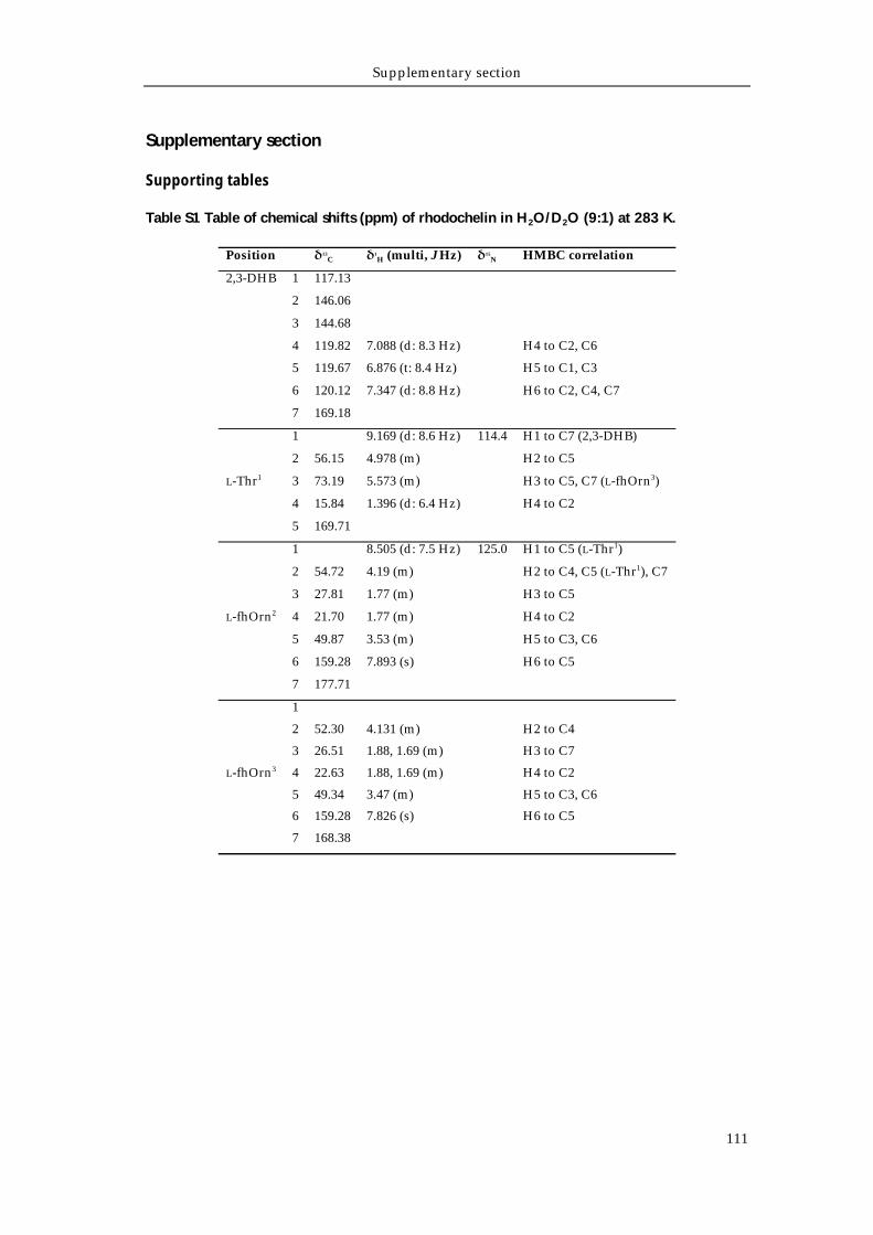
Supplementary section
111
Supplementary section
Supporting tables
Table S1 Table of chemical shifts (ppm) of rhodochelin in H2O/D2O (9:1) at 283 K.
Position δ13C δ1
H (multi, J Hz) δ15N HMBC correlation
2,3-DHB 1 117.13
2 146.06
3 144.68
4 119.82 7.088 (d: 8.3 Hz) H4 to C2, C6
5 119.67 6.876 (t: 8.4 Hz) H5 to C1, C3
6 120.12 7.347 (d: 8.8 Hz) H6 to C2, C4, C7
7 169.18
L-Thr1
1 9.169 (d: 8.6 Hz) 114.4 H1 to C7 (2,3-DHB)
2 56.15 4.978 (m) H2 to C5
3 73.19 5.573 (m) H3 to C5, C7 (L-fhOrn3)
4 15.84 1.396 (d: 6.4 Hz) H4 to C2
5 169.71
L-fhOrn2
1 8.505 (d: 7.5 Hz) 125.0 H1 to C5 (L-Thr1)
2 54.72 4.19 (m) H2 to C4, C5 (L-Thr1), C7
3 27.81 1.77 (m) H3 to C5
4 21.70 1.77 (m) H4 to C2
5 49.87 3.53 (m) H5 to C3, C6
6 159.28 7.893 (s) H6 to C5
7 177.71
L-fhOrn3
1
2 52.30 4.131 (m) H2 to C4
3 26.51 1.88, 1.69 (m) H3 to C7
4 22.63 1.88, 1.69 (m) H4 to C2
5 49.34 3.47 (m) H5 to C3, C6
6 159.28 7.826 (s) H6 to C5
7 168.38
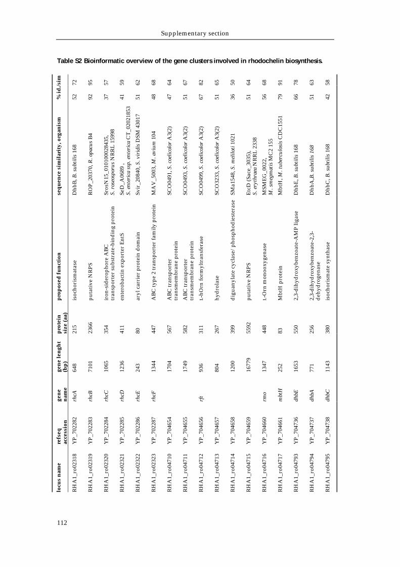
Supplementary section
112
Table S2 Bioinformatic overview of the gene clusters involved in rhodochelin biosynthesis. %
id./s
im
72
95
57
59
62
68
64
67
82
65
50
64
68
91
78
63
58
52
92
37
41
51
48
47
51
67
51
36
51
56
79
66
51
42
seq
uen
ce s
imil
arit
y, o
rgan
ism
Dhb
B, B
. sub
tilis
168
RO
P_2
0370
, R. o
pacu
s B
4
Sros
N15
_010
1000
2843
5,
S. r
oseo
spor
us N
RR
L 1
5998
SeD
_A06
89 ,
S. e
nter
ica
ssp.
ent
eric
a C
T_0
2021
853
Svir
_208
40, S
. vir
idis
DSM
430
17
MA
V_5
003,
M. a
vium
104
SCO
0491
, S. c
oelic
olor
A3(
2)
SCO
0493
, S. c
oelic
olor
A3(
2)
SCO
0499
, S. c
oelic
olor
A3(
2)
SCO
3233
, S. c
oelic
olor
A3(
2)
SMa1
548,
S. m
elilo
ti 1
021
Etc
D (S
ace_
3035
), S.
ery
thra
ea N
RR
L 2
338
MSM
EG
_002
2,
M. s
meg
mat
is M
C2
155
Mbt
H, M
. tub
ercu
losi
s C
DC
1551
Dhb
E, B
. sub
tilis
168
Dhb
A,B
. sub
tilis
168
Dhb
C, B
. sub
tilis
168
pro
pos
ed f
un
ctio
n
isoc
hori
smat
ase
pu
tati
ve N
RP
S
iron
-sid
erop
hore
AB
C
tran
spor
ter
subs
trat
e-bi
ndin
g p
rote
in
ente
roba
ctin
exp
orte
r E
ntS
aryl
car
rier
pro
tein
dom
ain
AB
C ty
pe
2 tr
ansp
orte
r fa
mily
pro
tein
AB
C tr
ansp
orte
r tr
ansm
embr
ane
pro
tein
AB
C tr
ansp
orte
r tr
ansm
embr
ane
pro
tein
L-h
Orn
form
yltr
ansf
eras
e
hyd
rola
se
dig
uan
ylat
e cy
clas
e/p
hosp
hod
iest
eras
e
pu
tati
ve N
RP
S
L-O
rn m
onoo
xyge
nase
Mbt
H p
rote
in
2,3-
dih
ydro
xybe
nzoa
te-A
MP
liga
se
2,3-
dih
ydro
xybe
nzoa
te-2
,3-
deh
ydro
gena
se
isoc
hori
smat
e sy
ntha
se
pro
tein
si
ze (a
a)
215
2366
354
411
80
447
567
582
311
267
399
5592
448
83
550
256
380
gen
e le
ngh
t (b
p)
648
7101
1065
1236
243
1344
1704
1749
936
804
1200
1677
9
1347
252
1653
771
1143
gen
e n
ame
rhcA
rhcB
rhcC
rhcD
rhcE
rhcF
rft
rmo
mbt
H
dhbE
dhbA
dhbC
refs
eq
acce
ssio
n
YP
_702
282
YP
_702
283
YP
_702
284
YP
_702
285
YP
_702
286
YP
_702
287
YP
_704
654
YP
_704
655
YP
_704
656
YP
_704
657
YP
_704
658
YP
_704
659
YP
_704
660
YP
_704
661
YP
_704
736
YP
_704
737
YP
_704
738
locu
s n
ame
RH
A1_
ro02
318
RH
A1_
ro02
319
RH
A1_
ro02
320
RH
A1_
ro02
321
RH
A1_
ro02
322
RH
A1_
ro02
323
RH
A1_
ro04
710
RH
A1_
ro04
711
RH
A1_
ro04
712
RH
A1_
ro04
713
RH
A1_
ro04
714
RH
A1_
ro04
715
RH
A1_
ro04
716
RH
A1_
ro04
717
RH
A1_
ro04
793
RH
A1_
ro04
794
RH
A1_
ro04
795
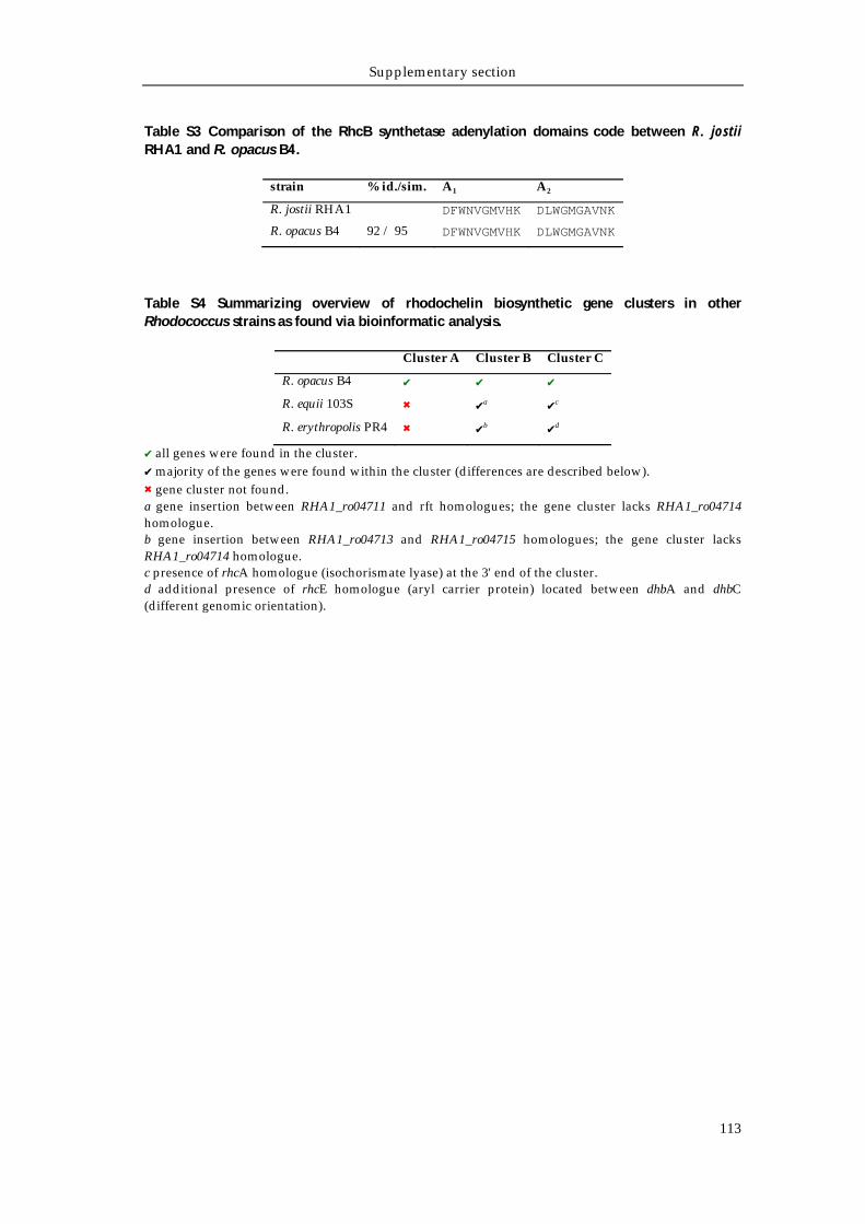
Supplementary section
113
Table S3 Comparison of the RhcB synthetase adenylation domains code between R. jostii RHA1 and R. opacus B4.
strain % id./sim. A1 A2
R. jostii RHA1 DFWNVGMVHK DLWGMGAVNK
R. opacus B4 92 / 95 DFWNVGMVHK DLWGMGAVNK
Table S4 Summarizing overview of rhodochelin biosynthetic gene clusters in other Rhodococcus strains as found via bioinformatic analysis.
Cluster A Cluster B Cluster C
R. opacus B4 ✔ ✔ ✔
R. equii 103S ✖ ✔a ✔c
R. erythropolis PR4 ✖ ✔b ✔d
✔ all genes were found in the cluster. ✔ majority of the genes were found within the cluster (differences are described below). ✖ gene cluster not found. a gene insertion between RHA1_ro04711 and rft homologues; the gene cluster lacks RHA1_ro04714 homologue. b gene insertion between RHA1_ro04713 and RHA1_ro04715 homologues; the gene cluster lacks RHA1_ro04714 homologue. c presence of rhcA homologue (isochorismate lyase) at the 3' end of the cluster. d additional presence of rhcE homologue (aryl carrier protein) located between dhbA and dhbC (different genomic orientation).
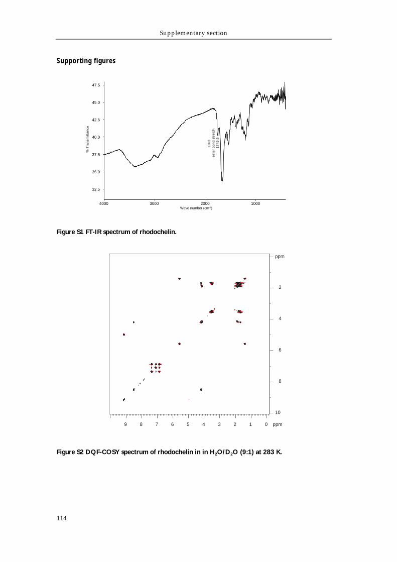
Supplementary section
114
Supporting figures
Figure S1 FT-IR spectrum of rhodochelin.
Figure S2 DQF-COSY spectrum of rhodochelin in in H2O/D2O (9:1) at 283 K.
1000200030004000
32.5
35.0
37.5
40.0
42.5
45.0
47.5
Wave number (cm-1)
% T
rans
mitt
ance
1749
.5
C=
O
este
r bo
nd s
tret
ch
ppm
9 8 7 6 5 4 3 2 1 0 ppm
10
8
6
4
2
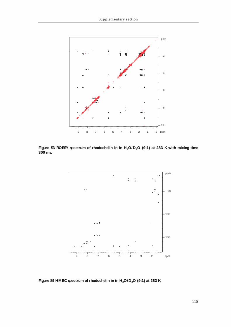
Supplementary section
115
Figure S3 ROESY spectrum of rhodochelin in in H2O/D2O (9:1) at 283 K with mixing time 300 ms.
Figure S4 HMBC spectrum of rhodochelin in in H2O/D2O (9:1) at 283 K.
ppm
9 8 7 6 5 4 3 2 1 0 ppm
10
8
6
4
2
ppm
23456789 ppm
50
100
150
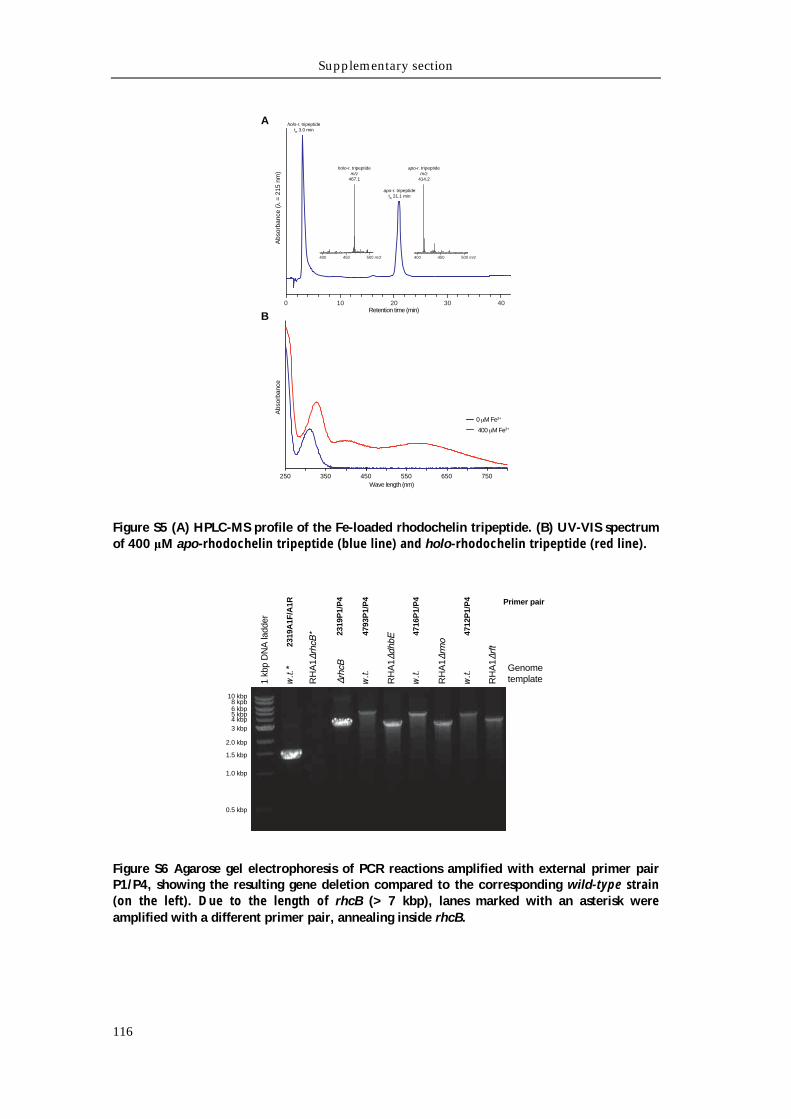
Supplementary section
116
Figure S5 (A) HPLC-MS profile of the Fe-loaded rhodochelin tripeptide. (B) UV-VIS spectrum of 400 μM apo-rhodochelin tripeptide (blue line) and holo-rhodochelin tripeptide (red line).
Figure S6 Agarose gel electrophoresis of PCR reactions amplified with external primer pair P1/P4, showing the resulting gene deletion compared to the corresponding wild-type strain (on the left). Due to the length of rhcB (> 7 kbp), lanes marked with an asterisk were amplified with a different primer pair, annealing inside rhcB.
A
B
250 350 450 550 650 750Wave length (nm)
0 M Fe3+
400 M Fe3+
0 10 20 30 40Retention time (min)
400 450 500 m/z 400 450 500 m/z
Abs
orba
nce
( =
215
nm
) A
bsor
banc
e
holo-r. tripeptidetR 3.0 min
holo-r. tripeptidem/z
467.1
apo-r. tripeptidetR 21.1 min
apo-r. tripeptidem/z
414.2
3 kbp
2.0 kbp
1.5 kbp
1.0 kbp
4 kbp5 kbp6 kbp8 kpb
10 kbp
w.t.
*
1 kb
p D
NA
ladd
er
RH
A1
w.t.
RH
A1
w.t.
RH
A1
w.t.
RH
A1
2319
A1F
/A1R
4793
P1/
P4
2319
P1/
P4
4716
P1/
P4
0.5 kbp
4712
P1/
P4 Primer pair
Genome template
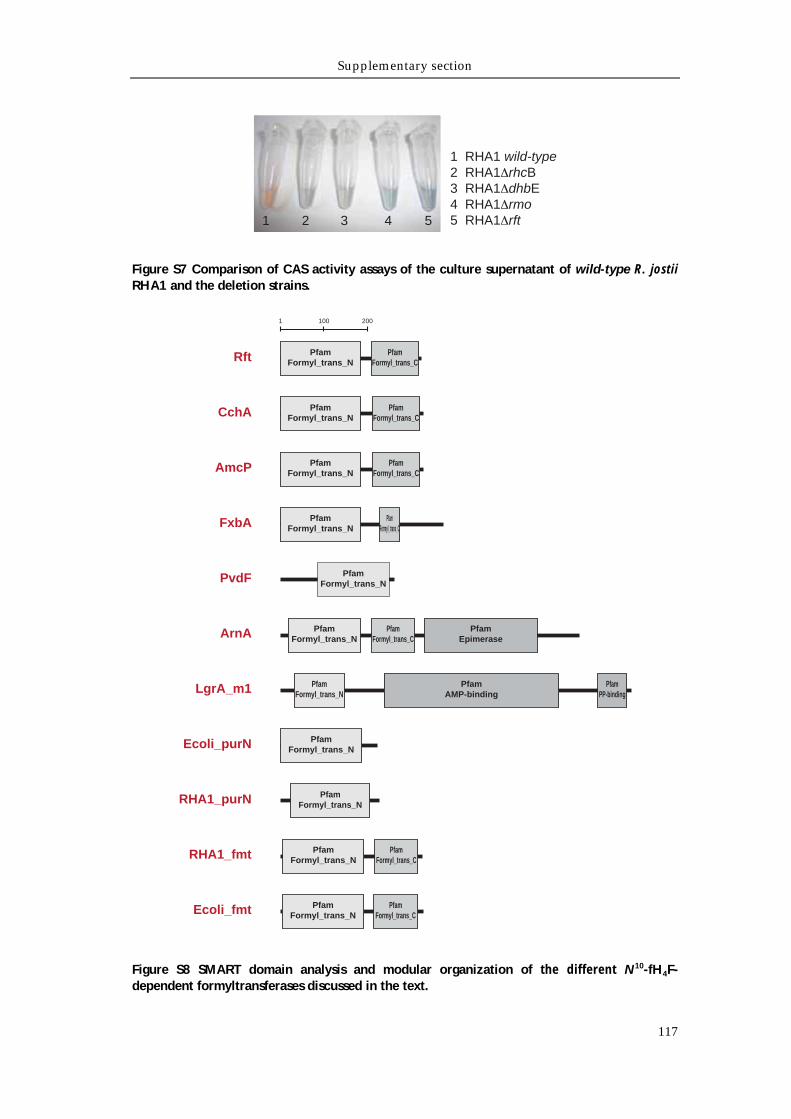
Supplementary section
117
Figure S7 Comparison of CAS activity assays of the culture supernatant of wild-type R. jostii RHA1 and the deletion strains.
Figure S8 SMART domain analysis and modular organization of the different N10-fH4F-dependent formyltransferases discussed in the text.
1 RHA1 wild-type2 RHA1 rhcB3 RHA1 dhbE4 RHA1 rmo5 RHA1 rft1 2 43 5
1 100 200
Rft PfamFormyl_trans_N
PfamFormyl_trans_C
FxbA PfamFormyl_trans_C
PfamFormyl_trans_N
PvdF PfamFormyl_trans_N
ArnA PfamFormyl_trans_N
PfamFormyl_trans_C
PfamEpimerase
RHA1_fmt PfamFormyl_trans_N
PfamFormyl_trans_C
CchA PfamFormyl_trans_N
PfamFormyl_trans_C
AmcP PfamFormyl_trans_N
PfamFormyl_trans_C
LgrA_m1 PfamAMP-binding
PfamPP-binding
PfamFormyl_trans_N
Ecoli_fmt PfamFormyl_trans_N
PfamFormyl_trans_C
Ecoli_purN PfamFormyl_trans_N
RHA1_purN PfamFormyl_trans_N

Acknowledgements
119
Acknowledgements
First and foremost, I would like to thank Prof. Dr. M. A. Marahiel for letting me join
his research group and for providing scientific guidance and constant support
during my Ph. D. thesis. I gratefully acknowledge his open‐mindedness towards
new ideas and enthusiasm about science, which was a constant source of motivation
and resulted in great benefit for my work. I am also grateful for the opportunity he
gave me to attend the “Annual Conference of the Association for General and
Applied Microbiology” in Karlsruhe and the “4th FEMS congress” in Geneve,
Switzerland. These meeting have truly been outstanding experiences that expanded
my scientific and personal horizons.
I gratefully thank Prof. Dr. M. Bölker for reviewing this thesis and, together with
Prof. Dr. E. Bremer and Dr. S.V. Albers, for being part of the thesis committee.
I would also like to thank the Max Planck Institute for Terrestrial Microbiology for
supporting my studies with the fellowship and the constructive activities held by
the International Max Planck Research School for Environmental, Cellular and
Molecular Microbiology. I am also grateful to Dr. C. van der Does for being part of
the IMRPS committee. I am thankful to Roswita Roller-Müller and Susanne Rommel
for their administrative help.
I would like to thank Dr. Uwe Linne for his excellent support with HPLC and MS
and for his expert advices. I am deeply indebt to Dr. Xiulan Xie for conducting
extensive and challenging NMR analyses for rhodochelin structure elucidation.
A special thank you goes to everyone in the Marahiel group for their support, the
fruitful discussions and the great time in the lab. I would like to thank current and
former colleagues of lab 4710 for the nice working atmosphere: Dr. Lars Robbel, Dr.
Florian Peuckert, Dr. Alexander Albrecht, Andreas Mielcarek, Mustafa Zeyadi,
Kamila Franke and Christin Schönfeld.
Antje Schäfer, Anke Botthof, Christiane Bomm and Gabriele Schimpff‐Weiland are
gratefully acknowledged for the technical assistance through the years and Tobias
Gießen and Andreas Mielcarek for the careful proofreading of this thesis.
Special thanks go to my friends Luca, Anita, Simone and Emiliano, as well as all for
anyone who supported myself from Italy.
No words can describe how thankful I am to Rossella: our friendship grew over
distance (and through the internet) since the early days of my stay in Germany and
helped us support each other in the choices we made day by day.
Acknowledgements
120
Finally, I am completely grateful to my parents, who have been a continuous
support throughout my whole life. Their constant encouragement has made all of
this possible. For this, I dedicate this thesis to them.
Erklärung
121
Erklärung
Ich versichere, dass ich meine Dissertation „Structural characterization of the
siderophore rhodochelin from Rhodococcus jostii RHA1 and elucidation of its
biosynthetic machinery” selbständig, ohne erlaubte Hilfe angefertigt und mich
dabei keiner als der von mir ausdrücklich bezeichneten Quellen und Hilfen bedient
habe.
Die Dissertation wurde in der jetzigen oder einer ähnlichen Form noch bei keiner
anderen Hochschule eingereicht und hat noch keinen sonstigen Prüfungszwecken
gedient.
Marburg, 19 Juni 2012