austrian ournal of cardiolog Österreichische …p.b.b. 02z03110m , verlagsort 00 ablitz...
TRANSCRIPT
P.b.b. 02Z031105M, Verlagsort : 3003 Gablitz, Linzerstraße 177A/21 Preis: EUR 10,–
Krause & Pachernegg GmbH • Verlag für Medizin und Wirtschaft • A-3003 Gablitz
KardiologieJournal für
Austrian Journal of CardiologyÖsterreichische Zeitschrift für Herz-Kreislauferkrankungen
Indexed in EMBASE/Excerpta Medica/SCOPUS
Offizielles Organ des Österreichischen Herzfonds
Homepage:
www.kup.at/kardiologie
Online-Datenbank mit Autoren-
und Stichwortsuche
Member of the
ESC-Editor‘s Club
Vienna-Mayo Contemporary Clinical
Cardiology 2006 - September 14-16
2006, Vienna (Abstracts)
Journal für Kardiologie - Austrian
Journal of Cardiology 2006; 13
(11-12), 365-377
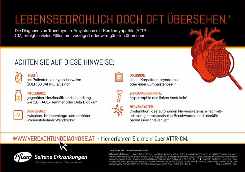
LebensbedrohLich doch oft übersehen.1
Die Diagnose von Transthyretin-Amyloidose mit Kardiomyopathie (ATTR-CM) erfolgt in vielen Fällen erst verzögert oder wird gänzlich übersehen.
HFpEF*: bei Patienten, die typischerweise übeR 60 JAhRe alt sind2
INTOLERANZ: gegenüber Herzinsuffizienzbehandlung wie z.b.: ACe-hemmer oder beta blocker3
DISKREPANZ: zwischen Niedervoltage und erhöhter linksventrikulärer Wanddicke4
HEART FAILURE WITH PRESERVED EJECTION FRACTION in pat ients typ ica l l y over 60 1-3
INTOLERANCE to s tandard hear t fa i lu re therap ies , such as ang io tens in-conver t ing enzyme inh ib i to rs , ang io tens in receptor b lockers , and beta b lockers 7-9
DISCORDANCE between QRS vo l tage on e lect rocard iography (ECG) and le f t vent r icu la r (LV ) wa l l th ickness seen on echocard iography 10,11
D iagnos is o f CARPAL TUNNEL SYNDROME or LUMBAR SPINAL STENOSIS8,14 ,16-22
Echocard iography showing INCREASED LV WALL THICKNESS2,13 ,22 ,25 ,26
AUTONOMIC NERVOUS SYSTEM dysfunction, inc lud ing gast ro in tes t ina l compla in ts or unexp la ined we ight loss 2,22,27 ,28
Achten sie Auf diese hinweise:
* heart failure with preserved ejection fraction
Pfizer Corporation Austria GmbH, Wien, www.pfizer.at
Referenzen: 1. Connors LH, Sam F, Skinner M, et al. Heart failure due to age-related cardiac amyloid disease associated with wild-type transthyretin: a pro-spective, observational cohort study. Circulation. 2016;133(3):282-290. 2. Maurer MS, Hanna M, Grogan M, et al. Genotype and phenotype of transthyretin cardiac amyloidosis: THAOS (Transthyretin Amyloid Outcome Survey). J Am Coll Cardiol. 2016;68(2):161-172. 3. Brunjes DL, Castano A, Clemons A, Rubin J, Maurer MS. Transthyretin cardiac amyloidosis in older Americans. J Card Fail. 2016;22(12):996-1003. 4. Carroll JD, Gaasch WH, McAdam KP. Amyloid cardiomyopathy: characterization by a distinctive voltage/mass relation. Am J Cardiol. 1982;49:9-13. PP-VYN-AUT-0207/01.2020
Pfizer Corporation Austria GmbH, Wien, www.pfizer.at
www.verdAchtunddiAgnose.At - hier erfahren sie mehr über Attr-cM.
DIAGNOSE: eines Karpaltunnelsyndroms oder einer Lumbalstenose1,3
ECHOKARDIOGRAPHIE: hypertrophie des linken Ventrikels2
NERVENSYSTEM: Dysfunktion des autonomen Nervensystems einschließ-lich von gastrointestinalen beschwerden und unerklär-barem Gewichtsverlust2
Cardiology 2006 – Abstracts
J KARDIOL 2006; 13 (11–12) 365
„Von der Forschung zur Klinik“
Vienna-Mayo Contemporary Clinical Cardiology 2006
September 14–16, 2006, Vienna
Abstracts
B-Type Natriuretic Peptide in Low-Flow Aortic Steno-sis: Relationship to Hemodynamics and Clinical Out-come. Results from the Multicenter TOPAS Study
J. Bergler-Klein1, G. Mundigler1, P. Pibarot2, I. Burwash3, C. Fuchs1, J. G. Dumesnil2,C. Blais2, R. Beanlands3, Z. Hachicha2, D. Mohty-Eichahidi2, N. Loho1, F. Rader1,P. Eickhoff1, H. Baumgartner1
1Department of Cardiology, Medical University of Vienna, Austria; 2Laval Hospital/Quebec Heart Institute, Laval University, Sainte Foy, Quebec, Canada; 3University ofOttawa Heart Institute, Ottawa, Ontario, CanadaBackground B-type natriuretic peptide (BNP) has been studied inaortic stenosis (AS), but no data have been reported for patients withlow-flow/low-gradient AS. Therefore, we studied the relationshipof BNP and Nt-BNP with rest and stress hemodynamics as well asclinical outcome in this group.Methods Plasma BNP and Nt-BNP were measured in 72 pts with ASundergoing dobutamine stress echocardiography (DSE). 63 pts hadlow-flow AS with indexed effective orifice area [EOA] < 0.6 cm2/m2, mean gradient [MG] < 40 mmHg and LV ejection fraction [EF]≤ 0.40 biplane Simpson technique. Nine pts with AS and normal EFserved as controls. Pts were classified as truly severe [TS] orpseudo-severe AS [PS] based on their projected EOA at a normalflow rate of 250 mL/s ≤ or > 1.0 cm2 in DSE, as previously proposedin the TOPAS study.Results BNP and Nt-BNP were markedly elevated in low-flowAS (BNP 991 ± 1115 vs. controls 190 ± 183 pg/mL, p = 0.025; Nt-BNP 7330 ± 16,261 vs. 193 ± 199 pg/mL), but varied widely. LogBNP was inversely related to EF at rest (r = 0.60; p < 0.0001) andpeak stress (r = 0.51; p < 0.0001), as well as to EOA at rest (r = 0.48,p < 0.0001) and peak stress (r = 0.47, p < 0.0001), stroke volume(BNP, r = 0.32, p = 0.012), mean transvalvular flow rate (r = 0.26,p = 0.04) and wall motion score index (r = 0.40, p = 0.001). Similarfindings were observed for Nt-BNP. BNP was significantly higherin 25 TS compared to 38 PS pts (1162 ± 1229 vs. 680 ± 866 pg/mL,p = 0.008). Similarly, BNP was higher in 23 vs. 40 pts with a peakstress EOA ≤ or > 1.0 cm2 (1466 ± 1448 vs. 530 ± 467 pg/mL,p < 0.001). In the subgroup of 24 patients who underwent aorticvalve replacement, BNP was higher in 6 pts who died postopera-tively compared to 18 pts surviving valve replacement (1975 ±2261 vs. 815 ± 492 pg/mL, p < 0.05). In the total cohort, cumula-tive 1-year survival of pts with BNP ≥ 550 pg/mL was signifi-cantly lower than of pts with BNP < 550 (51 ± 11 % vs. 92 ± 5 %,p = 0.04).Conclusion In pts with low-flow AS, BNP and Nt-BNP are mark-edly elevated and related to EF and EOA at rest and peak DSE. BNPis significantly higher in truly severe compared to pseudo-severe AS.BNP predicts poor postoperative outcome in the subset of patientsundergoing valve replacement. Overall one-year survival is poor inpts with BNP ≥ 550, but reasonable in pts with BNP < 550 pg/mL.
Exercise-Induced Pulmonary Hypertension in Patientsafter Successful Pulmonary Endarterectomy
D. Bonderman, R. Hitsch, A. Martischnig, N. Skoro-Sajer, M. Kneußl, W. Klepetko,I. M. LangDepartment of Internal Medicine II, Medical University of ViennaBackground Pulmonary endarterectomy (PEA) provides poten-tial cure for patients with chronic thromboembolic pulmonary hy-pertension (CTEPH). Successfully operated patients have been shownto normalize exercise capacity and hemodynamic parameters inlong-term studies.
Abstracts in alphabetical order based on first authors’ last names.
Methods To investigate whether pulmonary hypertension can beprovoked by exercise, we studied patients at least one year after suc-cessful PEA with documented (near) normalization of exercise ca-pacity and hemodynamics. Patients (n = 13) and age-matched non-pulmonary hypertensive controls (n = 14) underwent echocardio-graphy at submaximal treadmill exercise.Results Resting mean pulmonary arterial pressure was 25 ± 9 mmHg,mean pulmonary vascular resistance was 291 ± 148 dynes × s × cm–5,mixed venous saturation was 71 ± 5 % and mean cardiac output was5.2 ± 1.1 l/min at 63 ± 31 (range 16–120) months after PEA. Therewas no difference in age (61 ± 10 vs. 57 ± 13 years, p = 0.5) or6-minute walking distance (489 ± 114 vs. 456 ± 45 meters, p = 0.32)between patients and controls. While the difference in restingsystolic pulmonary arterial pressures (sPAP) reached only border-line significance (41 ± 18 vs. 30 ± 6 mmHg, p = 0.05), there was asignificant difference in exercise-sPAP (71 ± 23 vs. 46 ± 11 mmHg,p = 0.001), resting pulmonic valve acceleration time (102 ± 24vs. 132 ± 17 ms, p = 0.0008) and serum BNP levels (207 ± 134 vs.70 ± 77 pg/ml, p = 0.007).Conclusions Patients with normal exercise capacity and restinghemodynamics after PEA demonstrate significant pulmonary hy-pertension at exercise. There is a need for studies investigatingwhether this patient population does additionally benefit from va-sodilator therapies.
Bacterial Infection is a Mechanism Underlying a Failureof Thrombus Resolution in Chronic ThromboembolicPulmonary Hypertension
D. Bonderman1, B. Redwan1, J. Jakowitsch1, H. Bergmeister2, H. Panzenböck1,M. K. Renner1, W. Klepetko3, U. Losert3, A. Georgopolos4
1Department of Internal Medicine II; 2Institute of Biomedical Research; 3Departmentof Cardiothoracic Surgery, Division of Cardiology; 4Department of Internal Medicine I,Medical University of Vienna
Background Chronic thromboembolic pulmonary hypertension(CTEPH) results from single or recurrent pulmonary thromboemboliarising from sites of venous thrombosis. In patients with CTEPH,thromboemboli do not resolve but form endothelialized, fibroticobstructions of the pulmonary vascular bed. Mechanisms underly-ing thrombus organisation are poorly understood. Because of theobservation that infected intravenous leads enhance the likelihoodof CTEPH, we tested the hypothesis that bacterial infection causes afailure of thrombus resolution.Methods Human thromboendarterectomy specimens were sterillycollected during surgery and analyzed with a bacterial 16S ribo-somal DNA screening protocol. In a next step, a mouse model of venousthrombus formation was employed to investigate thrombus resolu-tion in the absence and presence of low doses of staphylococcusaureus (0.15 ml of 105/ml injected as a single bolus into the tailvein). On days 1, 3, 7, 14 and 28 after thrombus induction, animalswere sacrificed, thrombi were harvested, fixed and embedded inparaffin.Results 520 bp PCR products were obtained in 16 of 25 CTEPHthrombi, but in only 4 specimens derived from patients with acutepulmonary embolism. Cross-sectional area analysis demonstratedthat thrombi from infected animals were larger than control thrombi(day 7: median cross-sectional area (CSA) 0.431 vs. 0.279 mm2; day28: median CSA 0.128 vs. 0.018 mm2, n = 8, p < 0.05). Volumetryconfirmed significantly larger thrombus volumes on days 3 and 28(day 3: median thrombus volume 1.798 vs. 1.441 mm³; day 28 me-dian thrombus volume 0.427 vs. 0.056 mm³, n = 8, p < 0.05). Real-time PCR demonstrated increasing expression of connective tissuegrowth factor (CTGF) in the thrombi over the observation period,contrasting the decline of CTGF expression in controls.
For personal use only. Not to be reproduced without permission of Krause & Pachernegg GmbH.
366 J KARDIOL 2006; 13 (11–12)
Cardiology 2006 – Abstracts
Discussion The data demonstrate that infection with staphylococcusaureus enhances thrombus formation and persistence. CTGF expres-sion analysis suggests that abnormal thrombus organization occursafter bacterial infection.
Bosentan for the Treatment of Chronic Thromboem-bolic Pulmonary Hypertension – One-Year Experience
D. Bonderman, N. Skoro-Sajer, M. Kneußl, W. Klepetko, I. LangDepartment of Internal Medicine II, Medical University of Vienna
Background Bosentan, an oral endothelin ETA/ETB-receptor an-tagonist, is effective in the short-term treatment of inoperablechronic thromboembolic pulmonary arterial hypertension (CTEPH).We investigated hemodynamics, safety and efficacy of bosentantherapy at one year of therapy in 21 patients (13�/8�, mean age 71± 12 years) who were treated off-label over 16 ± 6 months.
Results After one year of treatment, NYHA functional class hadimproved by one class in 14 patients. Mean six-minute walking dis-tances increased from 299 ± 131 m at baseline to 387 ± 121 m(p = 0.04). In parallel, proBNP decreased from 3365 ± 2923 pg/mlto 1579 ± 2103 pg/ml (p = 0.02). Overall, mean pulmonary arterialpressure (mPAP) decreased from 48 ± 10 to 43 ± 12 mmHg (p = 0.17),pulmonary vascular resistance (PVR) changed from 653 ± 247 to468 ± 205 dynes × s × cm–5 (p = 0.04). If hemodynamic non-responders to therapy were excluded (n = 5), mPAP decreased from50 ± 10 to 42 ± 11 mmHg (p = 0.17), and PVR changed from757 ± 232 to 420 ± 137 dynes × s × cm–5 (p = 0.015). NeitherAST (25 ± 2 vs. 25 ± 2 U/l, p = 0.25) nor ALT (23 ± 12 vs.24 ± 9 U/l, p = 0.57) changed significantly. Two deaths occurredfrom causes unrelated to pulmonary hypertension.
Conclusions Our study suggests a beneficial long-term effect ofthe oral dual endothelin receptor antagonist, bosentan, in patientswith inoperable CTEPH. Non-responders to bosentan therapy mustbe further characterized.
Decreased Cardiac Remodeling after Combined (Intra-myocardial and Intracoronary) Autologous Stem CellTreatment in Chronic Heart Failure
S. Charwat, M. Gyöngyösi, R. Jacob, G. Beran, I. Lang, M. Dettke, S. Graf,N. Nyolczas, H. Sochor, D. GlogarDepartment of Cardiology, Medical University of Vienna
Background The aim of our prospective study was to assess theeffect of combined (intramyocardial and intracoronary) autologousbone-marrow stem cell (BM-SC) therapy on cardiac remodeling inpatients with severe coronary artery disease and chronic heart failure.
Methods Thirty-two no-option patients (94 % men, 55 ± 12 y)with congestive heart failure and left ventricular (LV) ejection frac-tion (EF) < 40 %, not amenable for conventional revascularization,underwent combined, NOGA-guided intramyocardial (3.8 ± 0.3 ml)and intracoronary (29 ± 14 ml) autologous BM-SC therapy. Base-line and 6-month follow-up (FUP) clinical symptoms (NYHA,CCS), LV systolic and diastolic functions (measured by contrastventriculography), myocardial viability and segmental wall motion(NOGA endocardial mapping) and stress-induced as well as restingperfusion defect sizes (99m-Tc-Sestamibi SPECT myocardialperfusion scintigraphy) were compared.
Results At FUP, a marked increase in LV EF (from 36.5 ± 8.0 %to 43.0 ± 10.4 %, p < 0.001) along with a significant (p < 0.05) de-crease in LV end-diastolic volume (from 240 ± 57 to 223 ± 60 ml),end-diastolic pressure (from 24.1 ± 7.9 to 20.8 ± 8.0 mmHg), LVend-diastolic diameter (from 57.9 ± 5.2 to 54.9 ± 4.6 mm) and dia-meter of the left atrium (from 46.6 ± 6.9 to 44.0 ± 8.1 mm) was found.This improvement was accompanied by a decrease (p < 0.01) inheart rate (from 72.3 ± 13.4 to 67.7 ± 12.6), CCS (from 2.4 ± 1.1 to1.3 ± 0.6) and NYHA (from 2.5 ± 0.8 to 1.5 ± 0.7). Combined stemcell therapy induced a reduction of stress-induced perfusion defectsize (from 26.9 ± 8.7 to 22.2 ± 10.1 % of the total myocardium, p <0.05), while a trend to smaller resting defect at FUP was measured.Myocardial viability (measured by NOGA mapping, from 7.7 ± 2.8to 8.6 ± 2.3 mV) and the local linear shortening (from 5.6 ± 1.4 to7.3 ± 1.5 %) of the treated area improved significantly.
Conclusions Combined application of stem cell therapy decreasescardiac remodeling in patients with chronic heart failure, improvingthe systolic and diastolic functions of the heart.
Rheumatoid Arthritis is Associated with SystemicArterial Stiffness
A. Cypiene¹, A. Laucevicius², A. Venalis¹, M. Kovaite², L. Ryliskyte², J. Dadoniene¹,Z. Petrulioniene², V. Dzenkeviciute²¹The Institute of Experimental and Clinical Medicine at Vilnius University, Lithuania;²Clinics of Heart Diseases, Vilnius University – Centre of Cardiology and Angiology,Vilnius University Hospital Santariskiu Klinikos, Vilnius, Lithuania
Background Rheumatoid arthritis (RA) is associated with prema-ture atherosclerosis. Chronic inflammation may impair arterial func-tion and lead to the increase of their stiffness.Aim of the Study was to assess whether RA, disease duration andincrease of high-sensitivity C-reactive protein (hsCRP) can influ-ence arterial stiffness in RA patients.Methods This study included 53 RA patients (40.1 ± 9.8 years)with moderate and high disease activities (DAS28 3.21–7.05) and55 controls (39.7 ± 8.1 years). Blood tests included serum lipid pro-file, glucose and hsCRP measurements. The augmentation index(AIx), a measure of systemic arterial stiffness, was assessed non-invasively by applanation tonometry (Sphygmocor v. 7.01, AtCorMedical).Results In RA patients, the AIx values adjusted for heart rate andlevel of CRP were significantly higher compared to controls (AIx21.3 ± 13.3 % vs. 12.7 ± 13.2, p = 0.001; CRP 31.32 ± 40.29 mg/l vs.1.58 ± 3.36 mg/l, p < 0.001, respectively). Significant influence ofdisease duration on AIx was observed by multiple regression analy-sis (adjusted r² = 0.559; p = 0.002). Correlation between hsCRP andAIx was not significant in RA patients (Pearson’s r = –0.044;p = 0.752) as well as in controls (Pearson’s r = 0.215; p = 0.121).Conclusion Duration of rheumatoid arthritis but not elevation ofserum hsCRP is related to the premature increase of systemic arte-rial stiffness.
Lp(a) Predicts Early Onset of Atherosclerosis
V. Dzenkeviciute, J. Badariene, L. Ryliskyte, Z. Petrulioniene, A. LauceviciusCenter of Cardiology and Angiology, Vilnius University Hospital SantariskiuKlinikos, Vilnius, Lithuania – Clinics of Heart Diseases, Vilnius University, Vilnius,Lithuania
Background A family history of premature coronary artery disease(CAD) is one of the main risk factors in middle-aged patients.Aim The aim of our study was to assess the relationship be-tween intima-media thickness (IMT), measured by B-mode ul-trasound, and conventional risk factors in families with prema-ture CAD.Methods The study population consisted of 32 families with pre-mature CAD. In total, 50 subjects were studied. Each family in thecohort has at least one affected sibling with premature CAD. Dataabout sex and other risk factors were obtained. Plasma levels ofhomocysteine, IL-6, CRP, ox-LDL and lipids were measured. Ca-rotid and femoral IMTs were assessed by high-resolution B-modecarotid ultrasound (GE, 13 MHz).Results Patients with premature CAD were more likely to havediabetes mellitus (5.3 % vs. 0 %, p = 0.001), arterial hypertension(69 % vs. 25 %, p = 0.017), dyslipidemia (96.9 % vs. 71.5 %,p = 0.006) and were male (97 % vs. 50 %, p = 0.001) with a higherBody Mass Index (31.7 ± 6.1 vs. 28.19 ± 3.2, p = 0.03). Advancedsub-clinical atherosclerosis was present in 69 % of family members,but prevalence of elevated IMT was higher in CAD patients(p = 0.001). Patients with premature CAD had major values of Lp(a)and lower values of total cholesterol, HDL-C, ApoA1, IL-6 and Ox-LDL-C. In a stepwise regression model, only gender (p < 0.036) in-dependently predicted the mean IMT. After controlling for gender,the independent predictor of mean IMT was Lp(a) (p < 0.015)(Table 1).Conclusion Lp(a) and gender showed a significant associationwith subclinical atherosclerosis. The present study demonstrates thatLp(a) is a strong predictor for early onset of atherosclerosis.
Table 1. V. Dzenkeviciute et al.
Variable Beta 95 % CI P βββββ SE
Gender 0.351 0.01–0.037 0.036 0.019 0.009After adjustment ofgender Lp(a) 0.398 0.0078–0.86 0.021 0.337 0.015
Cardiology 2006 – Abstracts
J KARDIOL 2006; 13 (11–12) 367
Assessment Of Plaque Composition in Cardiac Allo-graft Vasculopathy by Virtual HistologyTM
J. Fingernagel, C. Schukro, P. Pichler, S. Winkler, M. Vertesich, S. Ingerle,D. GlogarDept. of Internal Medicine II, Division of Cardiology, Medical University of Vienna
Background Previous pathological studies of coronary plaque inheart transplant patients showed a predominance of fibrous plaquecomponents. In this prospective study, we aimed to assess coronaryplaque composition in cardiac allograft vasculopathy by intravascularultrasound with Virtual HistologyTM (Volcano Therapeutics Inc.).Methods Intravascular ultrasound runs with automatic pullback(0.5 mm/s) were available for 20 heart transplant patients (trans-plantation was performed 8.6 ± 3.2 years ago). In each patient, onelesion of interest was defined at the site of maximal coronary plaqueburden. Analysis of plaque composition was performed with the VirtualHistologyTM software.Results Mean lesion length was 11.7 ± 4.8 mm. Three patientsshowed hemodynamically significant stenoses. Mean plaque burdenwas 33.7 ± 8.4 % (minimal lumen diameter: 3.0 ± 0.7 mm; minimallumen area: 9.4 ± 3.3 mm). Plaque composition as assessed by Vir-tual HistologyTM was predominantly fibrotic (66 %), whereas fibro-fatty, calcified and necrotic plaque fractions were present in 21 %,5 % and 8 %, respectively.Conclusions Intravascular ultrasound with Virtual HistologyTM
allows for differentiation of coronary plaque components in car-diac allograft vasculopathy. Comparable to previous ex vivo studies,plaque composition in heart transplant patients was predominantlyfibrotic.
Stenting of Coronary Bifurcations: One- vs. Two-StentStrategy in a Bench Model
B. Frey1, B. Pausa2, H. Mayr2, M. Zehetgruber1
1Department of Internal Medicine II, Medical University of Vienna; 2Central Clinic,St. Pölten
Background Bench-testing provides insights into complex stentingstrategies of bifurcations. A two-stent strategy to completely coverthe carina results in a higher sidebranch (SB) restenosis rate whencompared to a single-stent strategy of covering only the mainbranch(MB). This might be due to a smaller SB ostial lumen when usingcrushing techniques.Methods We compared a kissing-balloon- (KB) only strategy to ex-ternal-crush (EC) and internal-crush stenting (IC) in a bifurcationalsilicon model. Minimal lumen diameter (MLD) of the SB ostium aswell as the proximal carina (PC) and distal carina (DC) of the main-branch stent were measured by a nozzle gauge. Testing was performedusing Cypher Select (Cordis, NY) and Taxus Express (Boston Sci-entific, MA) stents in a 45-degree-angled 3.00 mm MB and 3.00 mmSB bifurcational model (n = 8 for all techniques). Cypher stents wereimplanted with 16 atm, Taxus Express stents were implanted with13 atm and postdilated with a Cypher balloon with 16 atm. Stentsoverlapped for > 5 mm in EC and IC. Final kissing was performedwith two Cypher balloons with 8 atm (Table 2).Results MLD of SB ostium was significantly larger in KB vs. EC andIC.Conclusions KB leads to a significantly larger MLD of the SB os-tium compared to EC and IC. This might explain in part the higherSB restenosis rate after a two-stent strategy.
Detection of Coronary Artery Fistulas in AsymptomaticPatients. The Role of Two-Dimensional Doppler Echo-cardiography
C. Ginghina, M. Rugina, B. A. Popescu, I. Coman, L. Zarma, M. Cozma,I. Craciunescu, C. Ceck, E. Apetrei“Prof. Dr. C. C. Iliescu” Institute of Cardiovascular Diseases, Bucharest, Romania
Background Coronary artery fistulas are occasionally found in pa-tients who undergo coronary angiography and they may involve anyepicardial coronary artery. The natural history in asymptomatic adultpatients is unknown.Aim The aim of the study was to assess the role of two-dimensionalechocardiography (TTE) complemented by pulsed Doppler ultrasoundand color flow imaging in the diagnosis of coronary artery fistulas (CAF)in asymptomatic patients, usually diagnosed by coronary angiographyand cardiac catheterization.Material and Methods In a retrospective study covering the years1985–2005, 19 patients (pts.) with silent CAF were identified. The pa-tients, aged 8–60 years, 12 of them men (63 %), were studied by two-dimensional TTE with pulsed Doppler and color flow imaging and byother noninvasive methods (ECG, chest X-ray, phonocardiogram, firstpass radionuclide). In all pts, the final diagnosis of CAF was made byselective coronary angiography.Results The clinical, echocardiographic, ECG and angiographic find-ings of clinically silent CAF were analyzed. Indications for echocardio-graphy were: continuous murmur in 15 pts (79 %) and ECG changes in4 pts (21 %). CAF was detected with two-dimensional TTE (Doppler andcolor flow) in 13 pts (70 %). The CAD originated from the left coronaryartery in 5 pts, the right coronary artery in 7 pts and bilaterally in 1 patient.The drainage sites were the right ventricle in 6 pts, the left ventricle in4 pts, the right atrium in 1 patient and the pulmonary artery in 3 pts. In allpatients, the final diagnosis of CAF was made by selective coronaryangiography. In one case, TTE showed the enlarged origin of a single leftcoronary artery with CAF communicating with the right ventricle.Conclusions Our study confirmed that two-dimensional TTE (withPW Doppler and color flow) is a useful non-invasive technique inidentifying asymptomatic pts with CAF.
Multiple Interatrial Septal Defects – Assessment ofMorphology and Pathological Associations
C. Ginghina, A. Teodorescu, C. Siminiceanu, B. A. Popescu, M. Rugina, I. Stoian,C. Calin, C. Ceck, E. Apetrei“Prof. Dr. C. C. Iliescu” Institute of Cardiovascular Diseases, Bucharest, Romania
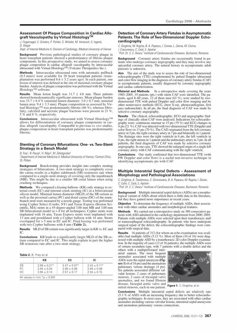
Background Multiple interatrial septal defects (ASDs) are a morpho-logical variant of ASDs about which there is little data in the literature,but they have gained more importance in recent years.Objective To determine the frequency of multiple ASDs, their associa-tion with other cardiac anomalies and morphological features.Methods We carried out a retrospective study on 389 consecutive pa-tients with ASD admitted in the cardiology department from 2000–2005.Patients with multiple ASDs were selected upon their transthoracic and/or transesophageal echocardiography. In patients who have undergonesurgical repair of the defect, the echocardiographic findings were com-pared with surgical data.Results 16 patients of 312 (for whom an echo examination was avail-able) had multiple ASDs (5.12 %). Most of them (10 of 16) were diag-nosed with multiple ASD by a transthoracic 2D color Doppler examina-tion. In the majority of cases (12 of 16 patients), the multiple ASDs wereof ostium secundum type, with 7 patients with a double defect and theothers with a multiperforated inter-atrial septum. The most frequentanomalies associated with multipleASDs were the septal aneurysm (Fig-ure 1) (6 of 16 pts) and the anomalouspulmonary venous drainage (4 pts).Six patients associated different val-vular lesions: 2 cases of pulmonarystenosis, 2 cases of tricuspid valveanomalies, and we found Ebsteindisease, bicuspid aortic valve andmitral stenosis, each in one patient.Conclusions Multiple interatrial septal defects are relatively rare(5.3 % of ASD) with an increasing frequency due to better echocardio-graphic techniques. In most cases, they are associated with other cardiacanomalies including various valvular lesions, interatrial septal aneurysmsand anomalous pulmonary venous connections.
Table 2. B. Frey et al.
KB EC IC
SB 2.69 ± 0.21* 2.47 ± 0.07* 2.42 ± 0.11*PC 2.94 ± 0.24 2.95 ± 0.08 2.94 ± 0.08DC 2.65 ± 0.14 2.57 ± 0.17 2.54 ± 0.15
All values in mm,*p < 0.001
Figure 1: C. Ginghina, et al.
368 J KARDIOL 2006; 13 (11–12)
Cardiology 2006 – Abstracts
FDG Gamma Camera PET Equipped with a 1-inch Crys-tal In Detection of Viable Myocardium: Comparisonwith Dedicated PET and Tc-Tetrofosmin
S. Graf1, A. Khorsand1, M. Behesti2, G. Dobrozemsky2, M. Wadsak2, K. Kletter2,R. Dudczak2, G. Porenta3, C. Pirich2
1Department of Cardiology; 2Department of Nuclear Medicine, Medical Universityof Vienna; 3Department of Nuclear Medicine, Rudolfinerhaus, Vienna
Background The purpose of this study was to compare FDG gammacamera PET (GCPET) equipped with one-inch NaI crystals and Tc-99-Tetrofosmin- (Tc) SPECT with and without attenuation correc-tion with FDG-dedicated PET (dPET) as validated referencemethod for detection of myocardial viability.Material and Methods GCPET, Tc and dPET were performed in11 patients (10 males, 1 female, age 63 ± 10 years) with coronaryartery disease and reduced left ventricular ejection fraction. Tcimaging was assessed with a dual-headed gamma camera (SiemensECAM). For GCPET imaging, a dual-headed gamma camera(GEMS Millennium VG with Hawkeye) equipped with 1-inch thickNaI crystals was used. PET studies were performed with a dedicatedPET camera (GE-Advance). For all three methods, polar maps weregenerated using the “Munich Heart” image analysis tool. For quantita-tive analysis of polar maps, each polar map was divided into 16myocardial segments (4 apical, 6 midventricular and 6 basal seg-ments). Segmental tracer uptake was normalized to the maximaluptake and expressed as percentage of the maximal segmental traceruptake.Results Regression analysis of averaged segmental activity of allmyocardial segments (n = 176) showed significant correlation betweenGCPET-ac and dPET (R = 0.82), GCPET-nc and dPET (R = 0.63),Tc-ac and dPET (R = 0.75), Tc-nc and dPET (R = 0.75). Cross-tableanalysis between different techniques and dPET for identificationof viable segments showed an agreement of 89 % (κ = 0.63) forGCPET-ac, 63 % (κ = 0.26) for GCPET-nc, 93 % (κ = 0.71) for Tc-ac and 89 % (κ = 0.64) for Tc-nc and dPET.Conclusion GCPET-ac, Tc-ac and Tc-nc show similar agreementwith dPET for identification of viable myocardium and comparablecorrelation with dPET. However, GCPET-nc is qualitatively infe-rior compared to dPET.
Reduced Coronary Flow Reserve in Patients withAngina and Normal Angiogram: Mechanism and In-fluencing Parameters
S. Graf1, A. Khorsand1, B. Fueger2, C. Pirich2, K. Kletter2, H. Sochor1, G. Porenta3,M. Zehetgruber1
1Department of Cardiology; 2Department of Nuclear Medicine, Medical Universityof Vienna; 3Department of Nuclear Medicine, Rudolfinerhaus, Vienna
Objectives Typical angina in patients with normal angiogram canbe caused by a reduction of coronary flow reserve (CFR), reflectingthe presence of microvascular disease. CFR is defined as the ratio ofhyperemic to resting blood flow. Consequently, a reduced CFR canresult from an increase of resting blood flow as well as from an im-pairment of vasodilator capacity. Accordingly, the present studywas undertaken to determine whether the altered CFR is due to in-creased resting or to reduced stress flow. In addition, the possibleinfluence of clinical parameters (age, sex, blood pressure, heart rateand left ventricular wall thickness using echocardiography) was in-vestigated.Methods In 65 patients (45�/20�, age 58 ± 10 years) with angina,normal angiogram and a positive stress test, myocardial blood flowwas measured at rest and after administration of intravenous dipyri-damole (0.6 mg/kg/5 min.). After injection of 800–900 MBq13N-ammonia (800–900 MBq), dynamic images were performed usingpositron emission tomography (PET). According to previous stud-ies, a CFR below 2.0 was considered abnormal.Results 34/65 patients had a normal CFR (3.4 ± 1.0), 31/65 pa-tients had a reduced CFR (1.6 ± 0.3, p < 0.0001). Patients with ab-normal CFR had both, higher resting (1.35 ± 0.4 vs. 1.0 ± 0.3,p < 0.0001) and lower hyperemic blood flow (2.1 ± 0.6 vs. 3.15 ± 1.0,p < 0.0001). Patients with left ventricular hypertrophy (interven-tricular septal thickness above 11 mm) had a significantly lowermean CFR value compared to patients with normal ventricles (2.1 ±0.9 vs. 2.9 ± 1.2, p < 0.01). Patients with reduced CFR were older(60 ± 11 vs. 55 ± 9, p < 0.03) and had higher systolic blood pressure(146 ± 25 vs. 129 ± 16, p < 0.002).
Conclusions A reduced CFR in patients with angina and normalangiogram is due to an impairment of coronary vasodilator capacityas well as to an increase of resting blood flow. Age and arterial hy-pertension are the main factors influencing the CFR.
Gender Differences in Patients with Acute STEMITreated with Primary PCI or Thrombolytic Therapy andImpact On In-Hospital Mortality
M. Gulesserian1, K. Kalla2, D. Gregor1, G. Christ3, H. D. Glogar3, R. Karnik4, R. Malzer5,G. Norman1, H. Pracher6, W. Schreiber7, G. Unger2, M. Penka2, K. Huber2, A. Kaff5,A. N. Laggner7, G. Maurer3, J. Mlczoch6, J. Slany4, H. Weber1 on behalf of theViennese Reperfusion Strategies in STEMI Registry Group11st Department of Medicine (Cardiology), Donauspital, Vienna; 23rd Department ofMedicine (Cardiology), Wilhelminenspital, Vienna; 32nd Department of Medicine(Cardiology), Medical University of Vienna; 42nd Department of Medicine (Cardio-logy), KH Rudolfstiftung, Vienna; 5Vienna Ambulance Service; 64th Department ofMedicine (Cardiology), KH Lainz, Vienna; 7Department of Emergency Medicine,Medical University of ViennaBackground and Aim Several studies have shown that among pa-tients (pts) with acute STEMI treated either with thrombolytictherapy (TT) or with primary PCI (PPCI), female gender is associ-ated with worse outcome. The aim of this analysis was to evaluategender differences in both reperfusion strategies and impact on in-hospital mortality.Patients and Methods In a period of 20 months, 912 pts (female[�] n = 247, 27.1 %) with acute STEMI of ≤ 12 hours duration weretreated with reperfusion therapy according to recent guidelines. 631(69.2 %; �: n = 171, 27.1 %) pts underwent PPCI and 281 (30.8 %;�: n = 76, 27 %) received TT, and gender differences were calcu-lated.Results As shown in table 3, female gender was associated withmore advanced age and higher in-hospital mortality in both treat-ment groups. In the TT group, female gender was additionally asso-ciated with prolonged time to reperfusion. No significant differencewas observed in terms of infarct location, incidence of shock atpresentation and times from onset of pain to hospital and to reper-fusion, respectively, in both groups.In a logistic regression analysis for prediction of in-hospital mortal-ity, female gender was no predictor of death in both treatmentgroups (PPCI: p = 0.558; OR 0.758 and TT: p = 0.430; OR 1.712). Inthe PPCI group, predictors for mortality were age (p < 0.001;OR 1.115), incidence of shock (p < 0.001; OR 62.5), time from on-set of pain to reperfusion (p = 0.003; OR 1.248) and infarct location(p = 0.059; OR 0.444), in the TT group predictors were age (p = 0.001;OR 1.092) and shock (p < 0.001; OR 53.71), respectively.Predictors for mortality in men in the PPCI group were age (p < 0.001;OR 1.131), shock (p < 0.001; OR 133.328), time from onset of painto arrival at hospital (p = 0.019; OR 0.531) and from onset of pain toreperfusion (p = 0.001; OR 2.137) and in the TT group, age (p =0.056; OR 1.062) and shock (p < 0.001; OR 33.507). Predictors formortality in women in the PPCI group were age (p = 0.004; OR 1.1),shock (p < 0.001; OR 33.678) and time from onset of pain to arrivalat hospital (p = 0.011; OR 1.342); and in the TT group age (p = 0.033;OR 1.136) and shock (p < 0.001; OR 176.598).Conclusion In pts with STEMI, women are associated with highermortality rates compared to men, either treated with PPCI or TT,mainly because of their more advanced age, but female gender didnot emerge as an independent predictor of death.
Table 3: M. Gulesserian et al.
Gender differences PPCI TT
� � p-value � � p-value
In-hospitalmortality (%) 6.5 12.3 0.018 5.4 15.8 0.005anterior wallinfarction (%) 50.3 49.4 0.954 49.3 47.9 0.846shock (%) 11.2 13.5 0.419 11.4 16.4 0.267age (y; mean ± SD) 59 ± 12 66 ± 14 < 0.001 59 ± 13 67 ± 14 < 0.001pain to hospital(h; mean ± SD) 2.9 ± 2.4 3.0 ± 2.6 0.58 2.7 ± 2.3 2.8 ± 2.3 0.653pain to reperfusion(h; mean ± SD) 4.2 ± 2.8 4.4 ± 2.8 0.553 2.4 ± 1.7 3 ± 2.1 0.039
Cardiology 2006 – Abstracts
J KARDIOL 2006; 13 (11–12) 369
Taxus Stents for Treatment of Multivessel Disease:6-Month Clinical and Angiographic Results of the Multi-center Austrian Taxus Multivessel Registry
M. Gyöngyösi, R. Badr-Eslam, N. Nyolczas, I. Lang, G. Kreiner, G. Christ, D. GlogarDepartment of Cardiology, Medical University of Vienna; Austrian Taxus Multi-vessel Registry GroupBackground Randomized trials reported similar or worse long-term outcome of PCI as compared with CABG in multivessel dis-ease. The aim of the multicenter Austrian Taxus Multivessel Regis-try was to investigate the long-term clinical and angiographic out-comes of patients with multivessel disease after implantations ofTaxus drug-eluting stents, in a real-world setting.Methods Between June 2004 and January 2005, 285 consecutivepatients (65 ± 12 y, 64 % male) with symptomatic multivessel coro-nary artery disease (CAD) were prospectively included in the Reg-istry in 8 Austrian high-volume PCI centres. Six-month clinicalcontrol was performed in 230 patients, while 202 patients under-went control coronary angiography (71 % control angiography rate).The primary clinical endpoint of the study was freedom from com-posite of major adverse cardiac events (MACE, defined as non-fatalMI, death and target vessel revascularization [TVR]). Baseline andfollow-up (FUP) quantitative angiographic parameters of in-stentand in-lesion (defined as lesion within 5 mm proximal or distal fromstent edge) were measured. Acute lumen gain (ALG) as well as in-stent and in-lesion late lumen loss (LLL) were calculated.Results Three-vessel disease was documented in 159 patients, 2-vessel disease in 285 patients. The stent/patient ratio was 3.6 ± 1.8.An acute lumen gain of 1.52 ± 0.43 mm was achieved, with a proce-dural success rate of 99 %. Two patients suffered from subacutestent thrombosis and were repeatedly revascularized. During thelong-term FUP, the incidence of AMI was 0.35 %, repeat TVR wasnecessary in 13 % of all patients, death occurred in 0.7 %. The 6-month overall freedom from MACE was 86.3 %. The binary in-stentrestenosis of the angiographically controlled lesions was 20.7 %; anin-stent LLL of 0.33 ± 0.63 mm was measured. No significant edgeeffect was demonstrated, as the proximal and distal in-lesion LLLswere 0.25 ± 0.26 and 0.38 ± 0.29 mm.Conclusions. Multivessel Taxus stent implantation can be safelyperformed on patients with complex coronary artery disease. Theneed for TVR increases because of the cumulative effect of targetlesion re-intervention on patients with multiple lesions.
Inhibition of IL-1-Beta Convertase and Caspase-1Reduces the Neointimal Development after BalloonInjury and Stenting of the Porcine Coronary Arteries
R. Hemetsberger1, W. Sperker2, P. Ferdinandy3, C. Csonka3, T. Csont3, I. Pavo jr2,K. Mauersberger2, D. Glogar2, M. Gyöngyösi21Center for Physiology and Pathophysiology, and 2Department of Cardiology,Medical University of Vienna, Austria; 3Department of Biochemistry, Universityof Szeged, Hungary
Purpose Intravenous application of interleukin1 (IL-1) receptorantagonists has been shown to be associated with a sustained, sig-nificant reduction of neointimal proliferation after vessel wall in-jury. The aim of our study was to investigate the effect of the irre-versible IL-1-beta convertase and caspase-1 inhibitor acetyl-tyro-sinyl-valyl-alanyl-aspartylchloromethyl-ketone (Ac-YVAD-cmk)on the development of neointima after oversized balloon injury andstenting of the porcine coronary arteries.Methods Sixteen pigs received intracoronary infusion of 50 mgAc-YVAD-cmk into the left coronary arteries before stenting(group 1, n = 8) or oversizing balloon injury (group 2, n = 8), while16 animals served as controls (group 3 with stenting, n = 7; andgroup 4 with balloon injury, n = 9). After 4 weeks, control coronaryangiography was performed. The degree of neointimal hyperplasiawas assessed by histomorphometry. Terminal transferase-mediateddUTP nick end labeling (TUNEL) was carried out to calculate thepercentage of the number of apoptotic cells in relation to the totalnumber of intimal cells. The tissue IL-1-beta concentration wasmeasured by porcine-specific ELISA.Results Histomorphometry revealed significantly (p < 0.05) smallerneointima in the Ac-YVAD-cmk treated groups compared with thecontrols: neointimal area in stent groups: 0.7 ± 0.2 vs. 1.73 ±0.76 mm2 in groups 1 vs. 3; and in balloon-groups: 0.5 ± 0.58 vs.0.93 ± 0.7 mm2 in groups 2 vs. 4. Similarly, the maximal % area ste-nosis was significantly (p < 0.05) smaller in treated groups: 31.2 ±11 % vs. 53.8 ± 12.6 % in groups 1 vs. 3 (stent groups); and 21.8 ±
21.3 % vs. 42.0 ± 22.8 % in groups 2 vs. 4 (balloon groups). Lowerapoptotic indices of the neointimal cells were observed in thetreated animals as compared with the controls: stent groups (group 1vs. 3): 3.5 ± 3.8 % vs. 13.4 ± 8 %, and balloon groups (group 2 vs. 4):4.7 ± 5.8 % vs. 13.8 ± 8.6 % of total intimal cells. The coronary ar-terial tissue IL-1-beta level was significantly (p < 0.05) decreased inthe animals treated with Ac-YVAD-cmk, as compared with the con-trols: stent groups (groups 1 vs. 3): 0.29 ± 0.13 vs. 0.6 ± 0.21 pg/mgprotein, and balloon groups (groups 2 vs. 4): 0.27 ± 0.15 vs. 0.54 ±0.18, 21 pg/mg protein. The tissue IL-1-beta level exhibited a posi-tive linear correlation (r = 0.68, p < 0.001) with the degree ofneointimal hyperplasia.Conclusions Pre-procedural intracoronary administration of IL-1-beta convertase and apoptosis inhibitor results in significantly de-creased neointimal hyperplasia in the animal model of coronarystenting or oversizing ballon injury.
Clinical Presentation and Abnormalities of the ECGin Patients with Acute Central and Peripheral Pulmo-nary Embolism
T. Höchtl1, C. Wenzel2, R. Jarai1, B. Fellner1, G. Brandl1, V. Havranek3, S. Hahne1,J. Wojta4, K. Huber1, K. Janata5
13rd Medical Department, Cardiology and Emergency Medicine, Wilhelminenspital,Vienna; 21st Department of Internal Medicine, Medical University of Vienna;32nd Medical Department, Pulmology, Wilhelminenspital, Vienna; Departments of4Cardiology and 5Emergency Medicine, Medical University of Vienna
Background and Aim As patients with acute pulmonary embo-lism (PE) present a high variability in clinical symptoms and ECGabnormalities, we tried to correlate clinical parameters and changesin the ECG at time of admission with the severity of disease (centralor peripheral PE).Methods 426 consecutive patients with acute pulmonary embo-lism (central/peripheral) in 3 specialized centers (1, 3, 5) were retro-spectively analyzed with respect to clinical symptoms at presenta-tion (dyspnoe on effort, dyspnoe at rest, tachypnoe, pleuritic pain,haemoptysis), abnormalities in the first documented 12-lead-ECG(P-pulmonale, S1Q3T3- and S1S2S3-types, clockwise rotation, in-complete or complete right BBB, ST-depression in all leads, ST-el-evation in leads V1 and aVR) and by therapeutic strategy (heparin orthrombolytic therapy), respectively. Data were compared betweenpatients with central (cPE) and peripheral PE (pPE). Statistic calcu-lations (χ-square tests and multivariate analyses) were performedby use of version 11.04 of SPSS software.
Results Compared to pPE (n = 275) patients with cPE (n = 151)suffered significantly more frequently (p < 0.001) from dyspnoe atrest (46.4 % vs. 25.8 %) and tachypnoe (29.1 % vs. 10.9 %), whereaspleuritic pain (33.1 % vs. 54.2 %) as well as haemoptysis (0.7 % vs.9.1 %) were more frequently observed in pPE (p = 0.001). Patientswith cPE also exhibited more abnormalities in the ECG (Fig. 2)among which especially an isolated ST-segment-elevation in aVR wasa significant predictor for cPE. In a multivariate analysis, ST-eleva-tion in V1 lost its significance, while tachypnoe, dyspnoe at rest, P-pul-monale and isolatedST-elevation in aVRwere independent sig-nificant predictors forcPE (Tab. 4). More-over, of the 65 pa-tients receiving throm-bolytic therapy (15 %)ST-elevation in aVRwas present in 67 %.
Discussion Patientswith cPE had moreclinical symptoms andECG abnormalitiescompared to patientswith pPE. The inde-pendent ECG parame-ter “isolated ST-ele-vation in aVR” seemsto predict more severecases and possiblymight indicate theneed for thrombolytictherapy in PE pa-tients.
Figure 2: T. Höchtl et al.
Table 4: T. Höchtl et al.
Multivariate analysis (p-value < 0.1 significant)
Clinic parameter p-value Odds ratio(OR)
ST-elevation aVR 0.02 2.1tachypnoe 0.03 2.1P-pulmonale 0.07 2.1dyspnoe at rest 0.07 1.6
370 J KARDIOL 2006; 13 (11–12)
Cardiology 2006 – Abstracts
Higher Risk of Myocardial Infarction in Young Patients(≤≤≤≤≤ 40 Years) from Former Yugoslavia
E. W. Holy1, H. Blessberger1, D. Azar1, G. Maurer1, K. Huber2, M. Schillinger3,G. Sodek4, F. Wiesbauer1
Departments of 1Cardiology, 3Angiology, and 4Emergency Medicine, MedicalUniversity of Vienna; 2Department of Cardiology and Emergency Medicine,Wilhelminenspital, Vienna, Austria
Background Myocardial infarction is the major killer in the West-ern society. Around ten percent of myocardial infarction patientsare below 45 years of age. A high proportion of young myocardialinfarction cases cannot be explained by established risk factors. Itcame to our attention that a high proportion of young infarction pa-tients treated at our institutions were born in former Yugoslavia. Itwas our aim to scientifically assess if „Yugoslavian descent“ was anindependent risk factor for developing myocardial infarction at ayoung age.Methods We performed a hospital-based case control study re-cruiting myocardial infarction patients 40 years of age or younger.Patients were recruited from two Viennese centers in the immediatepost-infarction period. We also recruited a random sample of hospi-tal controls matched on age, gender, time, and center. Logistic re-gression was used to assess associations between risk factors andmyocardial infarction.Results We recruited 57 myocardial infarction patients and 195controls. The mean age of infarction patients was 34.6 years. Ninety-one percent of them were male. Eleven infarction patients (19 %)but only eight control patients (4 %) were born in former Yugoslavia.The univariate odds ratio of the association between Yugoslaviandescent and myocardial infarction was 5.6 (95 % CI 2.13–14.7,p < 0.001). When we adjusted for other risk factors (elevated bloodpressure, BMI, smoking, physical activity, family history, HbA1c,total cholesterol, Lp(a), and triglycerides) this association remainedunchanged (odds ratio 5.78, 95 % CI 1.25–26.8, p-value 0.025).Conclusions We found that, in our collective, Yugoslavian de-scent was associated with the risk of developing myocardial infarc-tion at a young age. This association was independent of other es-tablished risk factors. Considering the fact that sixty percent of non-EU immigrants to Austria were born in former Yugoslavia, this is aconsiderable public health problem.
PCI-Outcome General Hospital of Vienna vs. Europe
S. Ingerle, H. Seybold, D. Glogar, G. ChristDepartment of Internal Medicine II, Medical University of Vienna
Background The Euro Heart Survey (EHS) on percutaneous coro-nary interventions (PCI) is part of the European Society of Cardi-ology (ESC) quality assurance programme to improve cardiac carein Europe.
Methods The registry included procedural and in-hospital out-come data of patients treated with PCI in the General Hospital,Vienna (AKH Wien). During 10 months, 976 consecutive PCIpatients were registered. Recorded data included the percentageof acute STEMI, left main stenosis ≥ 50 %, three-vessel disease≥ 50 %, previous CABG, previous PCI, diabetes, HLP, hyperten-sion. Interventional data such as the rates of left main PCI, multiple-vessel PCI and PCI in bypass were also evaluated as well as thecomplication rate (mortality, re-infarction, stent thrombosis, per-cutaneous arterial complications). Our center results are comparedto the European outcome (53 centers in 18 countries; 17,022 pa-tients).
Results Acute STEMI (29.51 % AKH Wien vs. 18.75 % ESC);left main stenosis ≥ 50 % (5.43 % vs. 4.64 %); three-vessel disease≥ 50 % (33.81 % vs. 23.25 %), previous CABG (9.12 % vs. 6.59 %);previous PCI (40.16 % vs. 21.97 %); diabetes (27.46 % vs. 25.34 %),HLP (74.39 % vs. 58.40 %); hypertension (78.28 % vs. 64.52 %);left main PCI (1.64 % vs. 2.49 %); multiple-vessel PCI (18.85 % vs.17.15 %); PCI in bypass (3.79 % vs. 1.96 %); mortality (3.89 % vs.1.50 %); re-infarction (2.97 % vs. 1.66 %); stent thrombosis (1.74 %vs. 0.65 %); percutaneous arterial complications (2.46 % vs. 1.78 %).
Conclusion In comparison to the other European centers, theGeneral Hospital of Vienna has a higher mortality rate (3.89 % vs.1.50 %) and complication rate after PCI. The reasons for this out-come might be the worse health condition of our patients and thefact that we evaluated all patients during a period of time. There-fore, not only a few of the “best” were selected.
Plasma Interleukin-6 and Nt-proBNP Levels are Strongand Independent Predictors of Outcome in PatientsWith Cardiogenic Shock
R. Jarai1, D. Haoula1, S. Farhan1, I. Tentzeris1, K. Kalla1, G. Zorn2, K. Huber1,A. Geppert1
1Department of Cardiology and Emergency Medicine, Wilhelminenspital, Vienna;2Department of Cardiology, Medical University of ViennaIntroduction High plasma levels of interleukin-6 (IL-6) have beenshown to be associated with multiple organ failure in cardiogenicshock (CS) but its relation to outcome has not yet been investigated.Recent studies reported massively elevated levels of N-terminalpro-B-type natriuretic peptide (Nt-proBNP) in critically ill patientsadmitted to an intensive care unit. At present, however, little isknown about prognostic significance of Nt-proBNP in patients withCS.Methods Plasma levels of IL-6 (R & D Systems, Germany) and Nt-proBNP (Roche Diagnostics, Austria) were determined in bloodsamples of 48 patients collected at admission to the coronary careunit.Results Both IL-6 and Nt-proBNP levels were significant predic-tors of mortality both in univariate (p = 0.005 for IL-6 and p = 0.009for Nt-proBNP) as well as in multivariate Cox-regression analyses(p = 0.01 and 0.009, respectively). According to ROC analyses, IL-6 of 200 pg/ml and Nt-proBNP of 12,782 pg/ml had the highest pre-dictive value of 30-day mortality. None of the patients with bothmarkers above these respective cut-offs survived more than 15days, while patients with lower levels of Nt-proBNP and/or IL-6had significantly better survival (p < 0.001; Figure 3).Conclusion Nt-proBNP and IL-6 levels are strong and independentpredictors of outcome in patients with CS. Simultaneous measure-ments of these markers in the intensive care unit could help developearly risk stratification of CS.
Influence of Pre-Hospital Delay on Door-to-BalloonTime and Impact on In-hospital Mortality in PatientsWith Acute STEMI Treated With Primary PCI
K. Kalla1, R. Jarai1, G. Christ2, H. D. Glogar2, R. Karnik3, R. Malzer4, G. Norman5,H. Pracher6, W. Schreiber7, G. Unger1, A. Kaff4, A. N. Laggner7, G. Maurer2,J. Mlczoch6, J. Slany3, H. Weber5, K. Huber1, on behalf of the VienneseReperfusion Strategies in STEMI Registry Group13rd Department of Medicine (Cardiology), Wilhelminenspital; 22nd Department ofMedicine (Cardiology), Medical University of Vienna; 32nd Department of Medicine(Cardiology), KH Rudolfstiftung; 4Vienna Ambulance Service; 51st Department ofMedicine (Cardiology), Donauspital , 64th Department of Medicine (Cardiology),KH Lainz, 7Department of Emergency Medicine, Medical University of Vienna
Background and Aim The purpose of this analysis was to evaluateif prolonged pre-hospital delay (PHD) influences door-to-balloon(DTB) times and in-hospital mortality in the Vienna STEMI registry.Patients and Methods In this registry, 631 consecutive patients(pts) with acute STEMI of < 12 hours (h) duration underwent pri-mary PCI (PPCI). PHD was calculated as the time from symptomonset to arrival at hospital and DTB time was calculated as the timefrom arrival at hospital to 1st balloon inflation. According to themedian PHD of 2 hours (h), pts where divided into 2 differentgroups, with PHD of ≤ 2 h and > 2 h, respectively.
Figure 3: R. Jarai et al.
Cardiology 2006 – Abstracts
J KARDIOL 2006; 13 (11–12) 371
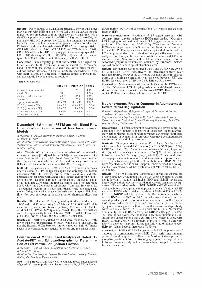
Results Pts with PHD of ≤ 2 h had significantly shorter DTB timesthan patients with PHD of > 2 h (p = 0.021). In a univariate logisticregression for prediction of in-hospital mortality, DTB time was asignificant predictor of death in the PHD ≤ 2 h group (p = 0.001) butnot in the PHD > 2 h group (p = 0.256). In a multivariate analysisincluding age, shock at presentation, gender, infarct location andDTB time, predictors of mortality in the PHD ≤ 2 h were age (p = 0.001;OR 1.078), shock (p < 0.001; OR 37.123) and DTB time (p = 0.088;OR 1.007), while in the PHD > 2 h group predictors were age (p < 0.001;OR 1.304) shock (p < 0.001; OR 1068.072) and infarct location(p = 0.068; OR 0.198) but not DTB time (p = 0.951; OR 1).Conclusion In this registry, pts with shorter PHD had a significantbenefit of short DTB in terms of in-hospital mortality. On the otherhand, in pts with prolonged PHD additional loss of time for PPCIdid not further influence in-hospital mortality. Accordingly, in ptswith short PHD (≤ 2 h) time from 1st medical contact to PPCI is cru-cial and should be kept a short as possible.
Dynamic N-13 Ammonia-PET Myocardial Blood FlowQuantification: Comparison of Two Tracer KineticModels
A. Khorsand1, S. Graf1, M. Beheshti2, H. Eidherr2, K. Kletter2, H. Sochor1,G. Porenta3, C. Pirich4
Departments of 1Cardiology and 2Nuclear Medicine, Medical University of Vienna;3Rudolfinerhaus, Vienna; 4Department of Nuclear Medicine, Private Medical Uni-versity of Salzburg
Aim The aim of the study was the comparison of two tracer-ki-netic models (two- [2CM] and three- [3CM] compartment models) forquantification of myocardial blood flow (MBF) under resting(MBFR) and stress conditions (MBFS) and coronary flow reserve(CFR) from dynamic N13-ammonia (NH3) PET images.Methods 26 patients (19�, 7�; age: 55 ± 13 yrs) with coronaryartery disease (n = 19) or typical angina and coronary risk factorsunderwent NH3-PET imaging during resting conditions and afterpharmacological stress with adenosine (140 µg/kg/min for 5 min.).Dynamic PET acquisition protocol consisted of 21 frames for a totalof 23 min. The 2CM used the first 12 frames (120 s) to determineMBF, while the 3CM used all 21 frames. Time-activity curves for12 sectional regions of 4 short-axis planes were calculated andmodel-fitting was applied to generate estimates of myocardial bloodflow. For both methods, an identical set of short-axis slices wasused.Results The calculated MBF (ml/g/min) by 2CM and 3CM were 0.9± 0.33 and 1 ± 0.36 under resting (p < 0.05), and 2.05 ± 0.98 and 2 ± 0.84under stress (p = n. s.) conditions, respectively. CFR was 2.29 ± 0.72 for2CM and 2.1 ± 0.9 for 3CM (p = n. s., paired t-test). The two methodscorrelated significantly for calculation of MBFR (r = 0.8, SEE = 0.22,p < 0.0001) and MBFS (r = 0.7, SEE = 0.61, p < 0.0001).Conclusion MBFR calculated by 2CM were found to be slightlylower than MBFR performed by 3CM, while MBFS and CFR didnot significantly differ between the two models. This differenceneeds to be considered for patient follow-up and in clinical trials.
Comparison of Model-Based Analysis of Gated 11C-Acetate-PET and Echocardiography for Determina-tion of Left Ventricular Ejection Fraction
A. Khorsand1, S. Graf1, M. Schütz2, M. Mitterhauser2, K. Kletter2, H. Sochor1,G. Maurer1, G. Porenta3
Departments of 1Cardiology and 2Nuclear Medicine, Medical University of Vienna,Austria; 3Rudolfinerhaus, Vienna, Austria
Aim The purpose of this study was to compare model-based analysisof gated 11C-acetate positron emission tomography (PET) and echo-
cardiography (ECHO) for determination of left ventricular ejectionfraction (EF).Material and Methods 9 patients (8�, 1�; age: 61 ± 9 years) withcoronary artery disease underwent ECG-gated cardiac 11C-acetatePET imaging for evaluation of myocardial oxygen consumption andperfusion. After injection of 500 MBq 11C-acetate, a 15-minuteECG-gated acquisition with 8 phases per heart cycle was per-formed. For PET images, endocardial and epicardial borders of theLV were generated on a set of short axis images with a model-basedanalysis tool. Endsystolic and enddiastolic volumes and EF weremeasured using Simpson’s method. EF was then compared to theechocardiographic measurements, obtained by Simpson’s methodapplied on the 4- and 2-apical chamber views.Results EF (mean ± SD) measured by PET and ECHO were 29.5 ±6.4 % and 32 ± 10.4 %, respectively. PET measured slightly lowerEFs than ECHO, however the difference was not significant (pairedt-test). A significant correlation was observed between PET andECHO for calculation of EF (r = 0.86, SEE = 5.5; p < 0.01).Conclusion Measurements of contractile function by ECG-gatedcardiac 11C-acetate PET imaging using a model-based methodshowed close agreement with results from ECHO. However, 11C-acetate PET measures slightly lower EFs than ECHO.
Neurohormones Predict Outcome in AsymptomaticSevere Mitral Regurgitation
U. Klaar1, J. Bergler-Klein1, M. Kapitan2, M. Heger1, R. Rosenhek1, H. Gabriel1,T. Szekeres3, K. Huber4, H. Baumgartner1
1Department of Cardiology; 2Core Unit for Medical Statistics and Informatics;3Clinical Institute of Medical and Chemical Laboratory Diagnostics, Medical Uni-versity of Vienna; 4Wilhelminenspital ViennaBackground The management of asymptomatic severe mitral re-gurgitation (MR) remains controversial. This study sought to evalu-ate whether plasma levels of neurohormones can predict short-termdevelopment of symptoms or left ventricular (LV) dysfunction and,therefore, improve timing of surgery.Methods 78 asymptomatic pts (age 57 ± 15 yrs; female n = 27)with severe MR, normal LV function (LVEF > 60 % [64 ± 5 %],LVESD < 45 mm [33 ± 5 mm]) and no severe pulmonary hyperten-sion (systolic pulmonary artery pressure [PAP] 36 ± 9 mmHg) werefollowed for 465 ± 221 days (range, 41–811). Clinical and echo-cardiographic evaluation as well as determination of plasma levelsof B-type natriuretic peptide (BNP) and N-terminal BNP (NtBNP)were repeated every 6 months. Endpoints were defined as develop-ment of symptoms or of LV dysfunction (LVEF ≤ 60 %, LVESD≥ 45 mm).Results 18 of 78 pts became symptomatic during FU whereas nopt developed LV dysfunction. Pts who developed symptoms withinthe following 6 months had higher BNP and NtBNP levels andhigher PAP at their previous visits than those who remained asymp-tomatic. By univariate analysis, BNP, NtBNP and PAP were signifi-cant predictors of symptom development whereas LV size and EFwere not. ROC-analysis yielded c-values of 0.874, 0.839 and 0.824for BNP, NtBNP and PAP, respectively. By multivariate analysis,neither BNP nor NtBNP nor PAP reached statistical significance asan independent predictor of symptom development. A BNP value≥ 65 pg/ml had a sensitivity of 84 % and specificity of 77 % forsymptom development within 6 months. Sensitivity/specificitywere 82 %/76 % for NtBNP ≥ 234 pg/ml and 80 %/60 % for PAP≥ 37 mmHg. Pts with BNP < 47 pg/ml, NtBNP < 182 pg/ml or PAP< 33 mmHg had a very low likelihood to become symptomatic (sen-sitivity for values beyond these cut-offs 95 %) whereas those withBNP > 93 pg/ml, NtBNP > 510 pg/ml or PAP > 44 mmHg were verylikely to develop symptoms during the following 6 months (speci-ficity for values beyond these cut-offs 90 %).Conclusion BNP and NtBNP together with PAP are predictors ofoutcome in asymptomatic severe MR. Their serial measurement(every 6 months) appears to allow stratification of these pts into agroup likely to benefit from elective surgery, a group that may safely befollowed conservatively and an intermediate group that requiresfurther evaluation.
Table 5. K. Kalla et al.
PHD ≤≤≤≤≤ 2 h PHD >>>>> 2 h p-value
in-hospital mortality (%) 7.9 7.6 0.91shock (%) 13 10.2 0.285anterior wall infarction (%) 49.1 50.7 0.691female (%) 26 28.4 0.504age (y; mean ± SD) 60 ± 13 61 ± 13 0.547PHD (h; mean ± SD) 1.3 ± 0.5 4.8 ± 2.5 < 0.001PHD (h; median ± SD) 1.3 ± 0.5 4 ± 2.5 < 0.001DTB (min; mean ± SD) 77 ± 46 86 ± 55 0.021DTB (min; median) 65 72 0.021
372 J KARDIOL 2006; 13 (11–12)
Cardiology 2006 – Abstracts
Flow-Mediated Dilatation is Associated With the CarotidIntima-Media Thickness and Presence of Carotid PlaquesIn Patients Without Cardiovascular Disease
M. Kovaite, Z. Petrulioniene, A. Cypiene, L. Ryliskyte, J. Badariene,V. Dzenkeviciute, A. LauceviciusClinic of Heart Diseases, Vilnius University, Centre of Cardiology and Angiology,Vilnius University Hospital Santariskiu Klinikos, VilniusBackground Brachial artery flow-mediated dilatation (FMD), ca-rotid intima-media thickness (IMT) and presence of carotid plaquesare used as surrogate markers of advanced subclinical atherosclero-sis. The relationship between carotid IMT, presence of carotidplaques and brachial FMD has not been well-established. The pur-pose of the study was to determine the relationship of brachial arteryFMD with carotid IMT and ultrasound-detected carotid plaques(CP) in individuals without cardiovascular (CV) disease.Methods We assessed traditional risk factors, carotid IMT andbrachial artery FMD in 160 individuals (69 males, aged 49.91 ± 7.16years) without clinical evidence of atherosclerotic disease andanalysed relationship of brachial artery FMD with carotid IMT andpresence of carotid plaques.Results Carotid IMT correlated with Body Mass Index (BMI)(p = 0.001), systolic blood pressure (SBP) (p < 0.001), diastolicblood pressure (DBP) (p < 0.001), total cholesterol (TC) (p = 0.001),high-density lipoprotein (HDL) cholesterol (p = 0.003), low-densitylipoprotein (LDL) cholesterol (p < 0.001), glucose (p < 0.001) andwith ten-year total fatal CV risk, estimated by SCORE (p < 0.001).Presence of CP was associated with SBP (p < 0.001), DBP (p < 0.001),BMI (p = 0.019), TC (p = 0.002), HDL cholesterol (p = 0.006), LDLcholesterol (p < 0.001) but not with the SCORE risk (p = 0.869).Brachial artery FMD inversely correlated with DBP (p < 0.001),carotid IMT (p < 0.001) and with SCORE risk (p = 0.005). Multi-variate analysis revealed that after adjustment for brachial arterydiameter and patients’ age carotid IMT and presence of carotidplaques remain significant predictors of FMD (p = 0.009 and p = 0.006,respectively).Conclusion Carotid IMT and brachial FMD correlated with ten-year total fatal CV risk, estimated by SCORE. Flow-mediated dilata-tion was associated with the carotid intima-media thickness and pres-ence of carotid plaques in patients without cardiovascular disease.
Stenting of Aortic Coarctation: a Ten-Year Follow-Up
R. Maier1, A. Gamillscheg2, J. I. Stein2, P. Ewert3, P. Zartner2, B. Heinzl2, A. Beitzke2
1Department of Medicine, Division of Cardiology; 2Department of Pediatrics, Divisionof Pediatric Cardiology, Medical University of Graz, Austria; 3German Heart Center,Berlin, GermanyBackground Stent implantation for the treatment of aortic coarc-tation is a field of increasing importance in adolescent and adultcongenital heart disease. While balloon coarctation angioplasty hasbeen considered an alternative to surgery, stent implantation isevolving now as the treatment of choice for coarctation in the ado-lescent and adult patient. Moreover, the availability of coveredstents offers new interventional perspectives, extending the tech-nique to long and extreme subatretic forms of coarctation. We reportour 10-year experience with stenting of aortic coarctation.Patients and Methods In our institution, 16 patients (13 male [81 %];mean age 29 ± 16 years, age range 13–64 years) underwent stentimplantation for the treatment of aortic coarctation between July1996 and December 2005. Six patients (37.5 %) underwent the pro-cedure for native coarctation of the aorta, another six patients pre-sented with recurrent coarctation after surgical repair, three patients(19 %) had developed re-coarctation after previous balloon angio-plasty, and one patient (6 %) had a history of surgical repair andballoon angioplasty. From 1996 until 2003, ten Palmaz stents (lengths40–50 mm) were implanted in nine patients. Since 2004, the coarc-tations of seven patients were treated with Cheatham platinum (CP)stents (lengths 28–39 mm; two covered stents). A bicuspid aorticvalve was the most frequent anomaly associated with aortic coarcta-tion.Results Stent implantation was successful in all cases. The mini-mal narrowing of the stenosis before stent implantation was 7 ± 2mm (range 3–10 mm). Prestenotic aortic diameter was 15 ± 5 mm(range 8–27 mm), poststenotic diameter was 21 ± 7 mm (range 13–37 mm). Stents could be dilated to a diameter of 15 ± 4 mm (range9–20 mm). Systolic aortic pressure gradients dropped from 35 ± 15mmHg (range 10–65 mmHg) to 6 ± 6 mmHg (range 0–19 mmHg),while mean pressure gradients dropped from 13 ± 8 mmHg (range
0–32 mmHg) to 2 ± 3 mmHg (range 0–9 mmHg). The postinterventio-nal course of one patient was complicated by a spurious aneurysmof the femoral artery requiring surgical repair. All other patients hadan uneventful course. Antihypertensive drug treatment could be re-duced in most patients after stent implantation. No reinterventionhas been necessary in any patient so far.Conclusions Transcatheter therapy of aortic coarctation withstent implantation is safe and effective. Thus, the procedure can beconsidered the treatment of choice for patients with coarctationfrom adolescence to adulthood. With the advent of covered stents,even long and extreme subatretic coarctations can be treated.Whether these promising mid-term results can be maintained needsto be determined.
Echocardiographic Phenotyping in a Transgenic MouseModel
R. Maier1, G. Haemmerle2, G. Gorkiewicz3, N. Watzinger1, A. Haumer4, H. Braunias4,R. Gasser1, H. Brussee1, G. Hoefler3, R. Zechner2
1Department of Medicine, Division of Cardiology, Medical University of Graz;2Institute of Molecular Biosciences, University of Graz; 3Institute of Pathology,Medical University of Graz; 4Siemens Medical Solutions, Ultrasound Division, ViennaBackground With the advent of transgenic technology, geneti-cally altered mice with remarkable cardiovascular phenotypes havebecome available. To benefit from the full potential of these geneti-cally engineered mice, it is crucial to have approaches to an accurateand reproducible assessment of cardiac anatomy and performance. Car-diac ultrasound is a proved and well-established technique for non-in-vasive evaluation of left ventricular (LV) morphology and function indifferent species. In this particular study, we used transthoracicechocardiography (TTE) for cardiac phenotyping in mice lacking adi-pose triglyceride lipase (ATGL). ATGL-deficient mice are supposed todie from lethal cardiomyopathy due to defective lipolysis.Materials and Methods TTE was performed in non-anesthetizedmice (12 knock-out, 11 wild-type, age range 59–136 days) using anAcuson Sequoia 512 equipped with a 15 MHz linear transducer(15L8). The heart was first imaged in the two-dimensional (2-D)mode in the parasternal long- and short-axis view. From short-axisview at the tip of the papillary muscle, motion-mode (M-mode) im-ages were obtained for measurement of LV enddiastolic and end-systolic diameter as well as interventricular septum (IVS) and leftventricular posterior wall (LVPW) thickness during diastole andsystole. From these M-mode dimensions, LV fractional shortening(FS), LV ejection fraction (EF), IVS and LVPW thickening, IVS/LVPW ratio, and LV myocardial mass (LVMM) were calculated.Results 2D-echocardiography revealed marked concentric LVhypertrophy with an abnormal myocardial texture in ATGL-defi-cient animals. LV hypertrophy, abnormal myocardial texture, andimpaired LV systolic function with asynchronous contraction pat-terns developed progressively with age. Additionally, in one olderanimal a large circumferential pericardial effusion could be clearlydetected. M-mode tracings confirmed pronounced LV hypertrophyin ATGL-deficient mice as indicated by increased diastolic wallthickness and LVMM, while systolic wall thickness as well as chamberdimensions were not significantly different between the two groups.Systolic thickening of the IVS and LVPW was markedly reduced inATGL-deficient mice, indicating a significantly reduced LV systo-lic function. This was also reflected by a significant reduction in LVFS and LV EF in ATGL-deficient mice compared to controls.Conclusions Echocardiography provides a powerful tool for study-ing cardiac morphology and function in mice. In this particularmouse model, TTE clearly allowed to discriminate knock-out ani-mals from controls and to follow the development of heart failure inATGL-deficient mice.
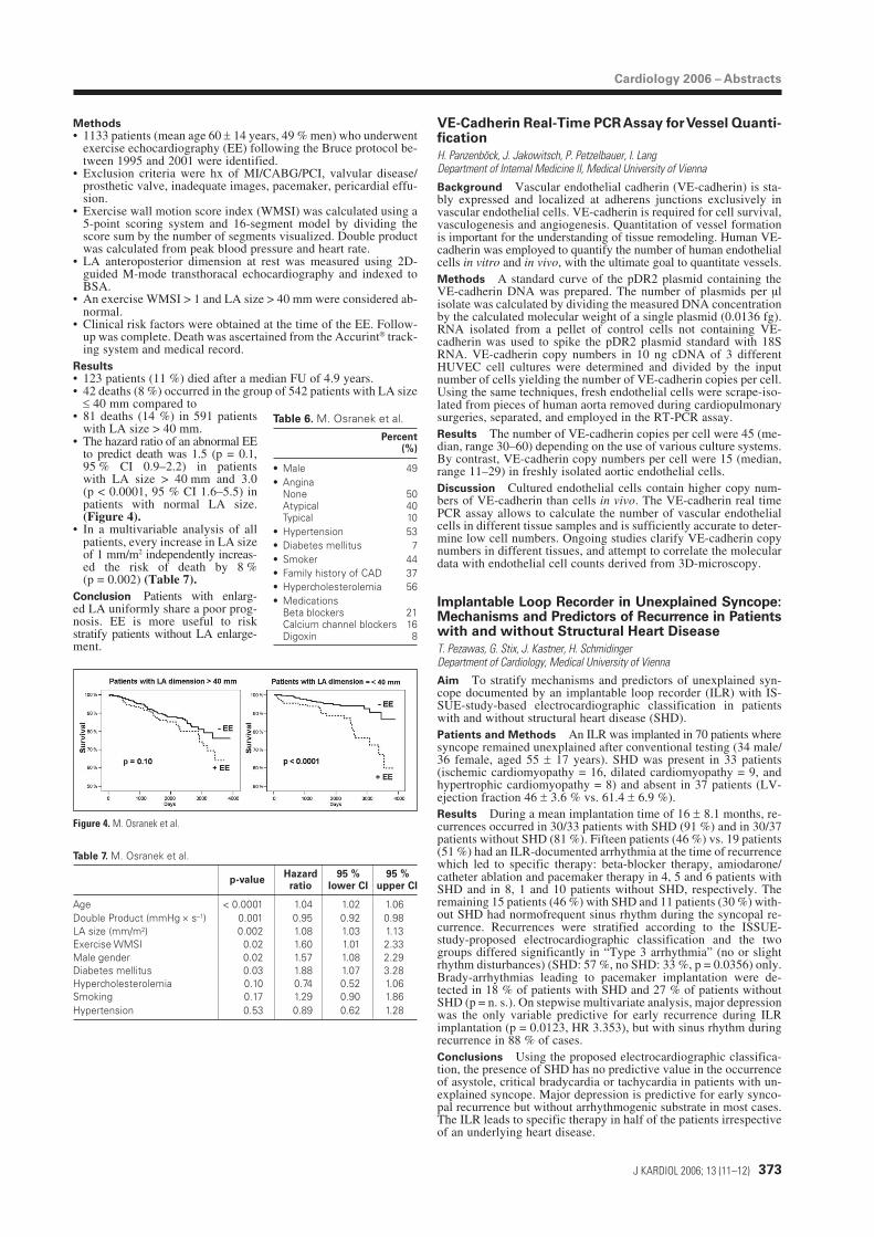
The Prognostic Value of Exercise Echocardiographyfor Long-Term Mortality is Greater in Patients with-out Left Atrial Enlargement
M. Osranek, J. B. SewardMayo Clinic, Rochester, MN, USABackground Exercise echocardiography (EE) is useful to detectcoronary artery disease (CAD) and a negative test result is associ-ated with an excellent prognosis. Left atrial (LA) enlargement is anindicator of longstanding diastolic dysfunction, associated withheart failure and other cardiovascular events including death. It isunknown if the predictive value of EE is independent of LA size.
Cardiology 2006 – Abstracts
J KARDIOL 2006; 13 (11–12) 373
Methods• 1133 patients (mean age 60 ± 14 years, 49 % men) who underwent
exercise echocardiography (EE) following the Bruce protocol be-tween 1995 and 2001 were identified.
• Exclusion criteria were hx of MI/CABG/PCI, valvular disease/prosthetic valve, inadequate images, pacemaker, pericardial effu-sion.
• Exercise wall motion score index (WMSI) was calculated using a5-point scoring system and 16-segment model by dividing thescore sum by the number of segments visualized. Double productwas calculated from peak blood pressure and heart rate.
• LA anteroposterior dimension at rest was measured using 2D-guided M-mode transthoracal echocardiography and indexed toBSA.
• An exercise WMSI > 1 and LA size > 40 mm were considered ab-normal.
• Clinical risk factors were obtained at the time of the EE. Follow-up was complete. Death was ascertained from the Accurint® track-ing system and medical record.
Results• 123 patients (11 %) died after a median FU of 4.9 years.• 42 deaths (8 %) occurred in the group of 542 patients with LA size
≤ 40 mm compared to• 81 deaths (14 %) in 591 patients
with LA size > 40 mm.• The hazard ratio of an abnormal EE
to predict death was 1.5 (p = 0.1,95 % CI 0.9–2.2) in patientswith LA size > 40 mm and 3.0(p < 0.0001, 95 % CI 1.6–5.5) inpatients with normal LA size.(Figure 4).
• In a multivariable analysis of allpatients, every increase in LA sizeof 1 mm/m2 independently increas-ed the risk of death by 8 %(p = 0.002) (Table 7).
Conclusion Patients with enlarg-ed LA uniformly share a poor prog-nosis. EE is more useful to riskstratify patients without LA enlarge-ment.
VE-Cadherin Real-Time PCR Assay for Vessel Quanti-fication
H. Panzenböck, J. Jakowitsch, P. Petzelbauer, I. LangDepartment of Internal Medicine II, Medical University of Vienna
Background Vascular endothelial cadherin (VE-cadherin) is sta-bly expressed and localized at adherens junctions exclusively invascular endothelial cells. VE-cadherin is required for cell survival,vasculogenesis and angiogenesis. Quantitation of vessel formationis important for the understanding of tissue remodeling. Human VE-cadherin was employed to quantify the number of human endothelialcells in vitro and in vivo, with the ultimate goal to quantitate vessels.Methods A standard curve of the pDR2 plasmid containing theVE-cadherin DNA was prepared. The number of plasmids per µlisolate was calculated by dividing the measured DNA concentrationby the calculated molecular weight of a single plasmid (0.0136 fg).RNA isolated from a pellet of control cells not containing VE-cadherin was used to spike the pDR2 plasmid standard with 18SRNA. VE-cadherin copy numbers in 10 ng cDNA of 3 differentHUVEC cell cultures were determined and divided by the inputnumber of cells yielding the number of VE-cadherin copies per cell.Using the same techniques, fresh endothelial cells were scrape-iso-lated from pieces of human aorta removed during cardiopulmonarysurgeries, separated, and employed in the RT-PCR assay.Results The number of VE-cadherin copies per cell were 45 (me-dian, range 30–60) depending on the use of various culture systems.By contrast, VE-cadherin copy numbers per cell were 15 (median,range 11–29) in freshly isolated aortic endothelial cells.Discussion Cultured endothelial cells contain higher copy num-bers of VE-cadherin than cells in vivo. The VE-cadherin real timePCR assay allows to calculate the number of vascular endothelialcells in different tissue samples and is sufficiently accurate to deter-mine low cell numbers. Ongoing studies clarify VE-cadherin copynumbers in different tissues, and attempt to correlate the moleculardata with endothelial cell counts derived from 3D-microscopy.
Implantable Loop Recorder in Unexplained Syncope:Mechanisms and Predictors of Recurrence in Patientswith and without Structural Heart Disease
T. Pezawas, G. Stix, J. Kastner, H. SchmidingerDepartment of Cardiology, Medical University of ViennaAim To stratify mechanisms and predictors of unexplained syn-cope documented by an implantable loop recorder (ILR) with IS-SUE-study-based electrocardiographic classification in patientswith and without structural heart disease (SHD).Patients and Methods An ILR was implanted in 70 patients wheresyncope remained unexplained after conventional testing (34 male/36 female, aged 55 ± 17 years). SHD was present in 33 patients(ischemic cardiomyopathy = 16, dilated cardiomyopathy = 9, andhypertrophic cardiomyopathy = 8) and absent in 37 patients (LV-ejection fraction 46 ± 3.6 % vs. 61.4 ± 6.9 %).Results During a mean implantation time of 16 ± 8.1 months, re-currences occurred in 30/33 patients with SHD (91 %) and in 30/37patients without SHD (81 %). Fifteen patients (46 %) vs. 19 patients(51 %) had an ILR-documented arrhythmia at the time of recurrencewhich led to specific therapy: beta-blocker therapy, amiodarone/catheter ablation and pacemaker therapy in 4, 5 and 6 patients withSHD and in 8, 1 and 10 patients without SHD, respectively. Theremaining 15 patients (46 %) with SHD and 11 patients (30 %) with-out SHD had normofrequent sinus rhythm during the syncopal re-currence. Recurrences were stratified according to the ISSUE-study-proposed electrocardiographic classification and the twogroups differed significantly in “Type 3 arrhythmia” (no or slightrhythm disturbances) (SHD: 57 %, no SHD: 33 %, p = 0.0356) only.Brady-arrhythmias leading to pacemaker implantation were de-tected in 18 % of patients with SHD and 27 % of patients withoutSHD (p = n. s.). On stepwise multivariate analysis, major depressionwas the only variable predictive for early recurrence during ILRimplantation (p = 0.0123, HR 3.353), but with sinus rhythm duringrecurrence in 88 % of cases.Conclusions Using the proposed electrocardiographic classifica-tion, the presence of SHD has no predictive value in the occurrenceof asystole, critical bradycardia or tachycardia in patients with un-explained syncope. Major depression is predictive for early synco-pal recurrence but without arrhythmogenic substrate in most cases.The ILR leads to specific therapy in half of the patients irrespectiveof an underlying heart disease.
Table 6. M. Osranek et al.
Percent(%)
• Male 49• Angina
None 50Atypical 40Typical 10
• Hypertension 53• Diabetes mellitus 7• Smoker 44• Family history of CAD 37• Hypercholesterolemia 56• Medications
Beta blockers 21Calcium channel blockers 16Digoxin 8
Figure 4. M. Osranek et al.
Table 7. M. Osranek et al.
p-valueHazard 95 % 95 %
ratio lower CI upper CI
Age < 0.0001 1.04 1.02 1.06Double Product (mmHg × s–1) 0.001 0.95 0.92 0.98LA size (mm/m2) 0.002 1.08 1.03 1.13Exercise WMSI 0.02 1.60 1.01 2.33Male gender 0.02 1.57 1.08 2.29Diabetes mellitus 0.03 1.88 1.07 3.28Hypercholesterolemia 0.10 0.74 0.52 1.06Smoking 0.17 1.29 0.90 1.86Hypertension 0.53 0.89 0.62 1.28

374 J KARDIOL 2006; 13 (11–12)
Cardiology 2006 – Abstracts
Multislice Computed Tomography in Detection ofCoronary Artery Disease in Heart Transplant Patients
P. Pichler1, C. Loewe2, S. Roedler3, B. Syeda1, T. Bader2, A. Aliabadi3, C. Schukro1,F. Wolf2, H. Imhof2, D. Glogar1
1Department of Internal Medicine II; 2Department of Radiology; 3Department ofSurgery, Medical University of ViennaObjectives We sought to assess whether multislice computed to-mography (MSCT) permits the exclusion of coronary artery diseasein asymptomatic heart transplant patients.Background Post-transplant follow-up of heart transplant patientsconsists of regular angiography (1, 3, 5, 7, 10 and 15 years) aftertransplantation and goes along with a significant amount of costs,discomfort and risk.Methods MSCT scans (Philips “CT MX 8000 IDT”) and invasivecoronary angiography were performed in 66 consecutive hearttransplant patients in sinus-rhythm scheduled for routine invasivecoronary angiography. 100 ml non-ionic iodinated contrast mediumwere applied for CT-angiography. All scans were performed duringone breath-hold. In axial MSCT images and multiplanar reconstruc-tions, all coronary arteries and side branches were assessed for thepresence of stenoses exceeding a 70 % diameter reduction. Coronarysegment analyses were performed according to a modified Ameri-can College of Cardiology/American Heart Association (ACC/AHA) classification in vessels with a diameter exceeding 1.5 mm.Results were compared with quantitative coronary angiographyanalysis.Results MSCT was successfully performed in 60 patients enrolledin our analysis; fourteen patients (21 %) had at least one significantstenosis, 9 (14 %) suffered from coronary artery disease without sig-nificance and the remaining 43 (65 %) were totally free from graftsclerosis. Forty-one out of 43 patients (95 %) who were estimated tobe fully evaluable in MSCT were correctly classified. On a per-seg-ment-basis, 10 out of 19 high-grade stenoses were assessed cor-rectly, 4 were unevaluable and 5 were incorrectly classified as non-significant (sensitivity: 53 %, specificity: 94 %). After exclusion of45 out of 764 coronary segments (6 %) due to motion artifacts orsevere calcification, sensitivity, specificity, positive and negativepredictive values were 67 %, 100 %, 91 % and 99 %, respectively.Conclusion Despite a low sensitivity, the high negative predictivevalue allows the exclusion of significant CAD with a high certaintyin a large number of patients. From the clinical point of view, thismight spare this patient cohort an additional invasive coronary angio-graphy.
Accuracy of Non-Invasive 16-Slice CT Angiographyin Patients with Stable Angina Pectoris Comparedwith Invasive Coronary Angiography
P. Pichler1, F. Wolf2 , C. Schukro1, C. Loewe2, B. Syeda1, T. Bader2, A. Stadler1,H. Sochor1, D. Glogar1
1Department of Internal Medicine II; 2Department of Radiology, Medical Universityof ViennaObjectives To assess whether 16-slice computed tomography (CT)permits detection or exclusion of coronary artery disease in patientswith stable angina.Background Non-invasive coronary CT angiography is a promis-ing coronary imaging technique.Methods 30 consecutive patients with stable angina pectoris un-derwent routine invasive and CT coronary angiography. Retrospec-tive ECG-gated CT coronary angiography was performed with a 16-slice CT-scanner (Philips MX 8000 IDT) after bolus-triggered i. v.administration of 100 ml non-ionic iodinated contrast medium. Betablockers were not administered routinely before the examination. Inaxial MSCT images and multiplanar reconstructions, all coronaryarteries and side branches exceeding 1.5 mm in diameter were as-sessed for the presence of stenoses using a 15-segment classifica-tion according to the American College of Cardiology/AmericanHeart Association (ACC/AHA). The consensus results of two blindedreaders were compared with quantitative coronary angiographyanalysis.Results MSCT was performed successfully in all patients enrolled inour analysis. According to invasive coronary angiography, 11 (37 %)patients had no significant coronary artery disease and 1VD, 2VDand 3VD where found in 7 (23 %), 9 (30 %) and 3 (10 %) patients,respectively. After exclusion of all unevaluable segments, the sensi-tivity of coronary CT-angiography for detection of significant le-sions was as high as 47 % (20 of 43 significant stenoses), specificity
was 95 % (204 of 214), positive predictive value (PPV) was 67 %(20 of 30) and negative predictive value (NPV) was 90 % (204 of 227).Conclusion Coronary MSCT is a sufficient method to excludecoronary artery disease. However, in the detection and quantifica-tion of significant coronary artery stenoses 16-slice CT cannot beregarded as a reliable diagnostic tool.
Gender Differences in Severe Aortic Stenosis Referredto Aortic Valve Surgery
R. Rosenhek, C. Fuchs, F. Rader, U. Klaar, C. Scholten, M. Heger, G. Maurer,H. BaumgartnerDepartment of Cardiology, Medical University of Vienna
Background Aortic stenosis has become the most frequent valvedisease in developed countries and has been extensively studied inrecent years. Although gender differences have drawn increasingattention in cardiology, little is known so far about gender differ-ences in aortic stenosis.Methods The clinical and echocardiographic characteristics of439 consecutive patients (age 70 ± 11 yrs) who were seen in our out-patient clinic and referred to surgery because of severe symptomaticaortic stenosis were analysed with regard to potential gender differ-ences.Results Patients were almost equally distributed between men(n = 205) and women (n = 234). Female pts were, however, signifi-cantly older at presentation with a mean age of 73 ± 10 yrs com-pared to 66 ± 11 yrs (p < 0.0001). They were more symptomatic thanmales with a New York Heart Association class 2.5 ± 0.7 vs. 2.1 ± 0.7,respectively (p < 0.0001). Females had smaller valve areas. How-ever, when adjusting for body surface area (BSA), this differencedisappeared (0.33 ± 0.1 vs. 0.34 ± 0.08 cm2/m2; p = 0.21). Never-theless, mean aortic valve gradients were significantly higher in fe-males with 66.6 ± 19.1 vs. 62.1 ± 20.2 mmHg (p = 0.017). LV sizedid not differ when adjusting for BSA but females presented withsignificantly less LV hypertrophy. Notably, 74 % of women pre-sented with hypertension compared to 58 % of men (p < 0.0005).However, coronary artery disease was significantly more frequentin male patients (39 % vs. 27 %; p = 0.01).Conclusion Female patients with aortic stenosis referred to aorticvalve replacement are older and more symptomatic compared tomale patients. Further research must address whether this is due todifferences in disease onset and progression or whether female pa-tients get delayed medical attention for some reasons.
Left Atrial Size Independently Predicts Outcome inAsymptomatic Severe Mitral Regurgitation
R. Rosenhek, F. Rader, M. Krejc, D. Kalbeck, H. Gabriel, U. Klaar, G. Maurer,H. BaumgartnerDepartment of Cardiology, Medical University of ViennaBackground Left atrial (LA) size is affected by left ventricular (LV)diastolic pressure, the degree of ventricular remodeling, the occur-rence of atrial fibrillation and the presence of mitral regurgitation(MR) itself. LA size has been proposed to be a predictor of outcomeafter mitral valve replacement with preserved LV function. How-ever, the predictive value of LA size for development of symptomsor LV dysfunction among pts with severe MR has not been studied.
Methods 132 consecutive asymptomatic pts (age 55 ± 15 yrs, 49female) with severe degenerative MR and normal LV function wereprospectively followed for a median of 69 months. Pts underwentserial clinical and echocardiographic exams. The following poten-tial predictors of outcome werestudied: LA size, endsystolic andenddiastolic LV diameter, pul-monary artery pressure and clini-cal risk factors.
Results Kaplan-Meier event-free survival for the entire ptgroup, with endpoints definedas development of symptomsor LV dysfunction (n = 38) anddeath related to MR (n = 0) was92 ± 2 % at 2 yrs, 78 ± 4 % at4 yrs, 65 ± 5 % at 6 yrs and 55 ±6 % at 8 yrs. LA size was thestrongest independent predic-tor of outcome: no events were Figure 5. R. Rosenhek et al.
Cardiology 2006 – Abstracts
J KARDIOL 2006; 13 (11–12) 375
observed in the group with a LA < 50 mm. Event-free survival forpatients with LA 50 to 69 mm was 94 ± 3 % at 2 yrs, 82 ± 5 % at 4and 51 ± 8 % at 8 yrs vs. 85 ± 8 % at 2 yrs, 47 ± 12 % at 4 yrs and40 ± 12 % at 8 yrs for patients with a LA ≥ 70 mm (p = 0.0001).None of the other studied parameters reached significance as pre-dictors of outcome in multivariate analysis (Figure 5).Conclusion LA size is a strong and independent predictor of out-come in patients with asymptomatic severe MR. Patients can bestratified by LA size in groups at low, intermediate and high risk forsubsequent symptom or LV dysfunction development requiringsurgery.
Core and Concomitant Components of the MetabolicSyndrome
L. Ryliskyte, J. Badariene, V. Dzenkeviciute, Z. Petrulioniene, A. LauceviciusCenter of Cardiology and Angiology, Vilnius University Hospital SantarisiuKlinikos, Vilnius, LithuaniaAim of the Study We sought to analyze what the core compo-nents of the metabolic syndrome are and which cardiovascular riskfactors are concomitant according to their relation to the insulin re-sistance indexes.Methods or Patients We studied 127 subjects (64 % men and 36 %women, aged 49.8 ± 9.4 years) suspected of having metabolic syn-drome (MS). All patients underwent detailed assessment of cardio-vascular risk factors, including a serum lipid profile measurementand an oral glucose tolerance test (OGTT). Serum insulin was meas-ured at baseline and 120 minutes after glucose load.Results The metabolic syndrome according to the National Cho-lesterol Education Program (NCEP) Adult Treatment Panel (ATP)III was diagnosed in 55 patients (43 %), according to the IDF classi-fication in 83 patients (65 %). Newly diagnosed diabetes, impairedglucose tolerance or fasting hyperglycaemia was present in 84 pa-tients (66 %). Normal fasting glucose (< 5.6 mmol/l) had 39 % ofpatients with impaired glucose tolerance (n = 9/23). Dyslipidemia,typical of the metabolic syndrome according to the IDF criteria, wasfound in 70 patients (55 %). Any type of dyslipidemia was presentin 91 patients (72 %). Insulin resistance indexes (HOMAIR and ISI)were correlated with Body Mass Index (r = 0.36, p < 0.0001 andr = –0.321, p = 0.003, respectively) and waist circumference (r = 0.40,p < 0.0001 for HOMAIR; r = –0.31, p = 0.004 for ISI), as well as withfasting and OGTT glycaemia. No relationship was found betweeninsulin resistance indexes and the presence of arterial hypertension(logistic regression coefficient beta = 0.17, p = 0.52 for HOMAIR
and beta = –1.23, p = 0.34 for ISI). Correlation between insulin re-sistance indexes (HOMAIR, ISI) and serum lipid profile was notfound, either.Conclusions Abdominal obesity and any type of glycaemic disor-der seem to be the core components of the metabolic syndrome. Anoral glucose tolerance test gives additional value for discriminationof subjects with glycaemic disorder.
Single-Center Analysis of a Randomized Trial Compar-ing PCI and CABG in Patients with Significant Three-Vessel and/or Left Main Disease
S. Scherzer1, C. Schukro1, G. Christ1, G. Kreiner1, I. Lang1, H. Sochor1, P. Simon2,D. Glogar1
1Department of Internal Medicine II; 2Department of Surgery, Medical Universityof ViennaBackground BARI and ARTS showed significant clinical improve-ment in patients with three-vessel disease after coronary artery by-pass graft (CABG) when compared to balloon angioplasty andstenting, respectively. Thus, the aim of the SYNTAX trial is to com-pare the clinical outcome of the percutaneous coronary intervention(PCI) with drug-eluting stents and CABG in patients with three-ves-sel disease and/or left main artery disease.Methods If patients accepted randomization, they were allocatedeither to PCI or to CABG. During PCI, they received Taxus-Ex-press2 drug-eluting stents. Follow-up visits comprised a clinical ex-amination, a specialized questionnaire and ECG. An angiographicsix-month follow-up was also planned.Results From November 2005 on, twenty patients have been rando-mized, of whom twelve were allocated to PCI and eight to CABG.One PCI patient with perforated LAD and following emergencyCABG died one week after the procedure due to multiorgan dis-
eases. One patient suffered from subacute stent thrombosis fourdays after PCI, which was successfully revascularized. On aver-age, patients indicated dyspnea NYHA I-II or angina CCS I-II atthe one-month follow-up visit. One patient was hospitalized due toan increase of enzymes. Angiography revealed an occlusion of adiagonal branch in a completely stented LAD. Between first andsecond follow-up, one patient died due to a septic shock. At thesix-month visit, the observed patients felt quite as well as afterfirst follow-up.Conclusion At short-term follow-up, there was no significant dif-ference between PCI with drug-eluting stent and CABG concerningthe clinical outcome, although PCI showed more post-proceduralevents.
L-arginine and Asymmetrical Dimethylarginine (ADMA)in Chronic Thromboembolic Pulmonary Hypertension(CTEPH)
N. Skoro-Sajer1, F. Mittermayer2, D. Bonderman1, M. Wolzt2, I. M. Lang1
Departments of 1Internal Medicine II and 2Clinical Pharmacology, MedicalUniversity of Vienna
Background Nitric oxide (NO) is synthesized from L-arginine byNO synthase, which can be inhibited by asymmetrical dimethyl-arginine (ADMA). ADMA is increased in idiopathic pulmonaryhypertension (iPAH) and associated with unfavorable outcome.Chronic thromboembolic pulmonary hypertension (CTEPH) showsvascular hypertensive changes similar to iPAH. Therefore, we hy-pothesized that plasma levels of ADMA may be increased in pa-tients with CTEPH.Methods We collected data from 10 patients with iPAH and 28patients with CTEPH (13 proximal and 15 distal). L-arginine andADMA plasma levels were measured by high-performance liquidchromatography. All patients underwent right heart catheterization.In addition, levels of ADMA were measured in 14 age- and sex-matched controls.Results All patients had severe pulmonary hypertension (iPAH:MPAP 56 ± 11 mmHg, CI 2.2 ± 0.0.5 L × min–1 × m–2; CTEPH:MPAP 51 ± 6 mmHg, CI 2.3 ± 0.6 L × min–1 × m–2). There was nodifference in haemodynamics between proximal and distal CTEPH.L-arginine plasma levels were significantly decreased in all patientswith pulmonary hypertension. L-arginine plasma levels correlated signi-ficantly with mixed-venous saturation (r = 0.48, p < 0.01) and car-diac index (r = 0.39, p < 0.05). ADMA plasma levels were significantlyelevated in distal CTEPH and iPAH patients (0.81 ± 0.22 µmol/l and0.69 ± 0.13 µmol/l) compared with controls (0.52 ± 0.14 µmol/l;p < 0.05, respectively). ADMA was significantly higher in distalCTEPH than in proximal CTEPH (p < 0.01).Conclusions ADMA plasma levels are increased in CTEPH pa-tients. The data confirm similar pathophysiologic mechanisms ofvascular remodeling in iPAH and CTEPH.
Survival in Patients with Inoperable CTEPH Treatedwith Subcutaneous Treprostinil
N. Skoro-Sajer1, F. Wiesbauer1, D. Bonderman1, W. Klepetko2, M. P. Kneussl3,G. Maurer1, I. M. Lang1
Departments of 1Cardiology and 2Cardiothoracic Surgery, Medical University ofVienna; 3Wilheminenspital, ViennaBackground Treprostinil improves exercise capacity, hemodyna-mics and signs and symptoms of pulmonary arterial hypertension(PAH). Inoperable CTEPH may be a target for vasodilator therapy.Aim In an open-label uncontrolled study, we investigated efficacyand survival in patients with severe inoperable CTEPH treated withsubcutaneous treprostinil.Methods Between September 1999 and 2005, 25 consecutive inoper-able CTEPH patients were treated with s. c. treprostinil. Inclusioncriteria were New York Heart Association (NYHA) functional classIII or IV and six-minute walking distance (6-MWD) ≤ 380 m. NYHAfunctional class, 6-MWD and B-type brain natriuretic peptide (BNP)served as primary efficacy endpoints. Right heart catheterizationwas performed at baseline and after at least 12 months of treatment.A historical group of 31 untreated patients with inoperable CTEPHmatched for NYHA functional class was used for comparative sur-vival analysis.Results Under treatment, patients demonstrated improvements in6-MWD, NYHA functional class, and decreased BNP levels. 12 pa-tients (age 55.4 ± 10 years) were catheterized after 25.4 ± 10 months
376 J KARDIOL 2006; 13 (11–12)
Cardiology 2006 – Abstracts
at a final mean treprostinil dose of 37.7 ± 9.6 ng/kg/min demonstrat-ing significant improvements of cardiac output +0.7 ± 1.0 L × min–1
(p = 0.03) and pulmonary vascular resistance –193 ± 287 dynes × s ×cm–5 (p = 0.02). Overall survival rates of treprostinil-treated patientsat one, two, three, and five years were 80 %, 80 %, 80 % and 53 %,respectively, compared with the untreated group (67 %, 43 %, 37 %and 16 %, p = 0.02). Treprostinil was an independent predictor ofimproved survival (p = 0.02).Conclusions Subcutaneous treprostinil improves exercise capac-ity, hemodynamics and survival in patients with severe inoperableCTEPH.
Microcirculation Changes after ACAT-Inhibitor, VULM1457, in Diabetic and Non-Diabetic HyperlipidemicHamsters
E. Vojtaššáková, M. Syneková, D. T∨∨∨∨∨
az∨∨∨∨∨ká, Š. Matyás, P. ŠvecDepartment of Pharmacology and Toxicology, Faculty of Pharmacy, ComeniusUniversity, Bratislava, SlovakiaAim of the Study The effect of the new ACAT-inhibitor, VULM1457, on velocity of red blood cells in hypercholesterolemic andhypercholesterolemic/streptozotocine-diabetic hamsters was stud-ied by videocapillaroscopy. It is a non-invasive examination of mi-crocirculation recently used in cardiological and diabetic patients.Methods Experimental studies were performed in 6 groups ofhamsters: a) fed on cholesterol lipid diet (CHL); b) streptozotocine-induced diabetes (D); c) streptozotocine-induced diabetes fed oncholesterol lipid diet (D CHL); d) fed on cholesterol-lipid diet re-ceiving VULM 1457 (CHL VULM 1457); e) streptozotocine-induceddiabetes fed on cholesterol-lipid diet receiving VULM 1457 (DCHL VULM 1457); f) control group. ACAT-inhibitor VULM 1457was given orally 50 mg/kg. The effect of treatment on hamster’spaw microcirculation was assessed using capillary microscopy bymeasuring red blood cell velocity after 3 months of treatment.Results The high cholesterol-level diet decreased significantlythe red blood cell velocity (6.2 %, p < 0.05) and this effect was evenhigher in diabetic animals (10.1 %, p < 0.01). Treatment of animalswith the new ACAT-inhibitor, VULM 1457, most significantly im-proved the velocity rate in all groups, most significantly in D CHLVULM 1457 in comparison to untreated groups of animals.Conclusion VULM 1457 positively improved red blood cell ve-locity reduced in diabetic and hypercholesterolemic hamsters. Sig-nificant effect was seen when VULM 1457 was given to diabetichamsters fed on cholesterol-lipid diet.This research was supported by grant VEGA 2/5129/25a and FaFUK/7/2006.
Percutaneous Coronary Intervention of UnprotectedLeft Main Stenosis: Safety and Long-Term Efficacy inStable Patients
S. Winkler, G. Christ, I. Lang, M. Gyöngyösi, G. Kreiner, H. Sochor, C. Schukro,D. GlogarDepartment of Cardiology, Medical University of ViennaBackground Percutaneous coronary intervention (PCI) of unpro-tected left main (LM) stenosis is still controversial. This retrospec-tive analysis evaluated the clinical and angiographic outcome ofthis patient cohort in a single high-volume PCI center.Methods 42 consecutive patients with LM stenosis and acute coro-nary syndrome (ACS) not eligible for acute coronary artery bypasssurgery and 50 selected patients with stable coronary artery disease(CAD) were treated by PCI with either bare metal stent (BMS, 44 %) ordrug-eluting stent (DES, 56 %) implantation between August 2001and July 2006.Results The cumulative survival rate up to one year (median 81days, range 0–365) of selected elective patients with LM stenosiswas 94 %, compared to 65.2 % in hemodynamically stable ACSpatients with LM stenosis and 36.8 % in ACS patients with cardio-genic shock and LM stenosis (p < 0.00001). Angiographic follow-up(median 194 days, range 1–1310 days) was performed in 39 % of allpatients, with a restenosis rate of 2.8 % after DES compared to11.1 % after BMS implantation (p = n. s.).Conclusion Whereas ACS patents with LM stenosis are still ahighest-risk patient cohort, especially in hemodynamically unstableconditions, LM PCI can be safely and efficaciously performed inselected stable patients preferably with DES implantation in an ex-perienced center.
“Oldest-Old” Patients In Intensive Care: Prognosis andTherapeutic Activity
S. Ziegler1,2, G. Heinze3, M. Ryffel3, M. Kompatscher3, J. Slany1, A. Valentin1
1Department of Internal Medicine II, Intensive Care Unit, KrankenanstaltRudolfstiftung, Vienna; 2Department of Internal Medicine II, Division of Angiology;3Core Unit for Medical Statistics and Informatics, Section of Clinical Biometrics,Medical University of ViennaObjective The present investigation aims at giving a descriptiveoverview on the prognosis and therapeutic activity for “oldest-old”patients, admitted to an intensive care unit (ICU) in comparison toyounger patients.Patients and Methods 3069 patients, admitted to the ICU during aseven-year study period, were assigned to one of 4 age categories:younger than 65 years (48 %), aged 65 to 74 years (26 %), aged 75to 85 years (22 %) and aged 85 years or older (5 %). Type and reasonfor ICU admission, length of ICU stay, severity of illness as meas-ured by the Simplified Acute Physiology Score (SAPS) -II, level ofprovided care as measured by the Simplified Therapeutic Interven-tion Scoring System (TISS) -28, vital status at and date of ICU dis-charge were recorded.Main Results The mortality rate of patients aged 85 years andolder was significantly higher than in patients younger than 65 years(OR for survival: 2.8, p < 0.001). Non-survivors had higher SAPS-II levels (even when age points were excluded) and daily averageTISS points. Daily average TISS score was negatively correlated toage (r = –0.03; p < 0.001) and was significantly lower in the “oldestold” when compared to all younger patient groups (p < 0.001).“Oldest-old” patients showed significantly shorter length of stay(median: 2; 1st–3rd quartiles: 1 to 3, p < 0.001) compared to theyounger patient groups.Conclusions Within the very elderly population, age is an impor-tant and independent predictor of mortality, but acute severity of ill-ness is even stronger associated with mortality. Consequently, agealone may be an inappropriate criterion for allocation of ICU re-sources.
Restenosis Caused by Stent Fracture of OverlappingZotarolimus-Eluting Stents
R. Badr Eslam, K. Katsaros, S. Ingerle, G. Delle-Karth, D. Glogar, G. ChristDepartment of Cardiology, Medical University of Vienna
Abstract The introduction of drug-eluting stents (DES) revolution-ized the field of interventional cardiology by dramatically reducingthe development of late in-stent restenosis (ISR). Stent fracture, how-ever, has recently been suggested as one possible mechanism ofrestenosis after DES implantation. We report the first case of ISRcaused by fracture of overlapping zotarolimus-eluting stents.Introduction Restenosis after intracoronary DES implantationmost commonly occurs in a localized pattern associated with eitherinjury outside the stent (edge restenosis) or discontinuity of stentcoverage (under expansion, gap or fracture) [1]. To date, variouscases of fractures of sirolimus-eluting stents (Cypher®, Cordis) [1–4]and one case of a paclitaxel-eluting stent (Taxus®, Boston Scientific)[5] have been described.To our knowledge, this is the first report on a case of delayed fractureof overlapping zotarolimus-eluting stents (Endeavor®, Medtronic).Case report A 66-year-old male with a history of hypertension andnon-insulin-dependent diabetes mellitus was admitted to anotherhospital without cardiac catheterization facilities due to acute infe-rior ST elevation myocardial infarction (STEMI). Despite immedi-ate thrombolytic therapy with tenecteplase (Metalyse®, BoehringerIngelheim) his clinical condition deteriorated with development ofpulmonary edema and cardiogenic shock. After intubation he wastransferred on high-dose catecholamine for rescue percutaneouscoronary intervention (PCI) to our institution.Coronary angiography showed a severe three-vessel disease with aproximally occluded right coronary artery (RCA, Figure 6a), twosignificant stenoses in the proximal left anterior descending artery(LAD, not shown) with diffuse disease in the mid and distal portion,as well as a high-grade stenosis in the mid-left circumflex artery(LCx, not shown) supplying the distal RCA via collaterals.Due to the severe hemodynamic instability of this critically ill pa-tient we intended to perform a complete percutaneous revascu-larization. Starting with the culprit vessel, two Endeavor® stents(3.5/24 mm and 3.0/18 mm, 16 atm) were implanted after balloonpredilatation in the mid a proximal RCA with a very generous over-
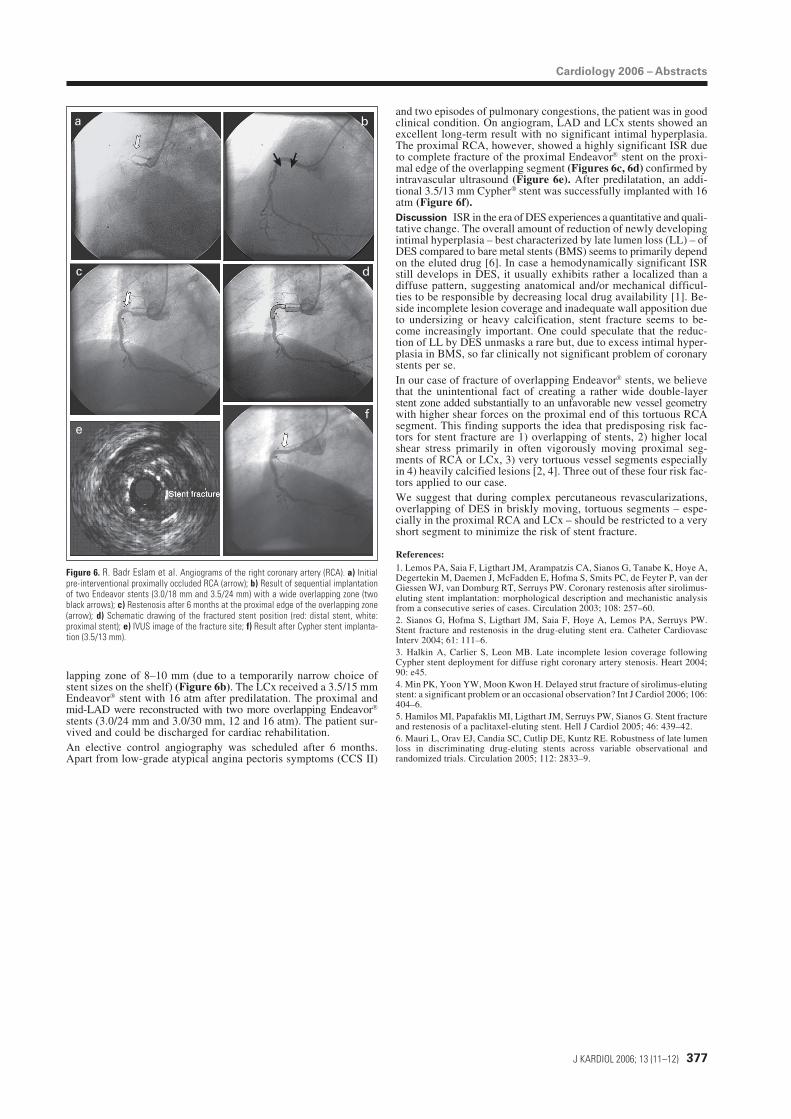
Cardiology 2006 – Abstracts
J KARDIOL 2006; 13 (11–12) 377
lapping zone of 8–10 mm (due to a temporarily narrow choice ofstent sizes on the shelf) (Figure 6b). The LCx received a 3.5/15 mmEndeavor® stent with 16 atm after predilatation. The proximal andmid-LAD were reconstructed with two more overlapping Endeavor®
stents (3.0/24 mm and 3.0/30 mm, 12 and 16 atm). The patient sur-vived and could be discharged for cardiac rehabilitation.An elective control angiography was scheduled after 6 months.Apart from low-grade atypical angina pectoris symptoms (CCS II)
a b
c d
ef
Figure 6. R. Badr Eslam et al. Angiograms of the right coronary artery (RCA). a) Initialpre-interventional proximally occluded RCA (arrow); b) Result of sequential implantationof two Endeavor stents (3.0/18 mm and 3.5/24 mm) with a wide overlapping zone (twoblack arrows); c) Restenosis after 6 months at the proximal edge of the overlapping zone(arrow); d) Schematic drawing of the fractured stent position (red: distal stent, white:proximal stent); e) IVUS image of the fracture site; f) Result after Cypher stent implanta-tion (3.5/13 mm).
and two episodes of pulmonary congestions, the patient was in goodclinical condition. On angiogram, LAD and LCx stents showed anexcellent long-term result with no significant intimal hyperplasia.The proximal RCA, however, showed a highly significant ISR dueto complete fracture of the proximal Endeavor® stent on the proxi-mal edge of the overlapping segment (Figures 6c, 6d) confirmed byintravascular ultrasound (Figure 6e). After predilatation, an addi-tional 3.5/13 mm Cypher® stent was successfully implanted with 16atm (Figure 6f).Discussion ISR in the era of DES experiences a quantitative and quali-tative change. The overall amount of reduction of newly developingintimal hyperplasia – best characterized by late lumen loss (LL) – ofDES compared to bare metal stents (BMS) seems to primarily dependon the eluted drug [6]. In case a hemodynamically significant ISRstill develops in DES, it usually exhibits rather a localized than adiffuse pattern, suggesting anatomical and/or mechanical difficul-ties to be responsible by decreasing local drug availability [1]. Be-side incomplete lesion coverage and inadequate wall apposition dueto undersizing or heavy calcification, stent fracture seems to be-come increasingly important. One could speculate that the reduc-tion of LL by DES unmasks a rare but, due to excess intimal hyper-plasia in BMS, so far clinically not significant problem of coronarystents per se.In our case of fracture of overlapping Endeavor® stents, we believethat the unintentional fact of creating a rather wide double-layerstent zone added substantially to an unfavorable new vessel geometrywith higher shear forces on the proximal end of this tortuous RCAsegment. This finding supports the idea that predisposing risk fac-tors for stent fracture are 1) overlapping of stents, 2) higher localshear stress primarily in often vigorously moving proximal seg-ments of RCA or LCx, 3) very tortuous vessel segments especiallyin 4) heavily calcified lesions [2, 4]. Three out of these four risk fac-tors applied to our case.We suggest that during complex percutaneous revascularizations,overlapping of DES in briskly moving, tortuous segments – espe-cially in the proximal RCA and LCx – should be restricted to a veryshort segment to minimize the risk of stent fracture.
References:1. Lemos PA, Saia F, Ligthart JM, Arampatzis CA, Sianos G, Tanabe K, Hoye A,Degertekin M, Daemen J, McFadden E, Hofma S, Smits PC, de Feyter P, van derGiessen WJ, van Domburg RT, Serruys PW. Coronary restenosis after sirolimus-eluting stent implantation: morphological description and mechanistic analysisfrom a consecutive series of cases. Circulation 2003; 108: 257–60.2. Sianos G, Hofma S, Ligthart JM, Saia F, Hoye A, Lemos PA, Serruys PW.Stent fracture and restenosis in the drug-eluting stent era. Catheter CardiovascInterv 2004; 61: 111–6.3. Halkin A, Carlier S, Leon MB. Late incomplete lesion coverage followingCypher stent deployment for diffuse right coronary artery stenosis. Heart 2004;90: e45.4. Min PK, Yoon YW, Moon Kwon H. Delayed strut fracture of sirolimus-elutingstent: a significant problem or an occasional observation? Int J Cardiol 2006; 106:404–6.5. Hamilos MI, Papafaklis MI, Ligthart JM, Serruys PW, Sianos G. Stent fractureand restenosis of a paclitaxel-eluting stent. Hell J Cardiol 2005; 46: 439–42.6. Mauri L, Orav EJ, Candia SC, Cutlip DE, Kuntz RE. Robustness of late lumenloss in discriminating drug-eluting stents across variable observational andrandomized trials. Circulation 2005; 112: 2833–9.
Haftungsausschluss
Die in unseren Webseiten publizierten Informationen richten sich ausschließlich an geprüfte und autorisierte medizinische Berufsgruppen und entbinden nicht von der ärztlichen Sorg-faltspflicht sowie von einer ausführlichen Patientenaufklärung über therapeutische Optionen und deren Wirkungen bzw. Nebenwirkungen. Die entsprechenden Angaben werden von den Autoren mit der größten Sorgfalt recherchiert und zusammengestellt. Die angegebenen Do-sierungen sind im Einzelfall anhand der Fachinformationen zu überprüfen. Weder die Autoren, noch die tragenden Gesellschaften noch der Verlag übernehmen irgendwelche Haftungsan-sprüche.
Bitte beachten Sie auch diese Seiten:
Impressum Disclaimers & Copyright Datenschutzerklärung
Mitteilungen aus der Redaktion
e-Journal-AboBeziehen Sie die elektronischen Ausgaben dieser Zeitschrift hier.
Die Lieferung umfasst 4–5 Ausgaben pro Jahr zzgl. allfälliger Sonderhefte.
Unsere e-Journale stehen als PDF-Datei zur Verfügung und sind auf den meisten der markt-üblichen e-Book-Readern, Tablets sowie auf iPad funktionsfähig.
Bestellung e-Journal-Abo
Haftungsausschluss
Die in unseren Webseiten publizierten Informationen richten sich ausschließlich an geprüfte und autorisierte medizinische Berufsgruppen und entbinden nicht von der ärztlichen Sorg-faltspflicht sowie von einer ausführlichen Patientenaufklärung über therapeutische Optionen und deren Wirkungen bzw. Nebenwirkungen. Die entsprechenden Angaben werden von den Autoren mit der größten Sorgfalt recherchiert und zusammengestellt. Die angegebenen Do-sierungen sind im Einzelfall anhand der Fachinformationen zu überprüfen. Weder die Autoren, noch die tragenden Gesellschaften noch der Verlag übernehmen irgendwelche Haftungs-ansprüche.
Bitte beachten Sie auch diese Seiten:
Impressum Disclaimers & Copyright Datenschutzerklärung
Mitteilungen aus der Redaktion
e-Journal-AboBeziehen Sie die elektronischen Ausgaben dieser Zeitschrift hier.
Die Lieferung umfasst 4–5 Ausgaben pro Jahr zzgl. allfälliger Sonderhefte.
Unsere e-Journale stehen als PDF-Datei zur Verfügung und sind auf den meisten der markt-üblichen e-Book-Readern, Tablets sowie auf iPad funktionsfähig.
Bestellung e-Journal-Abo
Besuchen Sie unsere Rubrik
Medizintechnik-Produkte
InControl 1050 Labotect GmbH
Aspirator 3 Labotect GmbH
Philips Azurion: Innovative Bildgebungslösung
Neues CRT-D Implantat Intica 7 HF-T QP von Biotronik
Artis pheno Siemens Healthcare Diagnostics GmbH