class iii phosphoinositide 3-kinase in melanoma
TRANSCRIPT
Class III Phosphoinositide 3-Kinase in Melanoma
Inauguraldissertation
zur
Erlangung der Würde eines Doktors der Philosophie
vorgelegt der
Philosophisch-Naturwissenschaftlichen Fakultät
der Universität Basel
von
Ann Christine Mertz Biro
aus Zürich, Schweiz
Basel, 2011

Index
2
Genehmigt von der Philosophisch-Naturwissenschaftlichen Fakultät
auf Antrag von
Prof. Dr. Matthias P. Wymann (Universität Basel)
Prof. Dr. Martin Spiess (Universität Basel)
Basel, 24. Mai 2011
Prof. Dr. Martin Spiess
Dekan
Index
3
1 Index
Class III Phosphoinositide 3-Kinase in Melanoma .......................................................................1 1 Index ....................................................................................................................................3 2 Abstract................................................................................................................................6 3 Introduction ..........................................................................................................................8
3.1 Early Publications on Phosphoinositide 3-kinases (PI3K)..............................................8 3.1.1 Yeast PI3K.............................................................................................................8 3.1.2 Mammalian PI3K isoforms .....................................................................................8 3.1.3 PI3K in Other Model Organisms: Early to Recent Publications.............................10
3.2 More Recent Knowledge of PI3K.................................................................................12 3.2.1 Class I PI3K .........................................................................................................12 3.2.2 Class II PI3K ........................................................................................................14 3.2.3 Class III PI3K .......................................................................................................15
3.3 PI3K and Cancer.........................................................................................................19 3.3.1 PI3K and Melanoma (Skin cancer) .......................................................................20
3.4 Nutrient regulation.......................................................................................................22 3.5 Autophagy...................................................................................................................27
3.5.1 Yeast Autophagy..................................................................................................27 3.5.2 Autophagy in Mammalian Cells and in other Organisms ......................................29 3.5.3 PI3K and Autophagy ............................................................................................34 3.5.4 Cancer Therapy- Pro or Contra Autophagy? ........................................................36
3.6 Drug Discovery and Inhibitors of PI3K.........................................................................37 4 Aim of Studies ....................................................................................................................47 5 Results ...............................................................................................................................49
5.1 Genetic Approach: Class III in Melanoma....................................................................49 5.1.1 Varying endogenous levels of hVps34 and Beclin1 were found in melanoma cell lines 49 5.1.2 Lentiviral stable hVps34 knockdown in HEK293 and A375 cell lines is tolerated.50 5.1.3 Loss of hVps34 leads to vacuolarization in A375 melanoma but not in HEK293...52 5.1.4 Proliferation upon hVps34 reduction is affected in both A375 and HEK293..........54 5.1.5 Cell size is only slightly increased in A375 and HEK293 upon hVps34 loss .........55 5.1.6 PI(3)P production is maintained in A375 depleted for hVps34 but abolished upon treatment with pan-PI3K inhibitor wortmannin ....................................................................57
5.2 Pharmacological Approach: Yeast Screening..............................................................58 5.2.1 Abstract................................................................................................................58 5.2.2 Materials and Methods .........................................................................................59 5.2.3 Results.................................................................................................................59 5.2.4 Discussion............................................................................................................62 5.2.5 Figures and Figure legends..................................................................................63
6 Peer-reviewed publications ................................................................................................78 6.1 “Targeting Melanoma with Dual PI3K/mTOR Inhibitors” ..............................................78 6.2 “(E,Z)-3-(3’,5’-dimethoxy-4’-hydroxy-benzylidene)-2-indolinone (Indolinone) blocks mast cell degranulation” .................................................................................................................78 6.3 “Separation and detection of all phosphoinositide isomers by ESI-MS” .......................78
7 Discussion..........................................................................................................................80 7.1 Vps34 in disease model systems ................................................................................80
7.1.1 Vps34 in melanoma .............................................................................................80 7.2 Autophagy in melanoma..............................................................................................81
Index
4
7.3 Phospholipids in melanoma.........................................................................................82 8 Materials and Methods .......................................................................................................84
8.1 Protocols .....................................................................................................................84 8.1.1 Yeast Cell Culture ................................................................................................84 8.1.2 Mammalian Cell Culture .......................................................................................84 8.1.3 Molecular Biology.................................................................................................84 8.1.4 Protein Methods...................................................................................................89 8.1.5 Microscopy...........................................................................................................92 8.1.6 Generating Stable Knockdown Cell lines..............................................................94 8.1.7 Proliferation Assays .............................................................................................95 8.1.8 Cell Size Assays ..................................................................................................96
8.2 Consumables ..............................................................................................................96 8.3 Antibodies ...................................................................................................................98 8.4 Plasmids .....................................................................................................................98
9 Abbreviations .....................................................................................................................99 10 Acknowledgments.........................................................................................................103 11 Curriculum Vitae ...........................................................................................................105

Introduction
5
Introduction
6
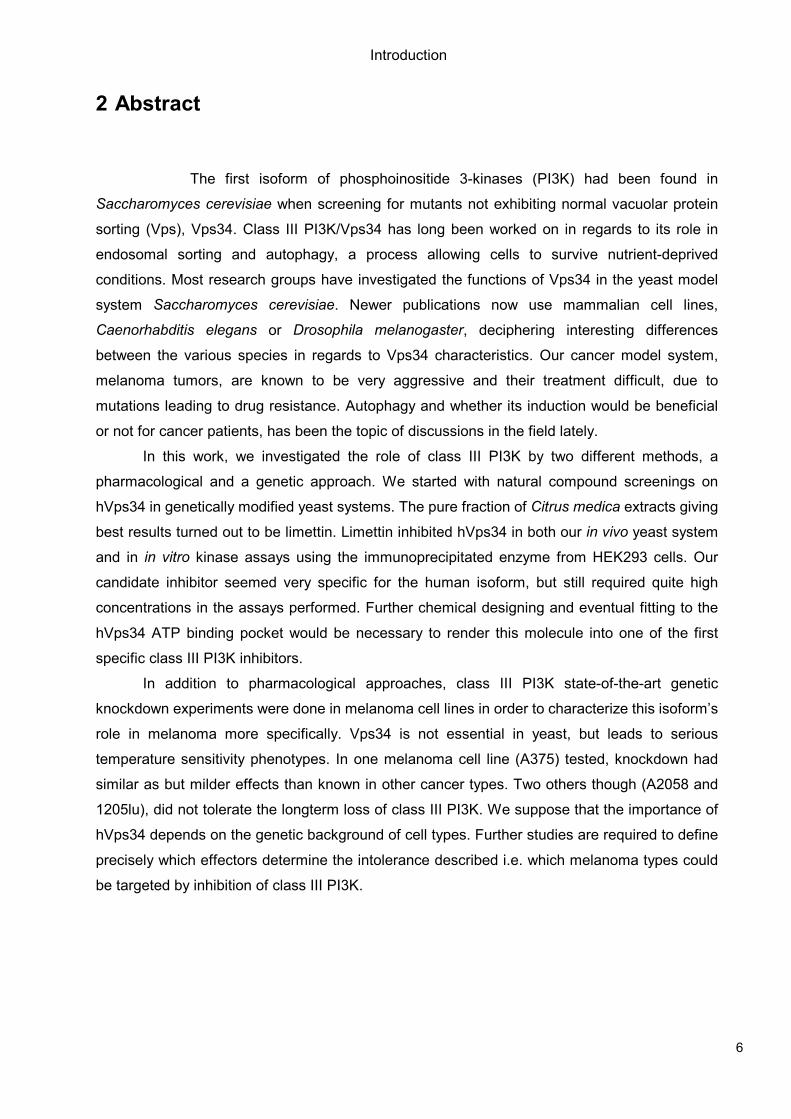
2 Abstract
The first isoform of phosphoinositide 3-kinases (PI3K) had been found in
Saccharomyces cerevisiae when screening for mutants not exhibiting normal vacuolar protein
sorting (Vps), Vps34. Class III PI3K/Vps34 has long been worked on in regards to its role in
endosomal sorting and autophagy, a process allowing cells to survive nutrient-deprived
conditions. Most research groups have investigated the functions of Vps34 in the yeast model
system Saccharomyces cerevisiae. Newer publications now use mammalian cell lines,
Caenorhabditis elegans or Drosophila melanogaster, deciphering interesting differences
between the various species in regards to Vps34 characteristics. Our cancer model system,
melanoma tumors, are known to be very aggressive and their treatment difficult, due to
mutations leading to drug resistance. Autophagy and whether its induction would be beneficial
or not for cancer patients, has been the topic of discussions in the field lately.
In this work, we investigated the role of class III PI3K by two different methods, a
pharmacological and a genetic approach. We started with natural compound screenings on
hVps34 in genetically modified yeast systems. The pure fraction of Citrus medica extracts giving
best results turned out to be limettin. Limettin inhibited hVps34 in both our in vivo yeast system
and in in vitro kinase assays using the immunoprecipitated enzyme from HEK293 cells. Our
candidate inhibitor seemed very specific for the human isoform, but still required quite high
concentrations in the assays performed. Further chemical designing and eventual fitting to the
hVps34 ATP binding pocket would be necessary to render this molecule into one of the first
specific class III PI3K inhibitors.
In addition to pharmacological approaches, class III PI3K state-of-the-art genetic
knockdown experiments were done in melanoma cell lines in order to characterize this isoform’s
role in melanoma more specifically. Vps34 is not essential in yeast, but leads to serious
temperature sensitivity phenotypes. In one melanoma cell line (A375) tested, knockdown had
similar as but milder effects than known in other cancer types. Two others though (A2058 and
1205lu), did not tolerate the longterm loss of class III PI3K. We suppose that the importance of
hVps34 depends on the genetic background of cell types. Further studies are required to define
precisely which effectors determine the intolerance described i.e. which melanoma types could
be targeted by inhibition of class III PI3K.

Introduction
7
Introduction
8
3 Introduction
3.1 Early Publications on Phosphoinositide 3-kinases (PI3K)
3.1.1 Yeast PI3K
In 1986, Rothman and Stevens published the discovery of a group of yeast mutants that
failed to properly sort CPY (carboxypeptidase Y) to the vacuole, the yeast’s lysosome. Their so
called eight “vpl mutants” defined a new class of proteins required for sorting of vacuolar
proteins during the secretory pathway (Rothman and Stevens 1986). Hence the first PI3K to be
described in yeast was Vps34, which was one of the genes involved in vacuolar protein sorting,
originally termed Vpt29 or Vpl7 (Robinson, Klionsky et al. 1988). At that time though, nothing
was known about its kinase function yet. One of the first publications on PI3K activity in yeast
was by Auger et al. in 1989. The authors claimed to have found PI3K lipid kinase activity in
Saccharomyces cerevisiae, similarly to an enzymatic reaction phosphorylating D-3 position of
the inositol ring which was at that time known in mammalian cells. Biochemical character of the
PI3K enzyme seemed different in yeast. Yet not only PI-4P, as originally thought, but PI(3)P
were found in liquid chromatography analysis of intact yeast cells labelled with (3H)inositol.
These results suggested an important and conserved functional role in cell cycle for PI3K in
eukaryotes (Auger, Carpenter et al. 1989).
3.1.2 Mammalian PI3K isoforms
One of the first publications on a kinase phosphorylating D-3 instead of D-4 position of
inositol in mammalian cells was a manuscript by Whitman M et al. in 1987 where they used
platelet-derived growth factor (PDGF)-stimulated murine fibroblasts. Phosphoinositide kinase
activity was found to associate with anti-phosphotyrosine immunoprecipitates from these cells.
A new class of lipids, phosphorylated at the D3-hydroxy-group of the inositol headgroup of
phosphoinositides was discovered (Whitman, Kaplan et al. 1987; Wymann and Pirola 1998).
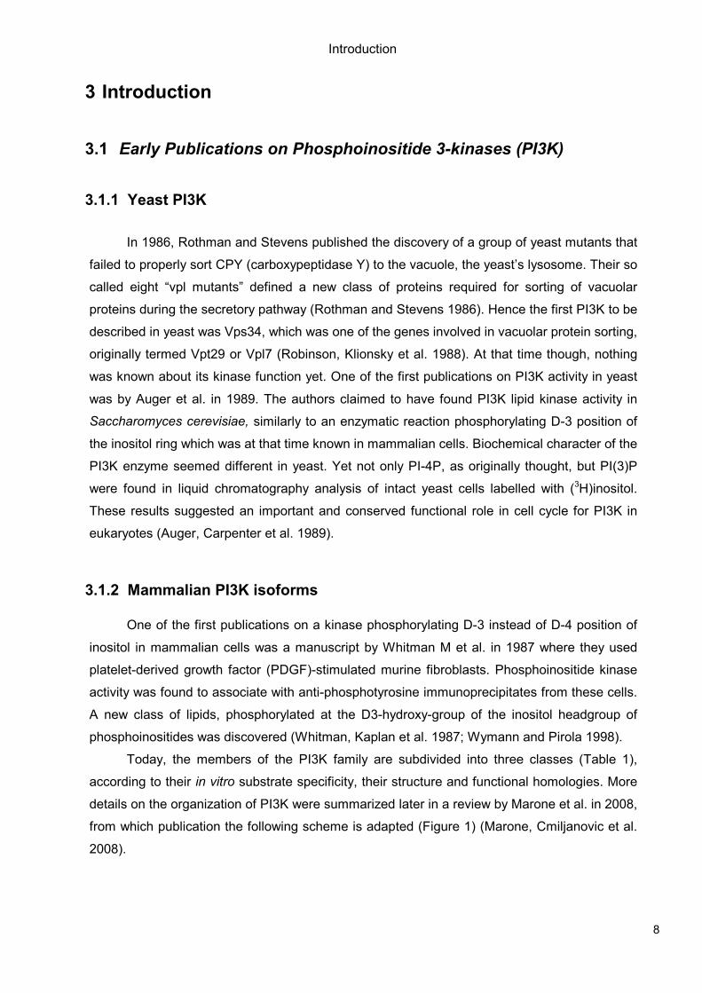
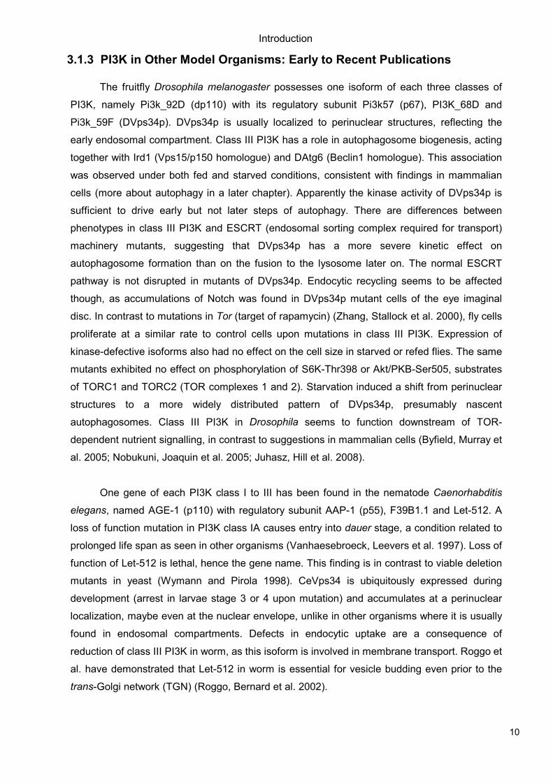
Today, the members of the PI3K family are subdivided into three classes (Table 1),
according to their in vitro substrate specificity, their structure and functional homologies. More
details on the organization of PI3K were summarized later in a review by Marone et al. in 2008,
from which publication the following scheme is adapted (Figure 1) (Marone, Cmiljanovic et al.
2008).
Introduction
9
Figure 1: Domain Structures of Mammalian PI3K isoforms
The members of the phosphoinositide 3-kinase (PI3K) family are subdivided into three classes, according to their in vitro substrate specificity, their structure and functional homologies. The upper panel (a) displays the catalytic subunits, the lower panel (b) shows their adaptors/regulatory subunits. a) The catalytic subunits contain a core catalytic domain (PI3Kc), including the ATP-binding site and a regulatory subunit binding domain at their N-terminus. Other known domains are a Ras-binding domain (RBD), a C2 domain (protein kinase C homology domain 2) and a PI3K accessory domain (helical, PI3Ka). Class II PI3K display several proline-rich (P) stretches, a PX (phox) and an additional C2 domain at their C-terminus. Class III PI3K, Vps34, only has a C2, PI3Ka and PI3Kc domain. b) The regulatory subunits/adaptors of class IA PI3K contain a proline-rich region and two SH2 (Src-homology 2) domains, an interSH2 region (iSH2) that binds to the catalytic subunit. SH3 (Src-homology 3) and BH (BCR homology) domains are found at the N-terminus of p85α and p85β. The structures of class IB adaptors are less well investigated. P150/hVps15, the adaptor of Vps34, contains a kinase domain, a HEAT (Huntingtin, Elongation Factor 3, protein phosphatase 2A and yeast TOR1) domain and several WD40 repeats. (Marone, Cmiljanovic et al. 2008)
Introduction
10
3.1.3 PI3K in Other Model Organisms: Early to Recent Publications
The fruitfly Drosophila melanogaster possesses one isoform of each three classes of
PI3K, namely Pi3k_92D (dp110) with its regulatory subunit Pi3k57 (p67), PI3K_68D and
Pi3k_59F (DVps34p). DVps34p is usually localized to perinuclear structures, reflecting the
early endosomal compartment. Class III PI3K has a role in autophagosome biogenesis, acting
together with Ird1 (Vps15/p150 homologue) and DAtg6 (Beclin1 homologue). This association
was observed under both fed and starved conditions, consistent with findings in mammalian
cells (more about autophagy in a later chapter). Apparently the kinase activity of DVps34p is
sufficient to drive early but not later steps of autophagy. There are differences between
phenotypes in class III PI3K and ESCRT (endosomal sorting complex required for transport)
machinery mutants, suggesting that DVps34p has a more severe kinetic effect on
autophagosome formation than on the fusion to the lysosome later on. The normal ESCRT
pathway is not disrupted in mutants of DVps34p. Endocytic recycling seems to be affected
though, as accumulations of Notch was found in DVps34p mutant cells of the eye imaginal
disc. In contrast to mutations in Tor (target of rapamycin) (Zhang, Stallock et al. 2000), fly cells
proliferate at a similar rate to control cells upon mutations in class III PI3K. Expression of
kinase-defective isoforms also had no effect on the cell size in starved or refed flies. The same
mutants exhibited no effect on phosphorylation of S6K-Thr398 or Akt/PKB-Ser505, substrates
of TORC1 and TORC2 (TOR complexes 1 and 2). Starvation induced a shift from perinuclear
structures to a more widely distributed pattern of DVps34p, presumably nascent
autophagosomes. Class III PI3K in Drosophila seems to function downstream of TOR-
dependent nutrient signalling, in contrast to suggestions in mammalian cells (Byfield, Murray et
al. 2005; Nobukuni, Joaquin et al. 2005; Juhasz, Hill et al. 2008).
One gene of each PI3K class I to III has been found in the nematode Caenorhabditis
elegans, named AGE-1 (p110) with regulatory subunit AAP-1 (p55), F39B1.1 and Let-512. A
loss of function mutation in PI3K class IA causes entry into dauer stage, a condition related to
prolonged life span as seen in other organisms (Vanhaesebroeck, Leevers et al. 1997). Loss of
function of Let-512 is lethal, hence the gene name. This finding is in contrast to viable deletion
mutants in yeast (Wymann and Pirola 1998). CeVps34 is ubiquitously expressed during
development (arrest in larvae stage 3 or 4 upon mutation) and accumulates at a perinuclear
localization, maybe even at the nuclear envelope, unlike in other organisms where it is usually
found in endosomal compartments. Defects in endocytic uptake are a consequence of
reduction of class III PI3K in worm, as this isoform is involved in membrane transport. Roggo et
al. have demonstrated that Let-512 in worm is essential for vesicle budding even prior to the
trans-Golgi network (TGN) (Roggo, Bernard et al. 2002).
Introduction
11
Class Type
Saccharomyces cerevisiae
Caenorhabditis elegans
Drosophila melanogaster
Mammals
Class I, catalytic
not present
AGE-1 (p110)
Pi3k_92D (dp110)
PIK3CA (p110α) PIK3CB (p110β) PIK3CD (p110δ) PIK3CG (p110γ)
Class I, regulatory
not present
AAP-1 (p55)
Pi3k57 (p67)
PIK3R1 (p85a/p55a/p50a) PIK3R2 (p85b) PIK3R3 (p55g) PIK3R5 (p101) PIK3R6 (p87/p84)
Class II
not present
F39B1.1
PI3K_68D
PIK3C2A (PI3K-C2α) PIK3C2B (PI3K-C2β) PIK3C2G (PI3K-C2γ)
Class III, catalytic
VPS34p
Let-512
Pi3k_59F (DVps34p)
PIK3C3 (hVps34 or Vps34)
Class III, regulatory
VPS15p
Vps15-like
Ird1
PIK3R4 (p150/vps15)
Table 1: The PI3K family members in different species
This table lists the homologs of PI3K isoforms in different species. Table adapated from (Vanhaesebroeck, Guillermet-Guibert et al.)
Introduction
12
3.2 More Recent Knowledge of PI3K
3.2.1 Class I PI3K
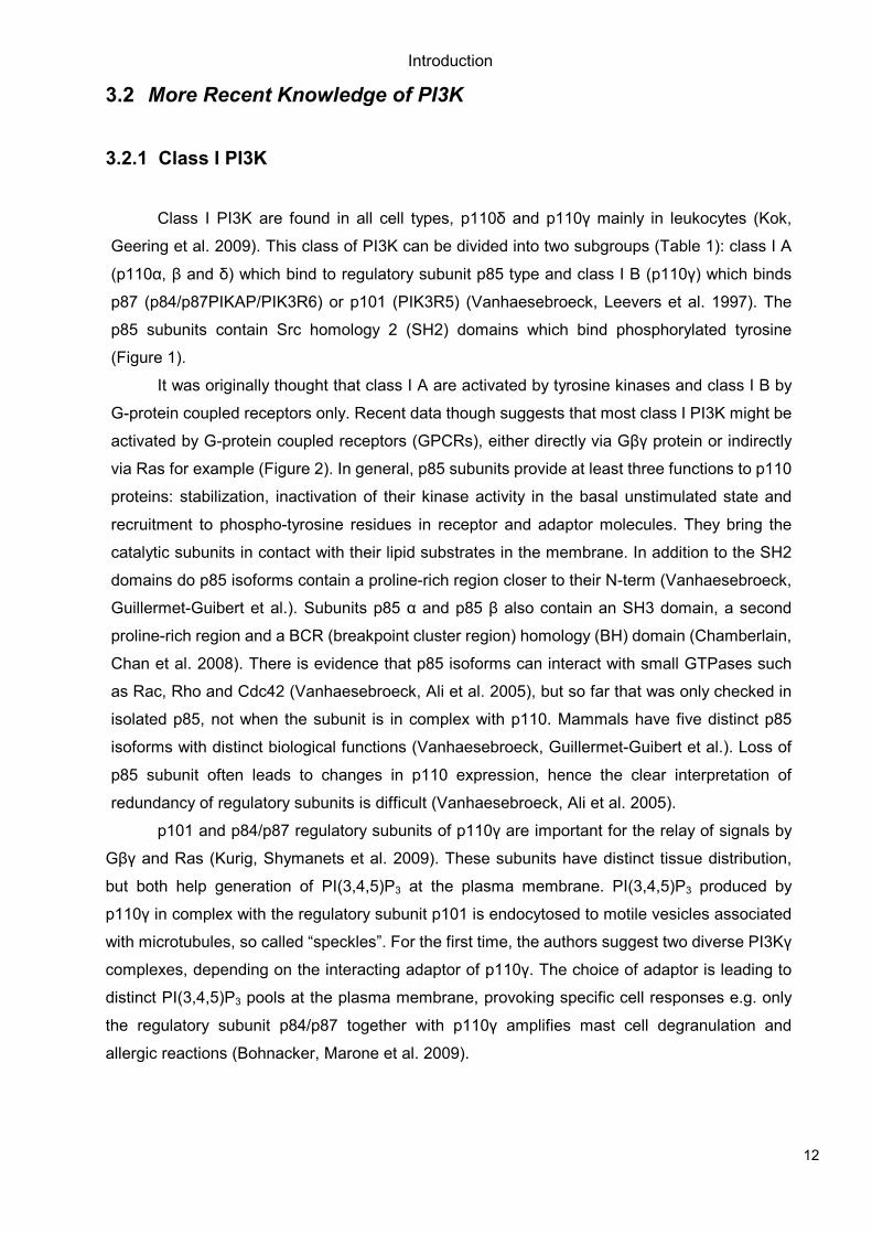
Class I PI3K are found in all cell types, p110δ and p110γ mainly in leukocytes (Kok,
Geering et al. 2009). This class of PI3K can be divided into two subgroups (Table 1): class I A
(p110α, β and δ) which bind to regulatory subunit p85 type and class I B (p110γ) which binds
p87 (p84/p87PIKAP/PIK3R6) or p101 (PIK3R5) (Vanhaesebroeck, Leevers et al. 1997). The
p85 subunits contain Src homology 2 (SH2) domains which bind phosphorylated tyrosine
(Figure 1).
It was originally thought that class I A are activated by tyrosine kinases and class I B by
G-protein coupled receptors only. Recent data though suggests that most class I PI3K might be
activated by G-protein coupled receptors (GPCRs), either directly via Gβγ protein or indirectly
via Ras for example (Figure 2). In general, p85 subunits provide at least three functions to p110
proteins: stabilization, inactivation of their kinase activity in the basal unstimulated state and
recruitment to phospho-tyrosine residues in receptor and adaptor molecules. They bring the
catalytic subunits in contact with their lipid substrates in the membrane. In addition to the SH2
domains do p85 isoforms contain a proline-rich region closer to their N-term (Vanhaesebroeck,
Guillermet-Guibert et al.). Subunits p85 α and p85 β also contain an SH3 domain, a second
proline-rich region and a BCR (breakpoint cluster region) homology (BH) domain (Chamberlain,
Chan et al. 2008). There is evidence that p85 isoforms can interact with small GTPases such
as Rac, Rho and Cdc42 (Vanhaesebroeck, Ali et al. 2005), but so far that was only checked in
isolated p85, not when the subunit is in complex with p110. Mammals have five distinct p85
isoforms with distinct biological functions (Vanhaesebroeck, Guillermet-Guibert et al.). Loss of
p85 subunit often leads to changes in p110 expression, hence the clear interpretation of
redundancy of regulatory subunits is difficult (Vanhaesebroeck, Ali et al. 2005).
p101 and p84/p87 regulatory subunits of p110γ are important for the relay of signals by
Gβγ and Ras (Kurig, Shymanets et al. 2009). These subunits have distinct tissue distribution,
but both help generation of PI(3,4,5)P3 at the plasma membrane. PI(3,4,5)P3 produced by
p110γ in complex with the regulatory subunit p101 is endocytosed to motile vesicles associated
with microtubules, so called “speckles”. For the first time, the authors suggest two diverse PI3Kγ
complexes, depending on the interacting adaptor of p110γ. The choice of adaptor is leading to
distinct PI(3,4,5)P3 pools at the plasma membrane, provoking specific cell responses e.g. only
the regulatory subunit p84/p87 together with p110γ amplifies mast cell degranulation and
allergic reactions (Bohnacker, Marone et al. 2009).
Introduction
13
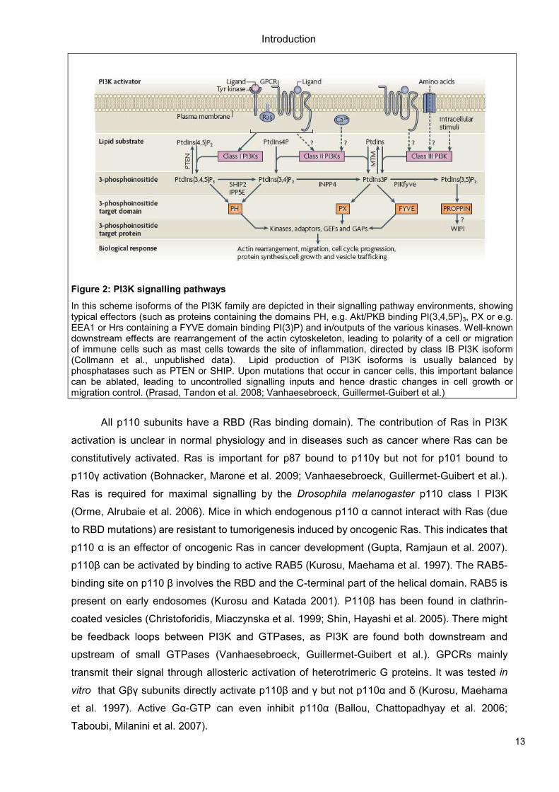
Figure 2: PI3K signalling pathways
In this scheme isoforms of the PI3K family are depicted in their signalling pathway environments, showing typical effectors (such as proteins containing the domains PH, e.g. Akt/PKB binding PI(3,4,5P)3, PX or e.g. EEA1 or Hrs containing a FYVE domain binding PI(3)P) and in/outputs of the various kinases. Well-known downstream effects are rearrangement of the actin cytoskeleton, leading to polarity of a cell or migration of immune cells such as mast cells towards the site of inflammation, directed by class IB PI3K isoform (Collmann et al., unpublished data). Lipid production of PI3K isoforms is usually balanced by phosphatases such as PTEN or SHIP. Upon mutations that occur in cancer cells, this important balance can be ablated, leading to uncontrolled signalling inputs and hence drastic changes in cell growth or migration control. (Prasad, Tandon et al. 2008; Vanhaesebroeck, Guillermet-Guibert et al.)
All p110 subunits have a RBD (Ras binding domain). The contribution of Ras in PI3K
activation is unclear in normal physiology and in diseases such as cancer where Ras can be
constitutively activated. Ras is important for p87 bound to p110γ but not for p101 bound to
p110γ activation (Bohnacker, Marone et al. 2009; Vanhaesebroeck, Guillermet-Guibert et al.).
Ras is required for maximal signalling by the Drosophila melanogaster p110 class I PI3K
(Orme, Alrubaie et al. 2006). Mice in which endogenous p110 α cannot interact with Ras (due
to RBD mutations) are resistant to tumorigenesis induced by oncogenic Ras. This indicates that
p110 α is an effector of oncogenic Ras in cancer development (Gupta, Ramjaun et al. 2007).
p110β can be activated by binding to active RAB5 (Kurosu, Maehama et al. 1997). The RAB5-
binding site on p110 β involves the RBD and the C-terminal part of the helical domain. RAB5 is
present on early endosomes (Kurosu and Katada 2001). P110β has been found in clathrin-
coated vesicles (Christoforidis, Miaczynska et al. 1999; Shin, Hayashi et al. 2005). There might
be feedback loops between PI3K and GTPases, as PI3K are found both downstream and
upstream of small GTPases (Vanhaesebroeck, Guillermet-Guibert et al.). GPCRs mainly
transmit their signal through allosteric activation of heterotrimeric G proteins. It was tested in
vitro that Gβγ subunits directly activate p110β and γ but not p110α and δ (Kurosu, Maehama
et al. 1997). Active Gα-GTP can even inhibit p110α (Ballou, Chattopadhyay et al. 2006;
Taboubi, Milanini et al. 2007).
Introduction
14
PI(3,4,5)P3 gets converted to PI(4,5)P2 and PI(3,5)P2 by PI-3- or PI-5-phosphatases. A
major PI-3-phosphatase is PTEN (phosphatase and tensin homologue deleted on chromosome
10), which is frequently inactivated in cancer and hence leading to PI3K activation. PTEN can
couple to p110β (Vanhaesebroeck, Guillermet-Guibert et al.). PI(3,4,5)P3 and PI(3,4)P2
coordinate the localization of multiple effector proteins which bind these lipids via their PH
(pleckstrin homology) domain, e.g. Akt/PKB or Btk. These lipids are mainly generated at the
plasma membrane (Vanhaesebroeck, Guillermet-Guibert et al.).
Class I PI3K do not have stable binding partners but can function as scaffolds, e.g.
p110γ binding to PKC (protein kinase C) (Hirsch, Braccini et al. 2009; Lehmann, Muller et al.
2009). PI3K can regulate small GTPases from Rac, Ras and Arf families by regulating their
GEFs (guanine nucleotide-exchange factors) and GAPs (GTPase activating proteins)
(Vanhaesebroeck, Leevers et al. 2001). Rac is positively regulated by all PI3K isoforms while
RhoA is negatively regulated by p110δ and p110α does not affect RhoA at all (Eickholt, Ahmed
et al. 2007; Papakonstanti, Ridley et al. 2007; Papakonstanti, Zwaenepoel et al. 2008).
Tumour-specific somatic mutations were only found in p110α first (Samuels, Wang et al.
2004). Later there were some mutations found in p110δ correlated to leukemia (Cornillet-
Lefebvre, Cuccuini et al. 2006). Most mutations in p110α are missense mutations that result in
an amino acid substitution in so called “hot-spot regions” in the kinase domain or helical
domain, while there are no mutations found so far in the RBD (Vogt, Kang et al. 2007; Zhao
and Vogt 2008). “Hot-spot region” mutations examples are His1047Arg or Glu545Lys of p110α
(Miled, Yan et al. 2007; Mandelker, Gabelli et al. 2009). Amplifications of the p110α gene are
also reported and have been found for the other isoforms as well (Shayesteh, Lu et al. 1999;
Kok, Geering et al. 2009).
3.2.2 Class II PI3K
Class II PI3Ks were discovered based on their sequence homology with class I and
class III PI3K. Their functional context is still poorly understood. Yeast does not have any
isoforms and only one isoform is found in both D. melanogaster and C.elegans. Mammals have
three class II PI3K isoforms: PI3K-C2α, PI3K-C2β and PI3K-C2γ. They are molecules of 166-
190kDa in size (Vanhaesebroeck, Guillermet-Guibert et al.). PI3K-C2α and PI3K-C2β are
broadly distributed in tissue, but PI3K-C2γ is more restricted to liver, breast and prostate tissue
(Elis, Triantafellow et al. 2008; Kok, Geering et al. 2009).
Pharmacological inhibitors have not been published so far. Sensitivity to pan-PI3K
inhibitors are slightly different than for the other classes though. Usually these inhibitors are
applied at high dosages that already affect class II PI3K, a possible side-effect that should be
kept in mind for interpretations of PI3K isoform-specific functions (Virbasius, Guilherme et al.
1996; Domin, Pages et al. 1997; Knight, Gonzalez et al. 2006).
Introduction
15
PX (phox) domain in the C-terminal end of PI3K-C2α can bind PI(4,5)P2, but today it is
not clear what function this represents (Song, Xu et al. 2001; Stahelin, Karathanassis et al.
2006). Class II PI3K do not have any regulatory subunits but contain extended N- and C-termini
(Vanhaesebroeck, Guillermet-Guibert et al.) and Ras binding domains (RBD).
Activation of class II PI3K might require the relocalization of a cytosolic pool to the
plasma membrane. Many stimuli have been discovered to activate PI3K-C2α and β, e.g.
insulin, EGF and PDGF, TNF-α, leptin and LPA (Figure 2). The exact mechanisms are often
still unclear though (Arcaro, Zvelebil et al. 2000; Elis, Triantafellow et al. 2008).
PI and PI(4)P are substrates of class II PI3K, whereas in vitro PI3K-C2α can produce
PI(3,4,5)P3 from PI(4,5)P2 if activated by clathrin (Domin, Pages et al. 1997). PI(3)P are mainly
localized in endosomes under unstimulated conditions and are bound by PX or FYVE domains.
Examples of proteins containing such domains are PIKfyve (Fab1), EEA1, HRS (Vps27) or Alfy
(Birkeland and Stenmark 2004; Di Paolo and De Camilli 2006; Hurley 2006; Lemmon 2008). It
is still not known to what extent class II PI3K contribute to basal level PI(3)P.
Downregulation of PI3K-C2α by RNAi does not affect the steady state levels of total
PI(3)P at the plasma membrane, as demonstrated in L6 muscle cell lines. Eventually do class II
PI3K contribute to the acute production of PI(3)P, e.g. PI3K-C2α generates the lipids at the
plasma membrane upon insulin stimulation while there is no increase in lipids at the
endosomes (Falasca, Hughes et al. 2007). Downregulation of PI3K-C2α but not PI3K-C2β
leads to a reduction in cell proliferation and viability by induced apoptosis in more than half of
23 cancer cell lines tested by Elis W. et al. (Elis, Triantafellow et al. 2008).
Genetic studies in fly (D. melanogaster, PI3K68D) and worm (C.elegans, F39B1.1)
provide evidence for a possible link between class II PI3Ks and Tyr kinase signalling pathways
but there is still investigation required for further details (Ashrafi, Chang et al. 2003;
MacDougall, Gagou et al. 2004). Links between class II PI3K and EGFR have been proposed
earlier in human carcinoma cell lines. (Arcaro, Zvelebil et al. 2000)
In summary, class II PI3Ks are involved in cell migration, glucose metabolism,
exocytosis, smooth muscle cell contraction and apoptosis (Falasca and Maffucci 2007). Viable
and fertile null-mice exist for PI3K-C2β but there are no obvious phenotypes in epidermal
differentiation, the only study done on this mouse strain so far (Harada, Truong et al. 2005).
3.2.3 Class III PI3K
Class III PI3K or Vps34, was originally identified in a screen for genes involved in
endosomal sorting to the yeast vacuole, the equivalent of the mammalian lysosome (Robinson,
Klionsky et al. 1988; Herman and Emr 1990). Vps34 is conserved from yeast to plants and
mammals. It fulfills multiple functions through association with distinct multiprotein complexes.
Vps34 forms a constitutive heterodimer with Vps15 (p150 in mammals) which is myristoylated
Introduction
16
and hence locates the complex to intracellular membranes (Vanhaesebroeck, Guillermet-
Guibert et al.).
In yeast, three complexes including Vps34 have been reported so far: The Vps34-
containing complex involved in autophagy consists of Vps34, Vps15, Atg14 and Vps30. The
vacuole protein sorting complex contains Vps38 and Vps30 besides class III PI3K and its
partner Vps15. In a third complex, Vps34 and Vps15 are interacting with Gprotein α subunit
activated with GTP and lead to a pheromone response upon mating factor binding
(Vanhaesebroeck, Guillermet-Guibert et al.).
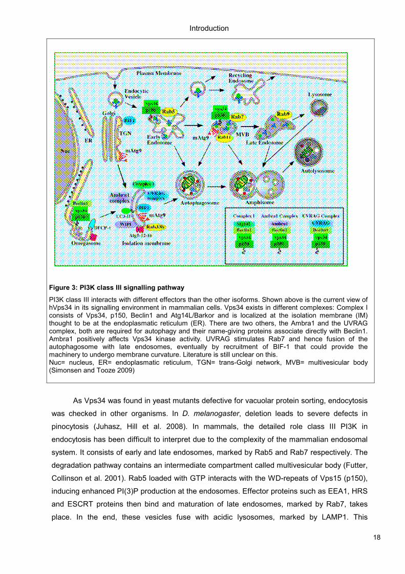
In mammals, there is also an autophagy complex containing Vps34 and p150 (Vps15),
ATG14L (also known as barkor), Beclin1, UVRAG, Ambra1, Rubicon and Bif1 (Figure 3). As for
endosomal trafficking there are different complexes, some consisting of UVRAG with Vps34
and p150, others containing MTM1 in addition to the PI3K. Effectors are FYVE domain bearing
proteins such as HRS or ESCRTII and III (Simonsen and Tooze 2009). Vps34-associated
effector proteins might also regulate the catalytic activity of Vps34, e.g. Bif1 (Bax-interacting
factor 1) which stimulates the kinase activity in the UVRAG-Beclin1 complex (Liang, Feng et al.
2006; Takahashi, Coppola et al. 2007).
Class III PI3K has a limited substrate specificity, generating PI(3)P from PI. It is not clear
today, which effectors class III and II PI3K share and to what extent their functions overlap
(Schu, Takegawa et al. 1993; Volinia, Dhand et al. 1995). Studies in C.elegans suggest that
Vps34-independent sources of PI(3)P exist, as the phenotype of a Vps34-null mutant can be
rescued by reducing PI(3)P-ase activity. Kinase dead versions could not rescue the deletion
phenotype in yeast or worm though, hinting that the lipid kinase activity of Vps34 is important
for its biological functions (Roggo, Bernard et al. 2002; Xue, Fares et al. 2003).
It is still controversary in the field, whether Vps34 is regulated by extracellular stimuli
(Figure 2). There is evidence for regulation by nutrients such as amino acids and glucose or
GPCRs (Byfield, Murray et al. 2005; Nobukuni, Joaquin et al. 2005) in human cell lines while
similar processes could not be confirmed in other model organisms such as Drosophila
melanogaster (Juhasz, Hill et al. 2008). GPCR stimulation was proposed to happen in yeast
during the pheromone response to mating factors. Pheromones lead to the activation and
dissociation of G protein α subunit from Gβγ at the plasma membrane. It then passes on to the
endosome where it binds and activates Vps34-Vps15 which leads to increase of PI(3)P
formation and hence recruitment of PI(3)P-binding effectors. Interestingly, here it is Gprotein α
subunit that activates the PI3K while it is Gβ γ that binds class I PI3K isoforms (Slessareva,
Routt et al. 2006). A closer look into this topic is given in the following chapter “Nutrient
Regulation”.
Vps34 might also work as a scaffold protein. Homozygous deletions in yeast are viable
but exhibit growth defects even under non-stressed conditions and, as mentioned before,
defects in vacuolar protein sorting (Backer 2008). The global deletion of Vps34 in yeast and fly
Introduction
17
is lethal and null-mice have not been reported so far (Vanhaesebroeck, Guillermet-Guibert et
al.). All known biological functions of Vps34 in mammals are related to regulation of vesicular
traffic, e.g. autophagy, endocytosis and phagocytosis. Some studies indicate that class III PI3K
is controlling amino acid-dependent activation of S6 kinase 1 (Byfield, Murray et al. 2005;
Nobukuni, Joaquin et al. 2005), this link is still under debate though.
Vps34’s involvement in autophagy was first shown in yeast (Kihara, Noda et al. 2001). In
flies, the role of class III PI3K in autophagy is strongly limited to the early formation steps of
autophagosomes (Juhasz, Hill et al. 2008). In mammals, its role is still not well-defined. The
origin of autophagosomes is still not clear, but there are links to the Golgi, plasma membrane
and ER (endoplasmatic reticulum). Omegasomes are cup shaped vesicles formed from the ER
membrane of amino acid starved mammalian cells, possibly representing nascent
autophagosomes (Axe, Walker et al. 2008).
Introduction
18
Figure 3: PI3K class III signalling pathway
PI3K class III interacts with different effectors than the other isoforms. Shown above is the current view of hVps34 in its signalling environment in mammalian cells. Vps34 exists in different complexes: Complex I consists of Vps34, p150, Beclin1 and Atg14L/Barkor and is localized at the isolation membrane (IM) thought to be at the endoplasmatic reticulum (ER). There are two others, the Ambra1 and the UVRAG complex, both are required for autophagy and their name-giving proteins associate directly with Beclin1. Ambra1 positively affects Vps34 kinase activity. UVRAG stimulates Rab7 and hence fusion of the autophagosome with late endosomes, eventually by recruitment of BIF-1 that could provide the machinery to undergo membrane curvature. Literature is still unclear on this. Nuc= nucleus, ER= endoplasmatic reticulum, TGN= trans-Golgi network, MVB= multivesicular body (Simonsen and Tooze 2009)
As Vps34 was found in yeast mutants defective for vacuolar protein sorting, endocytosis
was checked in other organisms. In D. melanogaster, deletion leads to severe defects in
pinocytosis (Juhasz, Hill et al. 2008). In mammals, the detailed role class III PI3K in
endocytosis has been difficult to interpret due to the complexity of the mammalian endosomal
system. It consists of early and late endosomes, marked by Rab5 and Rab7 respectively. The
degradation pathway contains an intermediate compartment called multivesicular body (Futter,
Collinson et al. 2001). Rab5 loaded with GTP interacts with the WD-repeats of Vps15 (p150),
inducing enhanced PI(3)P production at the endosomes. Effector proteins such as EEA1, HRS
and ESCRT proteins then bind and maturation of late endosomes, marked by Rab7, takes
place. In the end, these vesicles fuse with acidic lysosomes, marked by LAMP1. This
Introduction
19
endosomal trafficking is also used for receptor sorting, e.g. for receptor Tyr kinase or
transferring receptors. Upon Vps34 inhibition, recycling of transferring receptor or
internalization of PDGF (platelet-derived growth factor) or EGF (epidermal growth factor)
receptors are delayed (Siddhanta, McIlroy et al. 1998; Murray, Panaretou et al. 2002; Stein,
Feng et al. 2003; Cao, Laporte et al. 2007; Cao, Backer et al. 2008). Various waves of PI(3)P
accumulation happen during phagocytosis, probably through Vps34 (Ellson, Anderson et al.
2001). Contrary to the normal endocytic pathway, in phagocytosis class III PI3K seems to act
upstream of Rab5 by interaction with dynamin. Dynamin controls clathrin-mediated endocytosis
(Kinchen, Doukoumetzidis et al. 2008).
Many diseases are linked to mutations in enzymes that regulate the turnover of PI(3)P,
such as members of the myotubularin family and PIKfyve. A specific role for class III PI3K is
less clear, although processes like autophagy are certainly involved in infection and
inflammation mechanisms (Levine and Kroemer 2008). A role for Vps34 in proliferation has
been reported in cancer cell lines, but no deletions or inactivating mutations in the VPS34 gene
have been observed so far. Vps34 might be involved in schizophrenia though (Siddhanta,
McIlroy et al. 1998; Johnson, Overmeyer et al. 2006; Tang, Zhao et al. 2008).
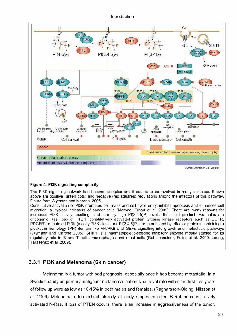
3.3 PI3K and Cancer
Cancer cells evolve from a benign non-invasive state to metastatic tumors which
proliferate aggressively out of their normal tissue context. Accumulation of genetic alterations
lead to multiple inputs to signal transduction pathways, e.g. the PI3K pathway (Fig.4).
Constitutive activation of PI3K promotes cell mass and cell cycle entry, inhibits apoptosis and
enhances cell migration, all typical indicaters of cancer cells (Marone, Erhart et al. 2009). There
are many reasons for increased PI3K activity resulting in abnormally high PI(3,4,5)P3 levels e.g.
oncogenic Ras, loss of PTEN, constitutively activated protein tyrosine kinase receptors such as
EGFR or PDGFR or mutated PI3K (mostly class Iα PI3K). PI(3,4,5)P3 are then bound by
effector proteins containing a pleckstrin homology (PH) domain like Akt/PKB and GEFs
signalling into growth and metastasis pathways (Wymann and Marone 2005) (Figure 4).
SHIP1 is a haematopoietic-specific inhibitory enzyme mostly studied for its regulatory
role in B and T cells, macrophages and mast cells (Rohrschneider, Fuller et al. 2000; Leung,
Tarasenko et al. 2009). Observations in mice suggest a mechanism in which SHIP1 negatively
regulates the PI3K pathway by hydrolysis of PI(3,4,5)P3 and might hence control cell
proliferation in a cancer context (Bunney and Katan).
Introduction
20
Figure 4: PI3K signalling complexity
The PI3K signalling network has become complex and it seems to be involved in many diseases. Shown above are positive (green dots) and negative (red squares) regulations among the effectors of this pathway. Figure from Wymann and Marone, 2005. Constitutive activation of PI3K promotes cell mass and cell cycle entry, inhibits apoptosis and enhances cell migration, all typical indicaters of cancer cells (Marone, Erhart et al. 2009). There are many reasons for increased PI3K activity resulting in abnormally high PI(3,4,5)P3 levels, their lipid product. Examples are oncogenic Ras, loss of PTEN, constitutively activated protein tyrosine kinase receptors such as EGFR, PDGFR) or mutated PI3K (mostly PI3K class I α). PI(3,4,5)P3 are then bound by effector proteins containing a pleckstrin homology (PH) domain like Akt/PKB and GEFs signalling into growth and metastasis pathways (Wymann and Marone 2005). SHIP1 is a haematopoietic-specific inhibitory enzyme mostly studied for its regulatory role in B and T cells, macrophages and mast cells (Rohrschneider, Fuller et al. 2000; Leung, Tarasenko et al. 2009).
3.3.1 PI3K and Melanoma (Skin cancer)
Melanoma is a tumor with bad prognosis, especially once it has become metastatic. In a
Swedish study on primary malignant melanoma, patients’ survival rate within the first five years
of follow up were as low as 10-15% in both males and females. (Ragnarsson-Olding, Nilsson et
al. 2009) Melanoma often exhibit already at early stages mutated B-Raf or constitutively
activated N-Ras. If loss of PTEN occurs, there is an increase in aggressiveness of the tumor,
Introduction
21
similar to what is seen upon up-regulation of PKB. Mutations in PI3K class I α happen very
rarely in cutaneous melanoma, it is more likely that the protein is upregulated (Curtin, Stark et
al. 2006; Omholt, Krockel et al. 2006; Stark and Hayward 2007).
The PI3K pathway was originally targeted with the potent steroidal fungal metabolite
wortmannin (Arcaro and Wymann 1993) or LY294002 (Vlahos, Matter et al. 1994), a pan-PI3K
inhibitor with low potency, low specificity and high toxicity. These inhibitors also inhibit related
proteins such as mTOR. More recent inhibitors like PI-103 and ZSTK474 led to improved
specificity. Newer dual inhibitors affecting both mTOR and PI3K are in clinical trials now,
treating patients with advanced malignancies. In a study by Marone et al. in 2009, we reported
our results on NVP-BEZ235 tested in mice and in vitro. This compound blocked proliferation of
melanoma cells by G1 cell cycle arrest and was well tolerated in mice. Both primary tumor (up
to 60% reduction) and metastases were greatly reduced upon treatment while no adverse
effects on the immune system was observed. Angiogenesis for the nutrient supply of the tumor
was shown to be sensitive to the inhibitor tested, as we could see a clear reduction of
neovascularization in mice treated with this compound. Most probably it was a combination of
effects on the PI3K/mTOR pathway in addition to the VEGF receptor (vascular endothelial
growth factor), as VEGFR inhibitors were not as efficient. NVP-BEZ235 not only reduced
proliferation of tumor cells in mice but also augmented tumor necrosis (Marone, Erhart et al.
2009).
Tormo et al. described the therapeutic induction of autophagy in melanoma cells in a
recent publication in Cancer Cell. The authors used three different mouse models: In the first
mouse model, B16 melanoma cells were inoculated in immunocompetent mice. In their second
model, they used GFP-labelled B16 or SK-Mel-103 cells in SCID-beige mice. Their third
approach was a disease model using Tyr::NRASQ61K; INK4a/ARF-/- mice, that exhibit features
similar to the human disease. These mouse models were used to show that melanoma cells
retain an innate immune system response to recognize cytosolic double-stranded RNA
(dsRNA) and start stress response programs to block tumor growth. The dsRNA was identified
to be an inducer of autophagy, requiring Atg5 for the cell death process. Knockdown of Beclin1
led to cellular senescence or cell death in many melanoma cell lines tested. As Atg5
knockdown was lethal, too, they used Atg5-/- MEFs which showed a similar reaction to dsRNA
as observed earlier in their melanoma cell lines. The authors conclude from their results that
their artificial dsRNA induced an early but persistent autophagy and a late apoptotic program,
hence killing the melanoma cells (Tormo, Checinska et al. 2009).
Another publication describing apoptosis and autophagy induction in melanoma cell lines
was written by Liu et al. They conclude that in a A375 melanoma cell line, application of
polygonatum cyrtonema (PC) lectin induces cell death by autophagy followed by apoptosis
through mitochondria-mediated ROS-p38-p53 pathways. Beclin1 levels were increased after
treatment with PC lectin and mature forms of LC-3 were observed, all representing symptoms
Introduction
22
of autophagy. ROS (reactive oxygen species) were generated and p38 (MAPK) and p53 (tumor
suppressor involved in cell cycle control) activated as well, evidence for increased apoptosis
(Liu, Cheng et al. 2009).
3.4 Nutrient regulation
There are two mTOR complexes (Figure 8), of which mTORC1 (containing mTOR,
raptor, PRAS40 and mLST8) is rapamycin-sensitive and controls cell growth, metabolism and
autophagy through various signals, among them amino acids. A typical readout is
phosphorylation of downstream substrate S6K1 and 4EBP1.
In the study of Byfield et al. in 2005, HEK293T cells overexpressing hVps34 exhibited a
two-fold increase in S6K activity, similar to what can be observed upon stimulation with insulin.
Blocking hVps34 function by overexpressing FYVE domain-construct did bind and hence block
PI(3)P signalling indicated by ablation of EEA1 endosomal localization. Other attempts to block
hVps34 by antibodies targeting the kinase or by application of siRNA gave similar results in
HeLa cells. Overall, Byfield et al. suggested that both hVps34 and its product PI(3)P are
required for both amino acid and insulin-stimulation of S6K1. They admit the possible
differences in mammalian and yeast pathways, as observed for AMPK’s role in autophagy
(Wang, Wilson et al. 2001; Byfield, Murray et al. 2005).
Another group claimed to have found a link between mTORC1 and hVps34 in the same
year, Nobukuni et al. working with siRNA technique in HEK293 and HeLa cells. Their results
showed that effects of amino acids on S6K1 Thr398 phosphorylation are not mediated by
TSC1/2. Wortmannin, a pan-PI3K inhibitor, did block the amino acid response though. It could
be shown that this did not happen via class I PI3K. Under amino acid deprivation, little PI(3)P
was detected in their cells. Upon knockdown of hVps34, they saw a coordinate reduction in
amino acid-induced phosphorylation of S6K1. Hence they suggested hVps34 to be an
upstream regulator of amino acid signal input towards mTORC1 (Nobukuni, Joaquin et al.
2005) (Figure 5). Amino acid activation of mTORC1 increases growth while it suppresses
autophagy. Hormones and growth factors activate the Tor complex through canonical signalling
via PI3K class I, in contrast, amino acids stimulate the complex via PI3K class III.
In a study by Gulati et al. in 2008, they claimed that amino acids induce an increase in
intracellular calcium and in response enhanced binding of calmodulin (CaM) to hVps34 (Figure
5). This binding then leads to activation of the mTORC1 signalling. Agents that increase
calcium levels within the cell have been reported to induce S6K1 phosphorylation without
inducing PKB activation. HeLa cells were treated with BAPTA-AM, a calcium chelator.
Treatment caused an attenuation of S6K1 activation under restimulation with amino acids.
Similar effects were seen after application of EGTA, another calcium chelator. To check
calcium levels within the cells, Gulati et al. used a calcium-sensitive indicator called Fluo-4.
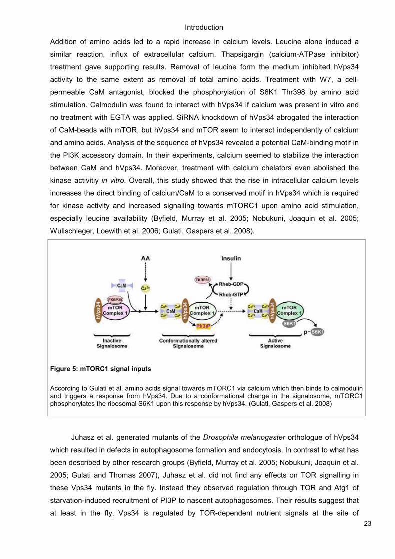
Introduction
23
Addition of amino acids led to a rapid increase in calcium levels. Leucine alone induced a
similar reaction, influx of extracellular calcium. Thapsigargin (calcium-ATPase inhibitor)
treatment gave supporting results. Removal of leucine form the medium inhibited hVps34
activity to the same extent as removal of total amino acids. Treatment with W7, a cell-
permeable CaM antagonist, blocked the phosphorylation of S6K1 Thr398 by amino acid
stimulation. Calmodulin was found to interact with hVps34 if calcium was present in vitro and
no treatment with EGTA was applied. SiRNA knockdown of hVps34 abrogated the interaction
of CaM-beads with mTOR, but hVps34 and mTOR seem to interact independently of calcium
and amino acids. Analysis of the sequence of hVps34 revealed a potential CaM-binding motif in
the PI3K accessory domain. In their experiments, calcium seemed to stabilize the interaction
between CaM and hVps34. Moreover, treatment with calcium chelators even abolished the
kinase activitiy in vitro. Overall, this study showed that the rise in intracellular calcium levels
increases the direct binding of calcium/CaM to a conserved motif in hVps34 which is required
for kinase activity and increased signalling towards mTORC1 upon amino acid stimulation,
especially leucine availability (Byfield, Murray et al. 2005; Nobukuni, Joaquin et al. 2005;
Wullschleger, Loewith et al. 2006; Gulati, Gaspers et al. 2008).
Figure 5: mTORC1 signal inputs
According to Gulati et al. amino acids signal towards mTORC1 via calcium which then binds to calmodulin and triggers a response from hVps34. Due to a conformational change in the signalosome, mTORC1 phosphorylates the ribosomal S6K1 upon this response by hVps34. (Gulati, Gaspers et al. 2008)
Juhasz et al. generated mutants of the Drosophila melanogaster orthologue of hVps34
which resulted in defects in autophagosome formation and endocytosis. In contrast to what has
been described by other research groups (Byfield, Murray et al. 2005; Nobukuni, Joaquin et al.
2005; Gulati and Thomas 2007), Juhasz et al. did not find any effects on TOR signalling in
these Vps34 mutants in the fly. Instead they observed regulation through TOR and Atg1 of
starvation-induced recruitment of PI3P to nascent autophagosomes. Their results suggest that
at least in the fly, Vps34 is regulated by TOR-dependent nutrient signals at the site of
Introduction
24
autophagosome formation. TOR mutants show a decrease in cell size, while neither Vps34
mutants nor cells overexpressing kinase-dead Vps34 did. Proliferation was not affected either,
indicating that in fly eye imaginal disc cells, TOR signalling is not compromised in Vps34
mutants. Readouts such as phosphorylated Thr398 of S6K1 were not affected in Vps34-
deficient cells. According to their data, Vps34 seems to be acting downstream of TOR-
dependent nutrient signalling in the fruit fly (Juhasz, Hill et al. 2008).
Figure 6: Schematic view of TOR’s in- and outputs
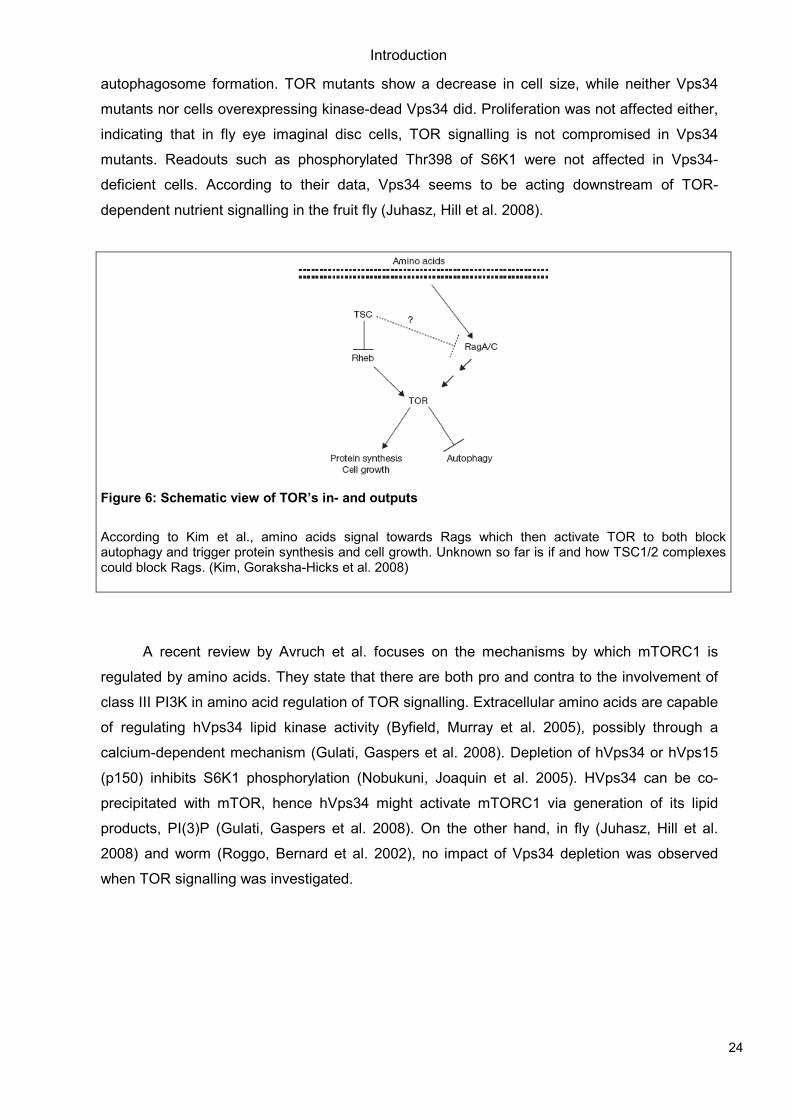
According to Kim et al., amino acids signal towards Rags which then activate TOR to both block autophagy and trigger protein synthesis and cell growth. Unknown so far is if and how TSC1/2 complexes could block Rags. (Kim, Goraksha-Hicks et al. 2008)
A recent review by Avruch et al. focuses on the mechanisms by which mTORC1 is
regulated by amino acids. They state that there are both pro and contra to the involvement of
class III PI3K in amino acid regulation of TOR signalling. Extracellular amino acids are capable
of regulating hVps34 lipid kinase activity (Byfield, Murray et al. 2005), possibly through a
calcium-dependent mechanism (Gulati, Gaspers et al. 2008). Depletion of hVps34 or hVps15
(p150) inhibits S6K1 phosphorylation (Nobukuni, Joaquin et al. 2005). HVps34 can be co-
precipitated with mTOR, hence hVps34 might activate mTORC1 via generation of its lipid
products, PI(3)P (Gulati, Gaspers et al. 2008). On the other hand, in fly (Juhasz, Hill et al.
2008) and worm (Roggo, Bernard et al. 2002), no impact of Vps34 depletion was observed
when TOR signalling was investigated.
Introduction
25
Figure 7: Nutrient signalling input towards mTORC1
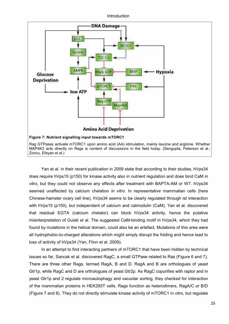
Rag GTPases activate mTORC1 upon amino acid (AA) stimulation, mainly leucine and arginine. Whether MAP4K3 acts directly on Rags is content of discussions in the field today. (Sengupta, Peterson et al.; Zoncu, Efeyan et al.)
Yan et al. in their recent publication in 2009 state that according to their studies, hVps34
does require hVps15 (p150) for kinase activity also in nutrient regulation and does bind CaM in
vitro, but they could not observe any effects after treatment with BAPTA-AM or W7. hVps34
seemed unaffected by calcium chelation in vitro. In representative mammalian cells (here
Chinese-hamster ovary cell line), hVps34 seems to be clearly regulated through ist interaction
with hVps15 (p150), but independent of calcium and calmodulin (CaM). Yan et al. discovered
that residual EGTA (calcium chelator) can block hVps34 activity, hence the putative
misinterpretation of Gulati et al. The suggested CaM-binding motif in hVps34, which they had
found by mutations in the helical domain, could also be an artefact. Mutations of this area were
all hydrophobic-to-charged alterations which might simply disrupt the folding and hence lead to
loss of activity of hVps34 (Yan, Flinn et al. 2009).
In an attempt to find interacting partners of mTORC1 that have been hidden by technical
issues so far, Sancak et al. discovered RagC, a small GTPase related to Ras (Figure 6 and 7).
There are three other Rags, termed RagA, B and D. RagA and B are orthologues of yeast
Gtr1p, while RagC and D are orthologues of yeast Gtr2p. As RagC copurifies with raptor and in
yeast Gtr1p and 2 regulate microautophagy and vacuolar sorting, they checked for interaction
of the mammalian proteins in HEK293T cells. Rags function as heterodimers, RagA/C or B/D
(Figure 7 and 8). They do not directly stimulate kinase activity of mTORC1 in vitro, but regulate
Introduction
26
the localization within the cell upon amino acid signalling. Sancak et al. propose that since both
mTOR and Rheb are present in Rab7-positive endosomes after amino acid stimulation, amino
acids might control the mTORC1 pathway via the Rag proteins in order for mTORC1 to meet its
activator Rheb. This hypothesis would explain why activators of Rheb, e.g. insulin, do not
stimulate the mTORC1 pathway when cells are in a nutrient deprived state (Sancak, Peterson
et al. 2008; Sengupta, Peterson et al.; Zoncu, Efeyan et al.).
Many signals, including amino acids, are known to activate mTORC1. Kim et al.
identified the small GTPases Rags as activators of TORC1 in response to amino acid signals
(Figure 6). In Drosophila melanogaster, knockdown of Rags led to suppression of stimulation of
TORC1 by amino acids, while expression of constitutively active Rags (GTP-loaded) activated
TORC1 when phosphorylation of dS6K at Thr398 was taken as readout. Rags are apparently
regulating cell size as expected of a TOR regulator, as well as autophagy and survival during
starvation. Especially dRagA could be shown to be involved in a nutrient response to amino
acids (Kim, Goraksha-Hicks et al. 2008).
Another player in the TOR nutrient sensing field has been discovered by Findlay et al. in
Drosophila melanogaster. MAP4K3 (CG7097 in the fly), a Ste20 family member, seems
required for maximal S6K/4EBP1 phosphorylation and regulates cell growth. It is itself
regulated by amino acids but not insulin and insensitive to rapamycin, suggesting that MAP4K3
is not downstream of TOR but upstream in this pathway. An almost complete knockdown of
MAP4K3 was required to get efficient suppression of S6K1 phosphorylation at Thr398, while
the effect seemed independent of TSC1/2 complexes. They picture MAP4K3 in a signalling
branch parallel to TSC1/2, passing on signals from amino acid input towards mTORC1
(Findlay, Yan et al. 2007).
Nicklin et al. showed in 2009 that cellular uptake of L-glutamine and ist subsequent rapid
efflux in the presence of essential amino acids (EEA) activates mTORC1. SLC1A5 (solute
carrier family 1 member 5 or ASCT2), a high-affinity transporter for L-glutamine, regulates this
uptake and loss of the sodium-dependent transporter results in reduction of cell growth and
activation of autophagy. SLC7A5 (LAT1)/SLC3A2, a bidirectional transporter, regulates the
simultaneous efflux of L-glutamine and transport of L-leucine/EEA into cells. The exchange of
L-glutamine for L-leucine happens before S6K1 activation as they showed in HeLa cells. They
claim that SLC1A5 and SLC7A5 may act upstream of TSC1/2 to mediate amino acid signals
towards mTOR. The findings seemed independent of cell types, as similar results were
obtained in MCF-7 breast cancer cells. SLC1A5 and L-glutamine suppressed autophagy in
RT112 cells, a human urinary bladder carcinoma cell line. Overall, Nicklin et al. presented
amino acid transporters upstream of mTORC1, which are involved in regulation of autophagy
by bidirectional transport of AA (amino acids) (Nicklin, Bergman et al. 2009).
Wang and Proud summarized today’s knowledge of nutrient control concerning TORC1
in a review in Trends in Cell Biology in 2009. In order to illustrate the above mentioned
Introduction
27
publications on the composition of mTOR complexes, Rags, upstream control of mTORC1 by
amino acids and putative links between mTORC1 and autophagy/hVps34, figures from this
article are pasted below (Figure 8) (Wang and Proud 2009).
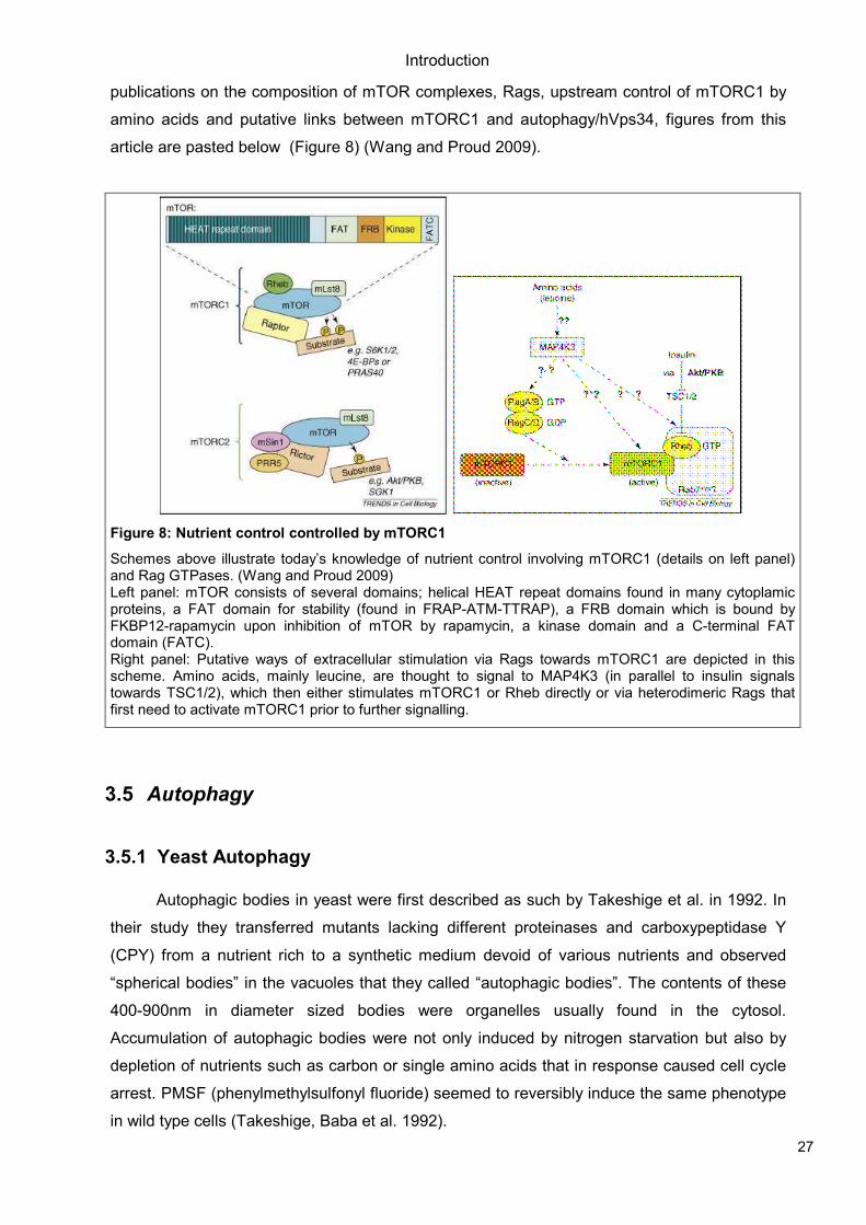
Figure 8: Nutrient control controlled by mTORC1
Schemes above illustrate today’s knowledge of nutrient control involving mTORC1 (details on left panel) and Rag GTPases. (Wang and Proud 2009) Left panel: mTOR consists of several domains; helical HEAT repeat domains found in many cytoplamic proteins, a FAT domain for stability (found in FRAP-ATM-TTRAP), a FRB domain which is bound by FKBP12-rapamycin upon inhibition of mTOR by rapamycin, a kinase domain and a C-terminal FAT domain (FATC). Right panel: Putative ways of extracellular stimulation via Rags towards mTORC1 are depicted in this scheme. Amino acids, mainly leucine, are thought to signal to MAP4K3 (in parallel to insulin signals towards TSC1/2), which then either stimulates mTORC1 or Rheb directly or via heterodimeric Rags that first need to activate mTORC1 prior to further signalling.
3.5 Autophagy
3.5.1 Yeast Autophagy
Autophagic bodies in yeast were first described as such by Takeshige et al. in 1992. In
their study they transferred mutants lacking different proteinases and carboxypeptidase Y
(CPY) from a nutrient rich to a synthetic medium devoid of various nutrients and observed
“spherical bodies” in the vacuoles that they called “autophagic bodies”. The contents of these
400-900nm in diameter sized bodies were organelles usually found in the cytosol.
Accumulation of autophagic bodies were not only induced by nitrogen starvation but also by
depletion of nutrients such as carbon or single amino acids that in response caused cell cycle
arrest. PMSF (phenylmethylsulfonyl fluoride) seemed to reversibly induce the same phenotype
in wild type cells (Takeshige, Baba et al. 1992).
Introduction
28
A year later, the same research group published a list of APG genes (later termed ATG
genes). These were supposed to be involved in autophagy in yeast, i.e. APG1-15. Mutants
defective for these genes did not accumulate autophagic bodies under nutrient-depleted
conditions, were defective in protein degradation in the vacuoles induced by nitrogen starvation
and lost viability much faster than wild type yeast if starved. Homozygous diploids of each APG
mutant did not sporulate (Tsukada and Ohsumi 1993).
“Autophagosomes” were first described by Baba et al. as double membraned structures
that apparently fused with vacuoles, hence they were thought to be precursors of autophagic
bodies (Figure 10 and 11). Electron microscopy was applied to investigate the process. They
often detected a cup-shaped structure next to clusters of autophagosomes which they
interpreted as newly forming autophagosomes. As for the origin of the membranes involved
they assumed it to be either smooth ER (Dunn 1990) or from post-Golgi (Baba, Takeshige et al.
1994).
In yeast there are various flavours of autophagy, two major forms are macroautophagy
and microautophagy. In macroautophagy, the initial sequestration takes place away from the
vacuole, resulting in cytoplasmic compartments called autophagosomes (Figure 10).
Microautophagy on the other hand describes a process where the limiting membrane of the
vacuole starts to invaginate, pinching off small vesicles that contain material from the cytosol. A
variation thereof is micropexophagy, where peroxisomes are taken up into the vacuole. Micro-
and macroautophagy are not mechanistically identical in S. cerevisiae. Yeast possess yet
another pathway to the Cvt-pathway, “cytoplasm-to-vacuole-transport”, which takes place
constitutively. Maturation of prApe1 (precursor of mApe1, mature Ape1) can be used as a
marker of autophagy in yeast strains defective for Cvt trafficking but not autophagy. A majority
of genes are shared by the two distinct pathways, e.g. Vps34 and Atg9 are involved in both
processes. On the other hand, Cvt9 is only part of the Cvt machinery and Atg17 only found
during autophagy. Proteasomes are another location for degrading proteins in yeast, but this
machinery requires specific polyubiquination signals on the proteins to be degraded or unfolded
proteins for the UPR, unfolded protein response. Mitochondria are recycled in a process called
mitophagy (Abeliovich and Klionsky 2001).
Introduction
29
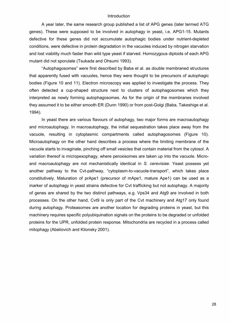
Figure 9: Autophagy and Endosomal trafficking complexes in yeast and mammals
Although the complexes for autophagy and endosomal trafficking differ slightly in yeast (panel a) and mammalian cells (panel b), many homologs have been found already. (Vanhaesebroeck, Guillermet-Guibert et al.)
3.5.2 Autophagy in Mammalian Cells and in other Organisms
In 1999, Liang et al. described Beclin1 (Figure 9), the human orthologue of yeast
Atg6/Vps30, to be a tumor suppressor. Beclin1 is a Bcl-2-interacting, coiled-coil protein and is
deleted in 40-75% of sporadic human breast cancers and ovarian cancers (Aita, Liang et al.
1999). It has structural similarity to the yeast protein Atg6p, so that Beclin1 expression can
promote autophagy in atg6 deletion mutants. The human cancer cell line MCF-7 does not
express detectable endogenous Beclin1 levels. Upon stable conditional transfection with
Beclin1, autophagic vacuoles marking nutrient deprivation-induced autophagy could be
observed in these cells. No evidence of apoptosis was found, the nuclei appeared normal.
Induced autophagy seemed to reduce proliferation rates and led to flatter appearance, larger
size and increased contact inhibition of the cells. Clonigenicity in vitro was highly impaired and
injection into nude mice gave rise to significantly less tumor formations. Out of eleven tested
human breast carcinoma cell lines, only three expressed endogenous Beclin1 while all 32
samples of normal breast epithelial tissue stained for strong Beclin1 immunoreactivity. The
authors suggest that Beclin1 is a key enzyme in inhibition of tumorigenesis through autophagy
(Liang, Jackson et al. 1999).
Alfy, was discovered by Simonsen et al. in 2004. It represents a 400kD protein that
contains a FYVE-domain and WD-40 repeats. Results in HeLa cells suggest that Alfy is indeed
binding PI(3)P and colocalizes with protein granules and autophagic membranes. Alfy-positive
structures accumulate in the cytoplasm after serum and amino acid starvation. These structure
at least partially costain with autophagy markers such as LC-3 and hAtg5. The authors imply a
scaffold role for Alfy in the autophagic machinery as it recognizes protein aggregates and
localizes close to ubiquitin-loaded structures (Simonsen, Birkeland et al. 2004).
Introduction
30
Zeng et al. described a new role for Beclin1 in 2006. In U-251 glioblastoma cells
immunoprecipitates contained not Bcl-2 but hVps34. Knockdown of Beclin1 blocked autophagy
induced by nutrient deprivation or C2-ceramide. Endocytosis on the other hand was not
impaired, as EGFR sorting or internalization of fluid phase markers such as HRP were not
affected. Their results suggested that Beclin1 was not a simple chaperone or adaptor of the
hVps34 complex in normal vesicular trafficking but mainly a positive regulator of the autophagic
pathway (Zeng, Overmeyer et al. 2006). These findings were in agreement with results in
Caenorhabditis elegans published earlier by another group (Melendez, Talloczy et al. 2003).
The group of Vojo Deretic showed in 2006 that autophagy can be used as a mechanism
to reduce bacterial load i.e. as defense against intracellular pathogens (Figure 13). In their
study, they had a closer look at murine Irgm1 of the family of immunity related GTPases (IRG).
IFN-γ (interferon γ) induces autophagy in macrophages, but expression of Irgm1 by itself
already leads to onset of autophagy in macrophages. Cells exhibited a MDC
(monodansylcadaverine)-positive profile, a preliminary marker for autophagic organelles.
Classical inhibitors of autophagy e.g. wortmannin and 3-MA (3-methyladenine) inhibited this
reaction. The autophagosome-like structures detected in their experiments showed double-
membrane character, known as phagophores i.e. nascent autophagosomes. Irgm1 seemed
important in early stages of autophagosome formation, in detail in LC-3 (human Atg8)
maturation. Further steps of such autophagic processes seemed to depend on Beclin1 and the
usual autophagy complex subunits. Similar results were obtained in human cell lines, U937,
HEK293T and HeLa. Human macrophages behaved the same way, IRGM (human orthologue
of murine Irgm1) inducing autophagy as seen earlier in murine cells. Unlike the IRG family in
mice, the human IRGM is not activated by IFN-γ. Nonetheless did IRGM participate in IFN-γ or
starvation induced autophagy in human macrophages, indicating a more general role for IRGM
in autophagy. More details on the role of IRGM in defense against intracellular pathogens
require further investigations (Singh, Davis et al. 2006).
In 2006, Liang et al. identified a novel coiled-coil UV irradiation resistance-associated
gene (UVRAG) that positively regulated the Beclin1-hVps34 complex. UVRAG is
monoallelically mutated at a high frequency in human colon cancers, hence it is a candidate to
be a tumor suppressor. UVRAG and Beclin1 interdependently induce autophagy. The new
player promotes autophagy and suppresses proliferation and tumorigenicity of human colon
cancer cells tested in their study (Liang, Feng et al. 2006). In a later publication, the same
author claimed that UVRAG binds Beclin1 and interacts with the endosomal fusion machinery.
Rab7 gets stimulated and autophagosomes then fuse with late endosomes, enhancing delivery
of autophagic cargo to the lysosome. UVRAG-endosomal sorting is genetically separate from
UVRAG-Beclin1-mediated autophagosome formation. UVRAG therefore functions as a
multivalent trafficking effector (Liang, Lee et al. 2008).
Introduction
31
Another positive regulator of autophagy directed by Beclin1 is Ambra1, discovered by
Fimia et al. in 2007. Ambra1 (Activating molecole in Beclin1-regulated autophagy) bears a
WD40-domain at its N-terminus and has a crucial role in embryogenesis. Ambra1
downregulation led to reduced capability of Beclin1 to interact with hVps34 and also kinase
activity of Vps34 was impaired in these cells. Upon functional deficiency, severe neural tube
defects were observed in mouse embryos as autophagy was impaired, ubiquitinated proteins
aggregated and cell proliferation became unbalanced while apoptosis was strongly increased
(Fimia, Stoykova et al. 2007).
To date it is not clear where the membranes of autophagosomes originate from. In 2008,
Takahashi et al. published a study in which they describe Bif-1 (Endophilin B1), a member of
the endophilin family. Bif-1 possesses membrane binding and liposome tubulation activities.
Under nutrient deprivation conditions, Bif-1 accumulates in punctate structures that colocalize
with LC-3, Atg5 and Atg9, markers of autophagosomes. Bif-1 positive vesicles were shown to
fuse with Atg9-positive small membranes to form autophagosomes in both HeLa and COS7
cells. As the N-BAR domain of Bif-1 interacts with Beclin1 through UVRAG and promotes
activation of hVps34, they suggest Bif-1 to be a potential regulator of autophagosome formation
(Takahashi, Coppola et al. 2007; Takahashi, Meyerkord et al. 2008).
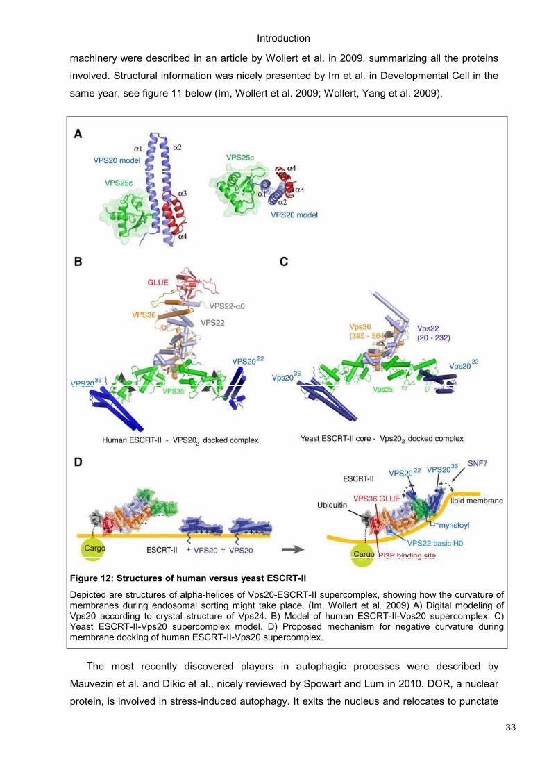
Axe et al. described autophagosome formation from membrane compartments
connected to the endoplasmatic reticulum (ER) (Figure 10 and 11). They applied a novel
protein termed DFCP1 (double-FYVE domain-containing protein 1) and observed PI(3)P lipids
translocating from unusual localizations on ER and Golgi membranes to autophagosomes upon
amino acid starvation. Translocation was dependent on hVps34 and Beclin1 in the HEK293
cells tested (Axe, Walker et al. 2008).
Introduction
32
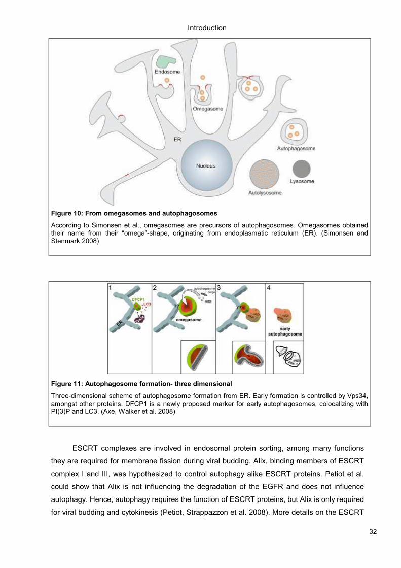
Figure 10: From omegasomes and autophagosomes
According to Simonsen et al., omegasomes are precursors of autophagosomes. Omegasomes obtained their name from their “omega”-shape, originating from endoplasmatic reticulum (ER). (Simonsen and Stenmark 2008)
Figure 11: Autophagosome formation- three dimensional
Three-dimensional scheme of autophagosome formation from ER. Early formation is controlled by Vps34, amongst other proteins. DFCP1 is a newly proposed marker for early autophagosomes, colocalizing with PI(3)P and LC3. (Axe, Walker et al. 2008)
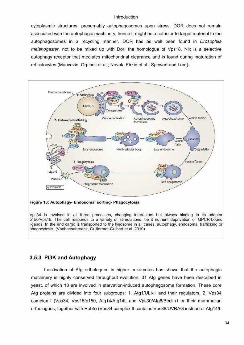
ESCRT complexes are involved in endosomal protein sorting, among many functions
they are required for membrane fission during viral budding. Alix, binding members of ESCRT
complex I and III, was hypothesized to control autophagy alike ESCRT proteins. Petiot et al.
could show that Alix is not influencing the degradation of the EGFR and does not influence
autophagy. Hence, autophagy requires the function of ESCRT proteins, but Alix is only required
for viral budding and cytokinesis (Petiot, Strappazzon et al. 2008). More details on the ESCRT
Introduction
33
machinery were described in an article by Wollert et al. in 2009, summarizing all the proteins
involved. Structural information was nicely presented by Im et al. in Developmental Cell in the
same year, see figure 11 below (Im, Wollert et al. 2009; Wollert, Yang et al. 2009).
Figure 12: Structures of human versus yeast ESCRT-II
Depicted are structures of alpha-helices of Vps20-ESCRT-II supercomplex, showing how the curvature of membranes during endosomal sorting might take place. (Im, Wollert et al. 2009) A) Digital modeling of Vps20 according to crystal structure of Vps24. B) Model of human ESCRT-II-Vps20 supercomplex. C) Yeast ESCRT-II-Vps20 supercomplex model. D) Proposed mechanism for negative curvature during membrane docking of human ESCRT-II-Vps20 supercomplex.
The most recently discovered players in autophagic processes were described by
Mauvezin et al. and Dikic et al., nicely reviewed by Spowart and Lum in 2010. DOR, a nuclear
protein, is involved in stress-induced autophagy. It exits the nucleus and relocates to punctate
Introduction
34
cytoplasmic structures, presumably autophagosomes upon stress. DOR does not remain
associated with the autophagic machinery, hence it might be a cofactor to target material to the
autophagosomes in a recycling manner. DOR has as well been found in Drosophila
melanogaster, not to be mixed up with Dor, the homologue of Vps18. Nix is a selective
autophagy receptor that mediates mitochondrial clearance and is found during maturation of
reticulocytes (Mauvezin, Orpinell et al.; Novak, Kirkin et al.; Spowart and Lum).
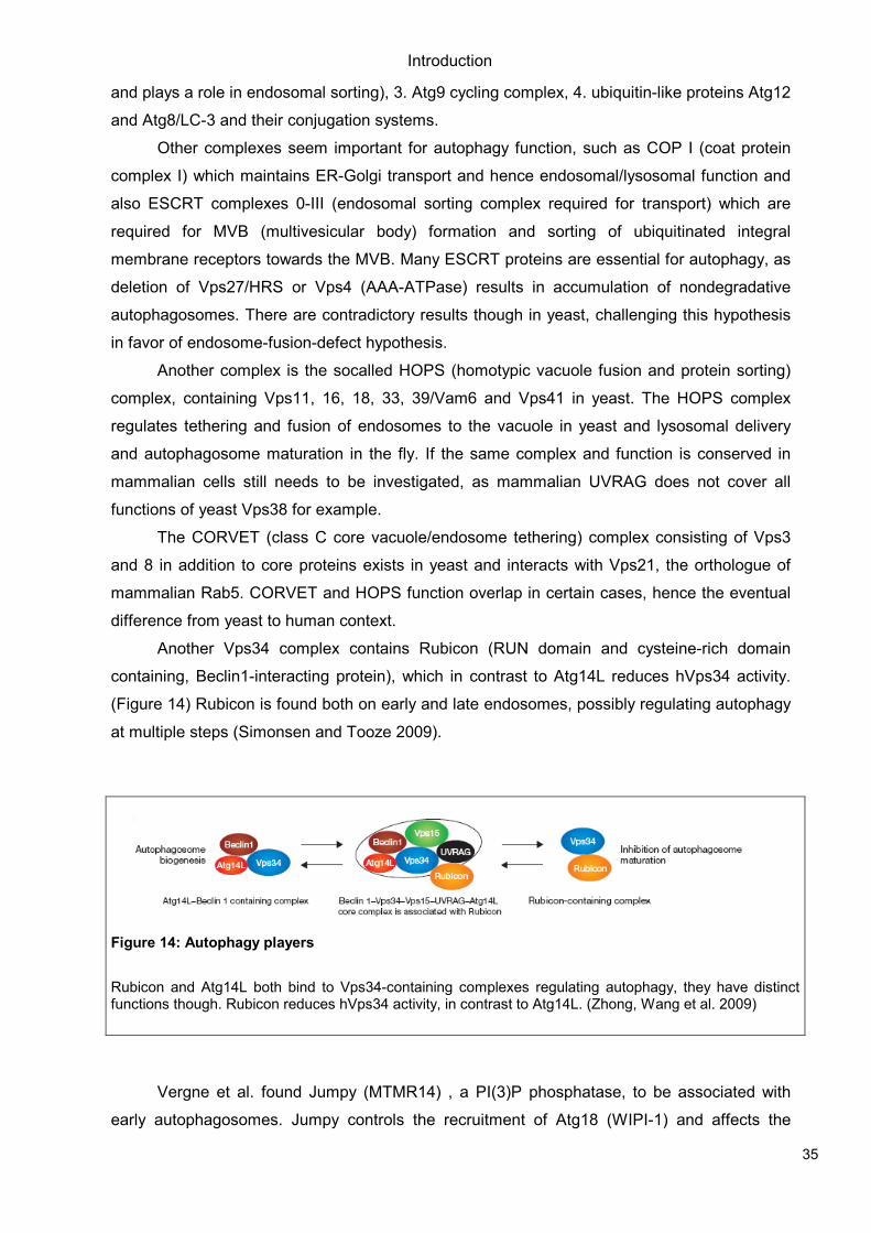
Figure 13: Autophagy- Endosomal sorting- Phagocytosis
Vps34 is involved in all three processes, changing interactors but always binding to its adaptor p150/Vps15. The cell responds to a variety of stimulations, be it nutrient deprivation or GPCR-bound ligands. In the end cargo is transported to the lysosome in all cases, autophagy, endosomal trafficking or phagocytosis. (Vanhaesebroeck, Guillermet-Guibert et al. 2010)
3.5.3 PI3K and Autophagy
Inactivation of Atg orthologues in higher eukaryotes has shown that the autophagic
machinery is highly conserved throughout evolution. 31 Atg genes have been described in
yeast, of which 18 are involved in starvation-induced autophagosome formation. These core
Atg proteins are divided into four subgroups: 1. Atg1/ULK1 and their regulators, 2. Vps34
complex I (Vps34, Vps15/p150, Atg14/Atg14L and Vps30/Atg6/Beclin1 or their mammalian
orthologues, together with Rab5) (Vps34 complex II contains Vps38/UVRAG instead of Atg14/L
Introduction
35
and plays a role in endosomal sorting), 3. Atg9 cycling complex, 4. ubiquitin-like proteins Atg12
and Atg8/LC-3 and their conjugation systems.
Other complexes seem important for autophagy function, such as COP I (coat protein
complex I) which maintains ER-Golgi transport and hence endosomal/lysosomal function and
also ESCRT complexes 0-III (endosomal sorting complex required for transport) which are
required for MVB (multivesicular body) formation and sorting of ubiquitinated integral
membrane receptors towards the MVB. Many ESCRT proteins are essential for autophagy, as
deletion of Vps27/HRS or Vps4 (AAA-ATPase) results in accumulation of nondegradative
autophagosomes. There are contradictory results though in yeast, challenging this hypothesis
in favor of endosome-fusion-defect hypothesis.
Another complex is the socalled HOPS (homotypic vacuole fusion and protein sorting)
complex, containing Vps11, 16, 18, 33, 39/Vam6 and Vps41 in yeast. The HOPS complex
regulates tethering and fusion of endosomes to the vacuole in yeast and lysosomal delivery
and autophagosome maturation in the fly. If the same complex and function is conserved in
mammalian cells still needs to be investigated, as mammalian UVRAG does not cover all
functions of yeast Vps38 for example.
The CORVET (class C core vacuole/endosome tethering) complex consisting of Vps3
and 8 in addition to core proteins exists in yeast and interacts with Vps21, the orthologue of
mammalian Rab5. CORVET and HOPS function overlap in certain cases, hence the eventual
difference from yeast to human context.
Another Vps34 complex contains Rubicon (RUN domain and cysteine-rich domain
containing, Beclin1-interacting protein), which in contrast to Atg14L reduces hVps34 activity.
(Figure 14) Rubicon is found both on early and late endosomes, possibly regulating autophagy
at multiple steps (Simonsen and Tooze 2009).
Figure 14: Autophagy players
Rubicon and Atg14L both bind to Vps34-containing complexes regulating autophagy, they have distinct functions though. Rubicon reduces hVps34 activity, in contrast to Atg14L. (Zhong, Wang et al. 2009)
Vergne et al. found Jumpy (MTMR14) , a PI(3)P phosphatase, to be associated with
early autophagosomes. Jumpy controls the recruitment of Atg18 (WIPI-1) and affects the
Introduction
36
distribution of Atg9 and LC-3. For the first time, the initiation of autophagy is controlled not only
by PI(3)P producing kinase but also by a specific PI(3)P phosphatase which apparently is even
linked to disease, e.g. congenital centronuclear myopathy (Vergne, Roberts et al. 2009).
3.5.4 Cancer Therapy- Pro or Contra Autophagy?
Beth Levine published a collection of questions and answers on autophagy and its role in
cancer in Nature Cell Biology in 2007. An important evolutionary conserved function of
autophagy is to help maintain the synthesis of essential proteins when nutrients are limited.
Through protein recycling, activated autophagy can ensure cell’s survival even if general
protein translation is shut down. In contrast to apoptosis that invariably leads to cell death,
autophagy usually contributes to cell survival. It can induce a cell death program if the apoptotic
machinery is impaired or when autophagy is strongly increased that the cells literally “eat
themselves to death”. Cancer cells often confer resistance to apoptosis and many
chemotherapeutic reagents induce autophagy. DNA damage is prevented by autophagy,
probably by removing sources of oxidative stress, e.g. defective mitochondria or endoplasmatic
reticulum. For these reasons, autophagic mechanisms can be considered beneficial for cancer
therapy. On the contrary, some autophagy-specific genes might promote survival of cancer
cells by allowing growth during poor nutrient conditions. When a tumor reaches 0.2-2mm in
diameter , nutrients, growth factors and oxygen cannot diffuse efficiently to the cells at the
center of the tumor mass due to inadequate vascularization. Hence autophagy might actually
facilitate a tumor’s survival in some cases (Folkman 2006). So far, there are contradictory
experimental results in the literature as for whether “autophagy is cancer’s friend or foe”. Most
probably it depends on the individual circumstances (such as vascularization in tumor
environment, stage of tumor development etc.) and genetic background of the cancer cells if
autophagy should be turned on or off by chemical compounds and which proteins are best to
target (Levine 2007).
In a review in 2008, Jin and White state that due to its protective role in maintaining
energy homeostasis and protein/organelle quality control, autophagy might be a tumor
suppressing mechanism managing metabolic stress. Cellular damage manifests in activation of
DNA damage response and may promote tumor initiation or drive cell-autonomous tumor
progression. Autophagic processes usually localize to regions in solid tumors that are
metabolically stressed. Defects in autophagy can then cause increased cell death and
inflammation. These cytokine reactions lead to even more accelerated tumor progression.
Hence, autophagy might be a tumor suppressor and future investigations should identify
human tumors with deficient autophagy and develop more rational cancer therapies making
use of the autophagy mechanisms (Jin and White 2008).
Introduction
37
Dalby et al. published a recent review focusing on autophagy as a novel therapeutic
strategy in cancer treatment. Autophagy has been described as programmed cell death type II
(PCD-type II), a non-apoptotic form which is caspase-independent. Proapoptotic Bcl-2 family
members such as Bak and Bax cause mitochondrial out membrane permeabilization and
cytochrome c release. In knockout fibroblasts, resistance to apoptosis and induction of
autophagic cell death was observed after starvation (Moretti, Attia et al. 2007). Several
compounds have also been found to stimulate PCD-type II, e.g. rottlerin (Akar, Ozpolat et al.
2007). Autophagy might therefore function as an alternative cell death program in cells which
are apoptosis defective or in which it is hard to induce. Examples of cancer types where
autophagy might be beneficial for treatment are pancreatic cancer and breast cancer. PKCδ
positively regulates the expression of TG 2 (tissue transglutaminase 2), which suppresses
autophagic cell death. Knockdown or inhibition of either PKCδ or TG2 induces autophagic cell
death with induction of apoptosis. TG2 is overexpressed in over 80% of pancreatic cancer cell
lines and increased levels have been implicated in drug resistance. Targetting PKCδ or TG2 to
induce autophagic cell death seems a promising therapeutic strategy for the treatment of
pancreatic cancer. This hypothesis was already tested in nude mice bearing xenografts of TG2-
expressing human pancreatic cancer cells. Knockdown of TG2 in these cells inhibited cancer
cell proliferation while no signs of apoptosis were observed.
Another example was found in breast cancer cell line MCF-7 which do not express
caspase 3. Hence they provide a higher threshold for the induction of apoptosis. 45-75% of all
breast cancer tissue samples tested in that study had no detectable levels of caspase-3. When
Bcl-2 was silenced in MCF-7 cells, autophagic but not apoptotic cell death was happening
(Devarajan, Chen et al. 2002; Devarajan, Sahin et al. 2002) (Dalby, Tekedereli et al.).
3.6 Drug Discovery and Inhibitors of PI3K
During the last few years, various small molecule inhibitors of PI3K have been
described. Most are not isoform-specific, as they target the catalytic site which exhibits a high
homology between the different isoforms of PI3K, at least for the ATP-binding pocket. The x-
ray coordinates of p110γ revealed that many inhibitors such as wortmannin, LY294002 etc.
bind to these conserved residues in a similar manner (Walker, Pacold et al. 2000). Studies on
the structure of each isoform would be required in order to find better optimized compounds
that selectively inhibit a specific isoform.
A site mutagenesis study was done by Frazzetto et al. in 2008, replacing residues found
in class I p110α by residues from p110β and vice versa. They claimed that certain inhibitors
show a preference for p110α because of blocked interaction with non-conserved regions of
p110β. The opposite was not observed though, challenging their hypothesis on the influence of
this non-conserved regions adjacent to the catalytic site on the potency of an inhibitor. Other
Introduction
38
groups support this view, suggesting that the non-conserved region of p110α (residues 852-59)
remains a potential target for future compounds exhibiting greater selectivity for a PI3K isoform
(Frazzetto, Suphioglu et al. 2008).
Earlier on, Knight et al. published a pharmacological map of the PI3K family defining
p110α’s role in insulin signalling. In order to investigate the unique roles of PI3K isoforms, the
authors synthesized a diverse panel of PI3K inhibitors and checked for their target selectivity.
Using crystal structures of three inhibitors (quinazolinone purine PIK-39, imidazoquinazoline
PIK-90 and phenylthiazole PIK-93) bound to p110γ, they identified a conformationally mobile
region that is uniquely exploited by selective inhibitors. Highly selective inhibitors projected out
of the plane occupied by ATP, the most potent ones targeted deeper into a binding pocket that
is not accessed by ATP (Ile 879, a residue structurally analogous to the gatekeeper residue in
protein kinasese). When they applied these compounds, they found p110α to be the primary
insulin-responsive PI3K isoform. P110β inhibitors had no effect on the other hand (Knight,
Gonzalez et al. 2006).
Apsel et al. published an article on the discovery of dual inhibitors of both tyrosine and
phopshoinositide kinases in 2008. In certain cases it might be beneficial to target different
kinases simultaneously. PP121 blocked proliferation of tumor cells by inhibition of oncogenic
tyrosine kinases and PI3K through a hydrophobic pocket conserved in both enzyme classes.
Related compounds had strong effects on mTOR and other kinases, such as PP242 blocking
mTOR quite efficiently (Apsel, Blair et al. 2008).
Another dual inhibitor was discovered by Maira et al., NVP-BEZ235. This imidazo(4,5-
c)quinoline derivative was shown to inhibit both PI3K and mTOR. Hence it exhibited in vivo
antitumor activity in various tumor cell lines and prostate tumor (PC3M, highly metastatic
human prostate tumor cell line) xenografts in mice respectively. BEZ235 possesses strong
antiproliferative capacity by G1-arrest, but showed no effects on cell viability. Application of the
compound was well tolerated in mice as body weight gain was still normal during/ 10 days after
treatment. When tested in increasing ATP concentrations, the compound’s IC50 values
increased in a linear manner. Results suggest that the compound binds to the ATP-binding
pocket that is present in both PI3K and mTOR, competing with ATP. The specificity of the
compound was tested in cell-based and by in vitro assays. The authors conclude that BEZ235
is a dual pan-PI3K/mTOR inhibitor, applicable for both cellular and in vivo settings. Phase I
clinical trials in cancer patients have started in 2008 (Maira, Stauffer et al. 2008).
Dual targeting of both PI3K and mTOR was first applied as therapy in various melanoma
cell lines and in xenografts in mice by Marone et al. in 2009 (see Publications), using the same
compound NVP-BEZ235 presented by Maira et al. (Maira, Stauffer et al. 2008) and similar
compounds. Exposure of the cells to these inhibitors resulted in complete G1-arrest shown by
flow cytometry, other markers of cell cycle arrest proven by western blotting, but only negligible
apoptosis levels. When the same cells were treated with pan-class I PI3K inhibitor ZSTK474
Introduction
39
(Yaguchi, Fukui et al. 2006) or mTORC1 inhibitor rapamycin, only minor effects on cell
proliferation were observed. A B16 mouse melanoma tumor model was used to test efficacy of
tumor growth attenuation in both primary and lymph node metastatic sites. No obvious toxicity
could be found, moreover, the mice displayed reduced numbers and significantly smaller tumor
cells. The authors claim that neovascularization was blocked and necrosis of the tumor
increased, suggesting that compounds targeting both PI3K and mTOR simultaneously could be
beneficial in melanoma therapy (Marone, Erhart et al. 2009).
A recent paper on the structure variation of class III PI3K was published by Miller et al.
The crystal structure of Vps34 (Figure 15) revealed an ATP-binding pocket distinct from class I
PI3K isoforms, explaining potency differences for many inhibitors. Vps34 seems to alternate
between a closed form of the phosphoinositide-binding loop and C-terminal helix in the cytosol
and an open form on the endo-membranes. 3-methyladenine (3-MA) is preferentially binding
class III PI3K (via the hinge) over the the other classes, even if the IC50 is very high at 10mM
in order to block autophagy. The ATP-binding pocket of Vps34 is more constricted than the
corresponding pocket of class I isoforms, e.g. p110γ. The P-loop curls inward and the hinge
between N-and C-lobes is shorter, too. Propeller-like inhibitors such as PIK-39 bind the other
isoforms much better due to the increased rigidity of class III PI3K. The authors came up with
PT210, a derivative of PIK93, with reversed kinase specificity. PT210 showed much higher
IC50 for p110γ (4uM) than for Vps34 (450nM). This way of designing small molecule inhibitors
according to their putative binding possibilities will be more and more applied in future drug
design, not only for PI3K but also for other enzymes (Miller, Tavshanjian et al.).
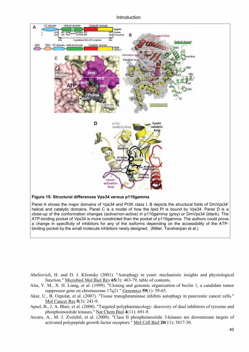
Introduction
40
D
Figure 15: Structural differences Vps34 versus p110gamma
Panel A shows the major domains of Vps34 and PI3K class I, B depicts the structural folds of DmVps34’ helical and catalytic domains. Panel C is a model of how the lipid PI is bound by Vps34. Panel D is a close-up of the conformation changes (active/non-active) in p110gamma (grey) or DmVps34 (black). The ATP-binding pocket of Vps34 is more constricted than the pocket of p110gamma. The authors could prove a change in specificity of inhibitors for any of the isoforms depending on the accessibility of the ATP-binding pocket by the small molecule inhibitors newly designed. (Miller, Tavshanjian et al.)
Abeliovich, H. and D. J. Klionsky (2001). "Autophagy in yeast: mechanistic insights and physiological
function." Microbiol Mol Biol Rev 65(3): 463-79, table of contents.
Aita, V. M., X. H. Liang, et al. (1999). "Cloning and genomic organization of beclin 1, a candidate tumor
suppressor gene on chromosome 17q21." Genomics 59(1): 59-65.
Akar, U., B. Ozpolat, et al. (2007). "Tissue transglutaminase inhibits autophagy in pancreatic cancer cells."
Mol Cancer Res 5(3): 241-9.
Apsel, B., J. A. Blair, et al. (2008). "Targeted polypharmacology: discovery of dual inhibitors of tyrosine and
phosphoinositide kinases." Nat Chem Biol 4(11): 691-9.
Arcaro, A., M. J. Zvelebil, et al. (2000). "Class II phosphoinositide 3-kinases are downstream targets of
activated polypeptide growth factor receptors." Mol Cell Biol 20(11): 3817-30.
Introduction
41
Ashrafi, K., F. Y. Chang, et al. (2003). "Genome-wide RNAi analysis of Caenorhabditis elegans fat regulatory
genes." Nature 421(6920): 268-72.
Auger, K. R., C. L. Carpenter, et al. (1989). "Phosphatidylinositol 3-kinase and its novel product,
phosphatidylinositol 3-phosphate, are present in Saccharomyces cerevisiae." J Biol Chem 264(34):
20181-4.
Avruch, J., X. Long, et al. (2008). "Amino Acid Regulation of TOR Complex 1." Am J Physiol Endocrinol
Metab.
Avruch, J., X. Long, et al. (2009). "Amino acid regulation of TOR complex 1." Am J Physiol Endocrinol
Metab 296(4): E592-602.
Axe, E. L., S. A. Walker, et al. (2008). "Autophagosome formation from membrane compartments enriched in
phosphatidylinositol 3-phosphate and dynamically connected to the endoplasmic reticulum." J Cell
Biol 182(4): 685-701.
Baba, M., K. Takeshige, et al. (1994). "Ultrastructural analysis of the autophagic process in yeast: detection of
autophagosomes and their characterization." J Cell Biol 124(6): 903-13.
Backer, J. M. (2008). "The regulation and function of Class III PI3Ks: novel roles for Vps34." Biochem J
410(1): 1-17.
Ballou, L. M., M. Chattopadhyay, et al. (2006). "Galphaq binds to p110alpha/p85alpha phosphoinositide 3-
kinase and displaces Ras." Biochem J 394(Pt 3): 557-62.
Birkeland, H. C. and H. Stenmark (2004). "Protein targeting to endosomes and phagosomes via FYVE and PX
domains." Curr Top Microbiol Immunol 282: 89-115.
Bohnacker, T., R. Marone, et al. (2009). "PI3Kgamma adaptor subunits define coupling to degranulation and
cell motility by distinct PtdIns(3,4,5)P3 pools in mast cells." Sci Signal 2(74): ra27.
Bunney, T. D. and M. Katan "Phosphoinositide signalling in cancer: beyond PI3K and PTEN." Nat Rev
Cancer 10(5): 342-52.
Byfield, M. P., J. T. Murray, et al. (2005). "hVps34 is a nutrient-regulated lipid kinase required for activation
of p70 S6 kinase." J Biol Chem 280(38): 33076-82.
Cao, C., J. M. Backer, et al. (2008). "Sequential Actions of Myotubularin Lipid Phosphatases Regulate
Endosomal PI(3)P and Growth Factor Receptor Trafficking." Mol Biol Cell 19(8): 3334-3346.
Cao, C., J. Laporte, et al. (2007). "Myotubularin lipid phosphatase binds the hVPS15/hVPS34 lipid kinase
complex on endosomes." Traffic 8(8): 1052-67.
Chamberlain, M. D., T. Chan, et al. (2008). "Disrupted RabGAP function of the p85 subunit of
phosphatidylinositol 3-kinase results in cell transformation." J Biol Chem 283(23): 15861-8.
Christoforidis, S., M. Miaczynska, et al. (1999). "Phosphatidylinositol-3-OH kinases are Rab5 effectors." Nat
Cell Biol 1(4): 249-52.
Cornillet-Lefebvre, P., W. Cuccuini, et al. (2006). "Constitutive phosphoinositide 3-kinase activation in acute
myeloid leukemia is not due to p110delta mutations." Leukemia 20(2): 374-6.
Curtin, J. A., M. S. Stark, et al. (2006). "PI3-kinase subunits are infrequent somatic targets in melanoma." J
Invest Dermatol 126(7): 1660-3.
Dalby, K. N., I. Tekedereli, et al. "Targeting the prodeath and prosurvival functions of autophagy as novel
therapeutic strategies in cancer." Autophagy 6(3): 322-9.
Devarajan, E., J. Chen, et al. (2002). "Human breast cancer MCF-7 cell line contains inherently drug-resistant
subclones with distinct genotypic and phenotypic features." Int J Oncol 20(5): 913-20.
Devarajan, E., A. A. Sahin, et al. (2002). "Down-regulation of caspase 3 in breast cancer: a possible
mechanism for chemoresistance." Oncogene 21(57): 8843-51.
Di Paolo, G. and P. De Camilli (2006). "Phosphoinositides in cell regulation and membrane dynamics."
Nature 443(7112): 651-7.
Domin, J., F. Pages, et al. (1997). "Cloning of a human phosphoinositide 3-kinase with a C2 domain that
displays reduced sensitivity to the inhibitor wortmannin." Biochem J 326 ( Pt 1): 139-47.
Dunn, W. A., Jr. (1990). "Studies on the mechanisms of autophagy: formation of the autophagic vacuole." J
Cell Biol 110(6): 1923-33.
Eickholt, B. J., A. I. Ahmed, et al. (2007). "Control of axonal growth and regeneration of sensory neurons by
the p110delta PI 3-kinase." PLoS One 2(9): e869.
Elis, W., E. Triantafellow, et al. (2008). "Down-regulation of class II phosphoinositide 3-kinase alpha
expression below a critical threshold induces apoptotic cell death." Mol Cancer Res 6(4): 614-23.
Ellson, C. D., K. E. Anderson, et al. (2001). "Phosphatidylinositol 3-phosphate is generated in phagosomal
membranes." Curr Biol 11(20): 1631-5.
Introduction
42
Falasca, M., W. E. Hughes, et al. (2007). "The role of phosphoinositide 3-kinase C2alpha in insulin
signaling." J Biol Chem 282(38): 28226-36.
Falasca, M. and T. Maffucci (2007). "Role of class II phosphoinositide 3-kinase in cell signalling." Biochem
Soc Trans 35(Pt 2): 211-4.
Fimia, G. M., A. Stoykova, et al. (2007). "Ambra1 regulates autophagy and development of the nervous
system." Nature 447(7148): 1121-5.
Findlay, G. M., L. Yan, et al. (2007). "A MAP4 kinase related to Ste20 is a nutrient-sensitive regulator of
mTOR signalling." Biochem J 403(1): 13-20.
Folkman, J. (2006). "Angiogenesis." Annu Rev Med 57: 1-18.
Frazzetto, M., C. Suphioglu, et al. (2008). "Dissecting isoform selectivity of PI3K inhibitors: the role of non-
conserved residues in the catalytic pocket." Biochem J 414(3): 383-90.
Futter, C. E., L. M. Collinson, et al. (2001). "Human VPS34 is required for internal vesicle formation within
multivesicular endosomes." J Cell Biol 155(7): 1251-64.
Gulati, P., L. D. Gaspers, et al. (2008). "Amino acids activate mTOR complex 1 via Ca2+/CaM signaling to
hVps34." Cell Metab 7(5): 456-65.
Gulati, P. and G. Thomas (2007). "Nutrient sensing in the mTOR/S6K1 signalling pathway." Biochem Soc
Trans 35(Pt 2): 236-8.
Gupta, S., A. R. Ramjaun, et al. (2007). "Binding of ras to phosphoinositide 3-kinase p110alpha is required
for ras-driven tumorigenesis in mice." Cell 129(5): 957-68.
Harada, K., A. B. Truong, et al. (2005). "The class II phosphoinositide 3-kinase C2beta is not essential for
epidermal differentiation." Mol Cell Biol 25(24): 11122-30.
Herman, P. K. and S. D. Emr (1990). "Characterization of VPS34, a gene required for vacuolar protein sorting
and vacuole segregation in Saccharomyces cerevisiae." Mol Cell Biol 10(12): 6742-54.
Hirsch, E., L. Braccini, et al. (2009). "Twice upon a time: PI3K's secret double life exposed." Trends Biochem
Sci 34(5): 244-8.
Hurley, J. H. (2006). "Membrane binding domains." Biochim Biophys Acta 1761(8): 805-11.
Im, Y. J., T. Wollert, et al. (2009). "Structure and function of the ESCRT-II-III interface in multivesicular
body biogenesis." Dev Cell 17(2): 234-43.
Itakura, E. and N. Mizushima (2009). "Atg14 and UVRAG: mutually exclusive subunits of mammalian
Beclin 1-PI3K complexes." Autophagy 5(4): 534-6.
Jin, S. and E. White (2008). "Tumor suppression by autophagy through the management of metabolic stress."
Autophagy 4(5): 563-6.
Johnson, E. E., J. H. Overmeyer, et al. (2006). "Gene silencing reveals a specific function of hVps34
phosphatidylinositol 3-kinase in late versus early endosomes." J Cell Sci 119(Pt 7): 1219-32.
Juhasz, G., J. H. Hill, et al. (2008). "The class III PI(3)K Vps34 promotes autophagy and endocytosis but not
TOR signaling in Drosophila." J Cell Biol 181(4): 655-66.
Kihara, A., T. Noda, et al. (2001). "Two distinct Vps34 phosphatidylinositol 3-kinase complexes function in
autophagy and carboxypeptidase Y sorting in Saccharomyces cerevisiae." J Cell Biol 152(3): 519-30.
Kim, E., P. Goraksha-Hicks, et al. (2008). "Regulation of TORC1 by Rag GTPases in nutrient response." Nat
Cell Biol 10(8): 935-45.
Kinchen, J. M., K. Doukoumetzidis, et al. (2008). "A pathway for phagosome maturation during engulfment
of apoptotic cells." Nat Cell Biol 10(5): 556-66.
Knight, Z. A., B. Gonzalez, et al. (2006). "A pharmacological map of the PI3-K family defines a role for
p110alpha in insulin signaling." Cell 125(4): 733-47.
Kok, K., B. Geering, et al. (2009). "Regulation of phosphoinositide 3-kinase expression in health and
disease." Trends Biochem Sci 34(3): 115-27.
Kurig, B., A. Shymanets, et al. (2009). "Ras is an indispensable coregulator of the class IB phosphoinositide
3-kinase p87/p110gamma." Proc Natl Acad Sci U S A 106(48): 20312-7.
Kurosu, H. and T. Katada (2001). "Association of phosphatidylinositol 3-kinase composed of p110beta-
catalytic and p85-regulatory subunits with the small GTPase Rab5." J Biochem 130(1): 73-8.
Kurosu, H., T. Maehama, et al. (1997). "Heterodimeric phosphoinositide 3-kinase consisting of p85 and
p110beta is synergistically activated by the betagamma subunits of G proteins and phosphotyrosyl
peptide." J Biol Chem 272(39): 24252-6.
Lehmann, K., J. P. Muller, et al. (2009). "PI3Kgamma controls oxidative bursts in neutrophils via interactions
with PKCalpha and p47phox." Biochem J 419(3): 603-10.
Lemmon, M. A. (2008). "Membrane recognition by phospholipid-binding domains." Nat Rev Mol Cell Biol
9(2): 99-111.
Introduction
43
Leung, W. H., T. Tarasenko, et al. (2009). "Differential roles for the inositol phosphatase SHIP in the
regulation of macrophages and lymphocytes." Immunol Res 43(1-3): 243-51.
Levine, B. (2007). "Cell biology: autophagy and cancer." Nature 446(7137): 745-7.
Levine, B. and G. Kroemer (2008). "Autophagy in the pathogenesis of disease." Cell 132(1): 27-42.
Liang, C., P. Feng, et al. (2006). "Autophagic and tumour suppressor activity of a novel Beclin1-binding
protein UVRAG." Nat Cell Biol 8(7): 688-99.
Liang, C., J. S. Lee, et al. (2008). "Beclin1-binding UVRAG targets the class C Vps complex to coordinate
autophagosome maturation and endocytic trafficking." Nat Cell Biol 10(7): 776-87.
Liang, X. H., S. Jackson, et al. (1999). "Induction of autophagy and inhibition of tumorigenesis by beclin 1."
Nature 402(6762): 672-6.
Liu, B., Y. Cheng, et al. (2009). "Polygonatum cyrtonema lectin induces apoptosis and autophagy in human
melanoma A375 cells through a mitochondria-mediated ROS-p38-p53 pathway." Cancer Lett 275(1):
54-60.
MacDougall, L. K., M. E. Gagou, et al. (2004). "Targeted expression of the class II phosphoinositide 3-kinase
in Drosophila melanogaster reveals lipid kinase-dependent effects on patterning and interactions with
receptor signaling pathways." Mol Cell Biol 24(2): 796-808.
Maira, S. M., F. Stauffer, et al. (2008). "Identification and characterization of NVP-BEZ235, a new orally
available dual phosphatidylinositol 3-kinase/mammalian target of rapamycin inhibitor with potent in
vivo antitumor activity." Mol Cancer Ther 7(7): 1851-63.
Mandelker, D., S. B. Gabelli, et al. (2009). "A frequent kinase domain mutation that changes the interaction
between PI3Kalpha and the membrane." Proc Natl Acad Sci U S A 106(40): 16996-7001.
Marone, R., D. Erhart, et al. (2009). "Targeting melanoma with dual phosphoinositide 3-kinase/mammalian
target of rapamycin inhibitors." Mol Cancer Res 7(4): 601-13.
Mathew, R., V. Karantza-Wadsworth, et al. (2007). "Role of autophagy in cancer." Nat Rev Cancer 7(12):
961-7.
Mauvezin, C., M. Orpinell, et al. "The nuclear cofactor DOR regulates autophagy in mammalian and
Drosophila cells." EMBO Rep 11(1): 37-44.
Melendez, A., Z. Talloczy, et al. (2003). "Autophagy genes are essential for dauer development and life-span
extension in C. elegans." Science 301(5638): 1387-91.
Miled, N., Y. Yan, et al. (2007). "Mechanism of two classes of cancer mutations in the phosphoinositide 3-
kinase catalytic subunit." Science 317(5835): 239-42.
Miller, S., B. Tavshanjian, et al. "Shaping development of autophagy inhibitors with the structure of the lipid
kinase Vps34." Science 327(5973): 1638-42.
Moretti, L., A. Attia, et al. (2007). "Crosstalk between Bak/Bax and mTOR signaling regulates radiation-
induced autophagy." Autophagy 3(2): 142-4.
Murray, J. T., C. Panaretou, et al. (2002). "Role of Rab5 in the recruitment of hVps34/p150 to the early
endosome." Traffic 3(6): 416-27.
Nicklin, P., P. Bergman, et al. (2009). "Bidirectional transport of amino acids regulates mTOR and
autophagy." Cell 136(3): 521-34.
Nobukuni, T., M. Joaquin, et al. (2005). "Amino acids mediate mTOR/raptor signaling through activation of
class 3 phosphatidylinositol 3OH-kinase." Proc Natl Acad Sci U S A 102(40): 14238-43.
Novak, I., V. Kirkin, et al. "Nix is a selective autophagy receptor for mitochondrial clearance." EMBO Rep
11(1): 45-51.
Omholt, K., D. Krockel, et al. (2006). "Mutations of PIK3CA are rare in cutaneous melanoma." Melanoma
Res 16(2): 197-200.
Orme, M. H., S. Alrubaie, et al. (2006). "Input from Ras is required for maximal PI(3)K signalling in
Drosophila." Nat Cell Biol 8(11): 1298-302.
Papakonstanti, E. A., A. J. Ridley, et al. (2007). "The p110delta isoform of PI 3-kinase negatively controls
RhoA and PTEN." Embo J 26(13): 3050-61.
Papakonstanti, E. A., O. Zwaenepoel, et al. (2008). "Distinct roles of class IA PI3K isoforms in primary and
immortalised macrophages." J Cell Sci 121(Pt 24): 4124-33.
Petiot, A., F. Strappazzon, et al. (2008). "Alix differs from ESCRT proteins in the control of autophagy."
Biochem Biophys Res Commun 375(1): 63-8.
Prasad, N. K., M. Tandon, et al. (2008). "Phosphoinositol phosphatase SHIP2 promotes cancer development
and metastasis coupled with alterations in EGF receptor turnover." Carcinogenesis 29(1): 25-34.
Introduction
44
Robinson, J. S., D. J. Klionsky, et al. (1988). "Protein sorting in Saccharomyces cerevisiae: isolation of
mutants defective in the delivery and processing of multiple vacuolar hydrolases." Mol Cell Biol
8(11): 4936-48.
Roggo, L., V. Bernard, et al. (2002). "Membrane transport in Caenorhabditis elegans: an essential role for
VPS34 at the nuclear membrane." Embo J 21(7): 1673-83.
Rohrschneider, L. R., J. F. Fuller, et al. (2000). "Structure, function, and biology of SHIP proteins." Genes
Dev 14(5): 505-20.
Rothman, J. H. and T. H. Stevens (1986). "Protein sorting in yeast: mutants defective in vacuole biogenesis
mislocalize vacuolar proteins into the late secretory pathway." Cell 47(6): 1041-51.
Samuels, Y., Z. Wang, et al. (2004). "High frequency of mutations of the PIK3CA gene in human cancers."
Science 304(5670): 554.
Sancak, Y., T. R. Peterson, et al. (2008). "The Rag GTPases bind raptor and mediate amino acid signaling to
mTORC1." Science 320(5882): 1496-501.
Schu, P. V., K. Takegawa, et al. (1993). "Phosphatidylinositol 3-kinase encoded by yeast VPS34 gene
essential for protein sorting." Science 260(5104): 88-91.
Shayesteh, L., Y. Lu, et al. (1999). "PIK3CA is implicated as an oncogene in ovarian cancer." Nat Genet
21(1): 99-102.
Shin, H. W., M. Hayashi, et al. (2005). "An enzymatic cascade of Rab5 effectors regulates phosphoinositide
turnover in the endocytic pathway." J Cell Biol 170(4): 607-18.
Siddhanta, U., J. McIlroy, et al. (1998). "Distinct roles for the p110alpha and hVPS34 phosphatidylinositol 3'-
kinases in vesicular trafficking, regulation of the actin cytoskeleton, and mitogenesis." J Cell Biol
143(6): 1647-59.
Simonsen, A., H. C. Birkeland, et al. (2004). "Alfy, a novel FYVE-domain-containing protein associated with
protein granules and autophagic membranes." J Cell Sci 117(Pt 18): 4239-51.
Simonsen, A. and H. Stenmark (2008). "Self-eating from an ER-associated cup." J Cell Biol 182(4): 621-2.
Simonsen, A. and S. A. Tooze (2009). "Coordination of membrane events during autophagy by multiple class
III PI3-kinase complexes." J Cell Biol 186(6): 773-82.
Singh, S. B., A. S. Davis, et al. (2006). "Human IRGM induces autophagy to eliminate intracellular
mycobacteria." Science 313(5792): 1438-41.
Slessareva, J. E., S. M. Routt, et al. (2006). "Activation of the phosphatidylinositol 3-kinase Vps34 by a G
protein alpha subunit at the endosome." Cell 126(1): 191-203.
Song, X., W. Xu, et al. (2001). "Phox homology domains specifically bind phosphatidylinositol phosphates."
Biochemistry 40(30): 8940-4.
Spowart, J. and J. J. Lum "Opening a new DOR to autophagy." EMBO Rep 11(1): 4-5.
Stahelin, R. V., D. Karathanassis, et al. (2006). "Structural and membrane binding analysis of the Phox
homology domain of phosphoinositide 3-kinase-C2alpha." J Biol Chem 281(51): 39396-406.
Stark, M. and N. Hayward (2007). "Genome-wide loss of heterozygosity and copy number analysis in
melanoma using high-density single-nucleotide polymorphism arrays." Cancer Res 67(6): 2632-42.
Stein, M. P., Y. Feng, et al. (2003). "Human VPS34 and p150 are Rab7 interacting partners." Traffic 4(11):
754-71.
Taboubi, S., J. Milanini, et al. (2007). "G alpha(q/11)-coupled P2Y2 nucleotide receptor inhibits human
keratinocyte spreading and migration." Faseb J 21(14): 4047-58.
Takahashi, Y., D. Coppola, et al. (2007). "Bif-1 interacts with Beclin 1 through UVRAG and regulates
autophagy and tumorigenesis." Nat Cell Biol 9(10): 1142-51.
Takahashi, Y., D. Coppola, et al. (2007). "Bif-1 interacts with Beclin 1 through UVRAG and regulates
autophagy and tumorigenesis." Nat Cell Biol 9(10): 1142-51.
Takahashi, Y., C. L. Meyerkord, et al. (2008). "BARgaining membranes for autophagosome formation:
Regulation of autophagy and tumorigenesis by Bif-1/Endophilin B1." Autophagy 4(1): 121-4.
Takeshige, K., M. Baba, et al. (1992). "Autophagy in yeast demonstrated with proteinase-deficient mutants
and conditions for its induction." J Cell Biol 119(2): 301-11.
Tang, R., X. Zhao, et al. (2008). "Investigation of variants in the promoter region of PIK3C3 in
schizophrenia." Neurosci Lett 437(1): 42-4.
Tormo, D., A. Checinska, et al. (2009). "Targeted activation of innate immunity for therapeutic induction of
autophagy and apoptosis in melanoma cells." Cancer Cell 16(2): 103-14.
Tsukada, M. and Y. Ohsumi (1993). "Isolation and characterization of autophagy-defective mutants of
Saccharomyces cerevisiae." FEBS Lett 333(1-2): 169-74.
Introduction
45
Vanhaesebroeck, B., K. Ali, et al. (2005). "Signalling by PI3K isoforms: insights from gene-targeted mice."
Trends Biochem Sci 30(4): 194-204.
Vanhaesebroeck, B., J. Guillermet-Guibert, et al. (2010). "The emerging mechanisms of isoform-specific
PI3K signalling." Nat Rev Mol Cell Biol 11(5): 329-41.
Vanhaesebroeck, B., S. J. Leevers, et al. (2001). "Synthesis and function of 3-phosphorylated inositol lipids."
Annu Rev Biochem 70: 535-602.
Vanhaesebroeck, B., S. J. Leevers, et al. (1997). "Phosphoinositide 3-kinases: a conserved family of signal
transducers." Trends Biochem Sci 22(7): 267-72.
Vergne, I., E. Roberts, et al. (2009). "Control of autophagy initiation by phosphoinositide 3-phosphatase
Jumpy." Embo J 28(15): 2244-58.
Virbasius, J. V., A. Guilherme, et al. (1996). "Mouse p170 is a novel phosphatidylinositol 3-kinase containing
a C2 domain." J Biol Chem 271(23): 13304-7.
Vogt, P. K., S. Kang, et al. (2007). "Cancer-specific mutations in phosphatidylinositol 3-kinase." Trends
Biochem Sci 32(7): 342-9.
Volinia, S., R. Dhand, et al. (1995). "A human phosphatidylinositol 3-kinase complex related to the yeast
Vps34p-Vps15p protein sorting system." Embo J 14(14): 3339-48.
Walker, E. H., M. E. Pacold, et al. (2000). "Structural determinants of phosphoinositide 3-kinase inhibition by
wortmannin, LY294002, quercetin, myricetin, and staurosporine." Mol Cell 6(4): 909-19.
Wang, X. and C. G. Proud (2009). "Nutrient control of TORC1, a cell-cycle regulator." Trends Cell Biol
19(6): 260-7.
Wang, Z., W. A. Wilson, et al. (2001). "Antagonistic controls of autophagy and glycogen accumulation by
Snf1p, the yeast homolog of AMP-activated protein kinase, and the cyclin-dependent kinase Pho85p."
Mol Cell Biol 21(17): 5742-52.
Whitman, M., D. Kaplan, et al. (1987). "Evidence for two distinct phosphatidylinositol kinases in fibroblasts.
Implications for cellular regulation." Biochem J 247(1): 165-74.
Wollert, T., D. Yang, et al. (2009). "The ESCRT machinery at a glance." J Cell Sci 122(Pt 13): 2163-6.
Wullschleger, S., R. Loewith, et al. (2006). "TOR signaling in growth and metabolism." Cell 124(3): 471-84.
Wymann, M. P. and R. Marone (2005). "Phosphoinositide 3-kinase in disease: timing, location, and
scaffolding." Curr Opin Cell Biol 17(2): 141-9.
Wymann, M. P. and L. Pirola (1998). "Structure and function of phosphoinositide 3-kinases." Biochim
Biophys Acta 1436(1-2): 127-50.
Xue, Y., H. Fares, et al. (2003). "Genetic analysis of the myotubularin family of phosphatases in
Caenorhabditis elegans." J Biol Chem 278(36): 34380-6.
Yaguchi, S., Y. Fukui, et al. (2006). "Antitumor activity of ZSTK474, a new phosphatidylinositol 3-kinase
inhibitor." J Natl Cancer Inst 98(8): 545-56.
Yan, Y., R. J. Flinn, et al. (2009). "hVps15, but not Ca2+/CaM, is required for the activity and regulation of
hVps34 in mammalian cells." Biochem J 417(3): 747-55.
Zeng, X., J. H. Overmeyer, et al. (2006). "Functional specificity of the mammalian Beclin-Vps34 PI 3-kinase
complex in macroautophagy versus endocytosis and lysosomal enzyme trafficking." J Cell Sci 119(Pt
2): 259-70.
Zhang, H., J. P. Stallock, et al. (2000). "Regulation of cellular growth by the Drosophila target of rapamycin
dTOR." Genes Dev 14(21): 2712-24.
Zhao, L. and P. K. Vogt (2008). "Helical domain and kinase domain mutations in p110alpha of
phosphatidylinositol 3-kinase induce gain of function by different mechanisms." Proc Natl Acad Sci
U S A 105(7): 2652-7.
Zhong, Y., Q. J. Wang, et al. (2009). "Distinct regulation of autophagic activity by Atg14L and Rubicon
associated with Beclin 1-phosphatidylinositol-3-kinase complex." Nat Cell Biol 11(4): 468-76.

Introduction
46
Aim of Studies
47
4 Aim of Studies
Class III PI3K is involved in many cellular processes, among them autophagy (Kihara,
Noda et al. 2001). Autophagy helps the cell to survive nutrient-deprived conditions by “self-
eating” and recycling of organelles and metabolic waste. Especially cancer cells are confronted
with lack of nutrients before angiogenesis is increased in their cellular surroundings. Whether
autophagy is beneficial or not for cell survival in a cancer context and at what stage or for which
cancer cells this might apply, is content of current discussions in the field (Folkman 2006;
Levine 2007).
To answer our main question on the importance of Vps34 and its involvement in cancer
cell survival, we started both a genetic and a pharmacological approach:
“What substance of natural origin will inhibit human Vps34? How specific? At what
concentrations?”
Our first approach was to screen a library of natural compounds for specific human
Vps34 inhibitors in a genetically modified yeast model system. We made use of the temperature
sensitive phenotype of Vps34 deletion mutants. Yeast PI3K deletion mutants were
complemented with a hybrid human/yeast Vps34 construct which allowed us to screen for
substances that abrogated growth at higher temperature. The crude plant extracts that showed
hits in this first round screen were then fractionated and retested on yeast mutants. To avoid
misleading side effects, we then tested the putative inhibitors on yeast Vps34 and rechecked for
human PI3K class III specificity in mammalian cell lines. In vitro kinase assays were applied in
the end to define the effective inhibitor concentration.
“How important is hVps34 for melanoma cell survival? What effects are observed upon
temporary or longterm reduction or loss of the kinase? In which cell lines is it essential or is its
loss tolerated? Could hVps34 be used as a drug target in patients?”
The second approach, this time genetic, was done in human melanoma cell lines. We
made use of knockdown methods by state-of-the-art siRNA or stable shRNA techniques in
melanoma cell lines. Our aim was to characterize different melanoma cell lines upon hVps34
loss, comparing our results to published data on other cell types. We asked whether targeting
hVps34 might affect melanoma survival and hence the kinase might serve as a putative drug
target in these cancer model systems.

Results
48
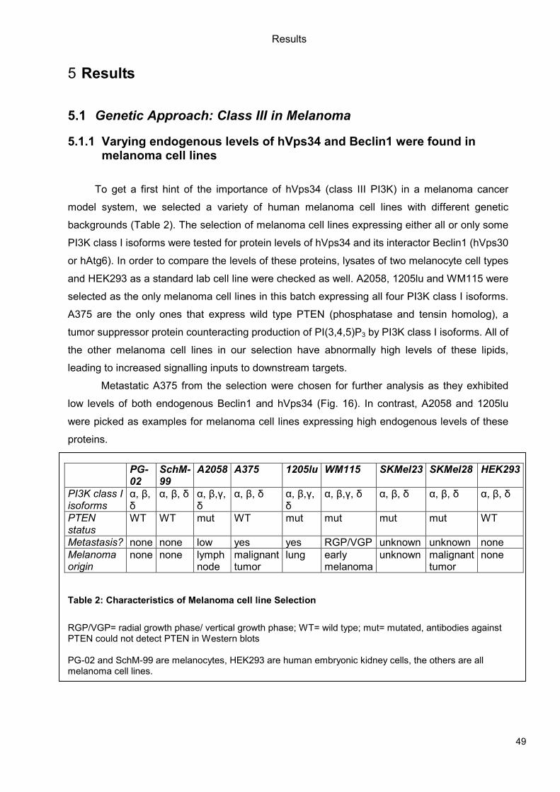
Results
49
5 Results
5.1 Genetic Approach: Class III in Melanoma
5.1.1 Varying endogenous levels of hVps34 and Beclin1 were found in melanoma cell lines
To get a first hint of the importance of hVps34 (class III PI3K) in a melanoma cancer
model system, we selected a variety of human melanoma cell lines with different genetic
backgrounds (Table 2). The selection of melanoma cell lines expressing either all or only some
PI3K class I isoforms were tested for protein levels of hVps34 and its interactor Beclin1 (hVps30
or hAtg6). In order to compare the levels of these proteins, lysates of two melanocyte cell types
and HEK293 as a standard lab cell line were checked as well. A2058, 1205lu and WM115 were
selected as the only melanoma cell lines in this batch expressing all four PI3K class I isoforms.
A375 are the only ones that express wild type PTEN (phosphatase and tensin homolog), a
tumor suppressor protein counteracting production of PI(3,4,5)P3 by PI3K class I isoforms. All of
the other melanoma cell lines in our selection have abnormally high levels of these lipids,
leading to increased signalling inputs to downstream targets.
Metastatic A375 from the selection were chosen for further analysis as they exhibited
low levels of both endogenous Beclin1 and hVps34 (Fig. 16). In contrast, A2058 and 1205lu
were picked as examples for melanoma cell lines expressing high endogenous levels of these
proteins.
PG-02
SchM-99
A2058 A375 1205lu WM115 SKMel23 SKMel28 HEK293
PI3K class I isoforms
α, β, δ
α, β, δ α, β,γ, δ
α, β, δ α, β,γ, δ
α, β,γ, δ α, β, δ α, β, δ α, β, δ
PTEN status
WT WT mut WT mut mut mut mut WT
Metastasis? none none low yes yes RGP/VGP unknown unknown none
Melanoma origin
none none lymph node
malignant tumor
lung early melanoma
unknown malignant tumor
none
Table 2: Characteristics of Melanoma cell line Selection
RGP/VGP= radial growth phase/ vertical growth phase; WT= wild type; mut= mutated, antibodies against PTEN could not detect PTEN in Western blots PG-02 and SchM-99 are melanocytes, HEK293 are human embryonic kidney cells, the others are all melanoma cell lines.
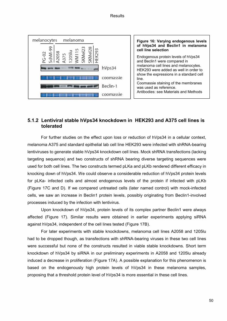
Results
50
5.1.2 Lentiviral stable hVps34 knockdown in HEK293 and A375 cell lines is tolerated
For further studies on the effect upon loss or reduction of hVps34 in a cellular context,
melanoma A375 and standard epithelial lab cell line HEK293 were infected with shRNA-bearing
lentiviruses to generate stable hVps34 knockdown cell lines. Mock shRNA transfections (lacking
targeting sequence) and two constructs of shRNA bearing diverse targeting sequences were
used for both cell lines. The two constructs termed pLKa and pLKb rendered different efficacy in
knocking down of hVps34. We could observe a considerable reduction of hVps34 protein levels
for pLKa- infected cells and almost endogenous levels of the protein if infected with pLKb
(Figure 17C and D). If we compared untreated cells (later named control) with mock-infected
cells, we saw an increase in Beclin1 protein levels, possibly originating from Beclin1-involved
processes induced by the infection with lentivirus.
Upon knockdown of hVps34, protein levels of its complex partner Beclin1 were always
affected (Figure 17). Similar results were obtained in earlier experiments applying siRNA
against hVps34, independent of the cell lines tested (Figure 17B).
For later experiments with stable knockdowns, melanoma cell lines A2058 and 1205lu
had to be dropped though, as transfections with shRNA-bearing viruses in these two cell lines
were successful but none of the constructs resulted in viable stable knockdowns. Short term
knockdown of hVps34 by siRNA in our preliminary experiments in A2058 and 1205lu already
induced a decrease in proliferation (Figure 17A). A possible explanation for this phenomenon is
based on the endogenously high protein levels of hVps34 in these melanoma samples,
proposing that a threshold protein level of hVps34 is more essential in these cell lines.
Figure 16: Varying endogenous levels of hVps34 and Beclin1 in melanoma cell line selection
Endogenous protein levels of hVps34 and Beclin1 were compared in melanoma cell lines and melanocytes. HEK293 were added as well in order to show the expressions in a standard cell line. Coomassie staining of the membranes was used as reference. Antibodies: see Materials and Methods
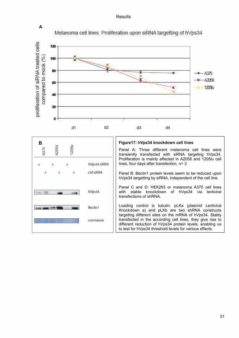
Results
51
A
d1 d2 d3 d4
B
Figure17: hVps34 knockdown cell lines
Panel A: Three different melanoma cell lines were transiently transfected with siRNA targeting hVps34. Proliferation is mainly affected in A2058 and 1205lu cell lines, four days after transfection. n= 3 Panel B: Beclin1 protein levels seem to be reduced upon hVps34 targetting by siRNA, independent of the cell line. Panel C and D: HEK293 or melanoma A375 cell lines with stable knockdown of hVps34 via lentiviral transfections of shRNA. Loading control is tubulin. pLKa (plasmid Lentiviral Knockdown a) and pLKb are two shRNA constructs targeting different sites on the mRNA of hVps34. Stably transfected in the according cell lines, they give rise to different reduction of hVps34 protein levels, enabling us to test for hVps34 threshold levels for various effects.
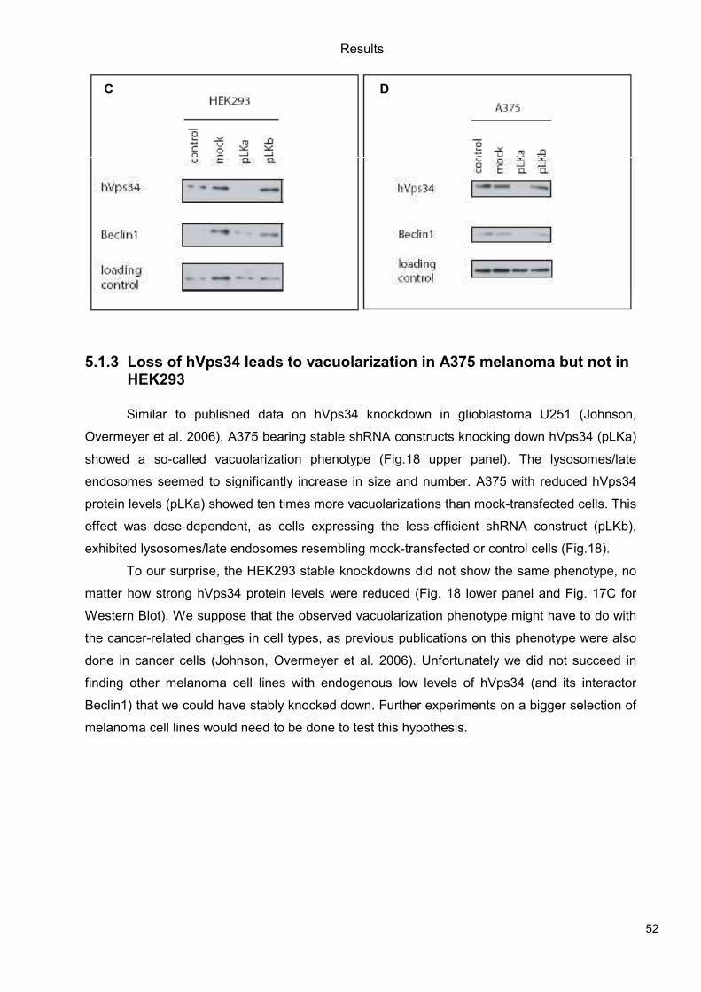
Results
52
5.1.3 Loss of hVps34 leads to vacuolarization in A375 melanoma but not in HEK293
Similar to published data on hVps34 knockdown in glioblastoma U251 (Johnson,
Overmeyer et al. 2006), A375 bearing stable shRNA constructs knocking down hVps34 (pLKa)
showed a so-called vacuolarization phenotype (Fig.18 upper panel). The lysosomes/late
endosomes seemed to significantly increase in size and number. A375 with reduced hVps34
protein levels (pLKa) showed ten times more vacuolarizations than mock-transfected cells. This
effect was dose-dependent, as cells expressing the less-efficient shRNA construct (pLKb),
exhibited lysosomes/late endosomes resembling mock-transfected or control cells (Fig.18).
To our surprise, the HEK293 stable knockdowns did not show the same phenotype, no
matter how strong hVps34 protein levels were reduced (Fig. 18 lower panel and Fig. 17C for
Western Blot). We suppose that the observed vacuolarization phenotype might have to do with
the cancer-related changes in cell types, as previous publications on this phenotype were also
done in cancer cells (Johnson, Overmeyer et al. 2006). Unfortunately we did not succeed in
finding other melanoma cell lines with endogenous low levels of hVps34 (and its interactor
Beclin1) that we could have stably knocked down. Further experiments on a bigger selection of
melanoma cell lines would need to be done to test this hypothesis.
C D
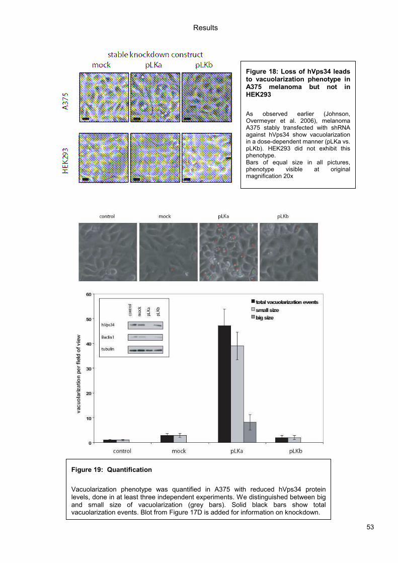
Results
53
Figure 18: Loss of hVps34 leads to vacuolarization phenotype in A375 melanoma but not in HEK293
As observed earlier (Johnson, Overmeyer et al. 2006), melanoma A375 stably transfected with shRNA against hVps34 show vacuolarization in a dose-dependent manner (pLKa vs. pLKb). HEK293 did not exhibit this phenotype. Bars of equal size in all pictures, phenotype visible at originalmagnification 20x
Figure 19: Quantification
Vacuolarization phenotype was quantified in A375 with reduced hVps34 protein levels, done in at least three independent experiments. We distinguished between big and small size of vacuolarization (grey bars). Solid black bars show total vacuolarization events. Blot from Figure 17D is added for information on knockdown.

Results
54
5.1.4 Proliferation upon hVps34 reduction is affected in both A375 and HEK293
a)
b)
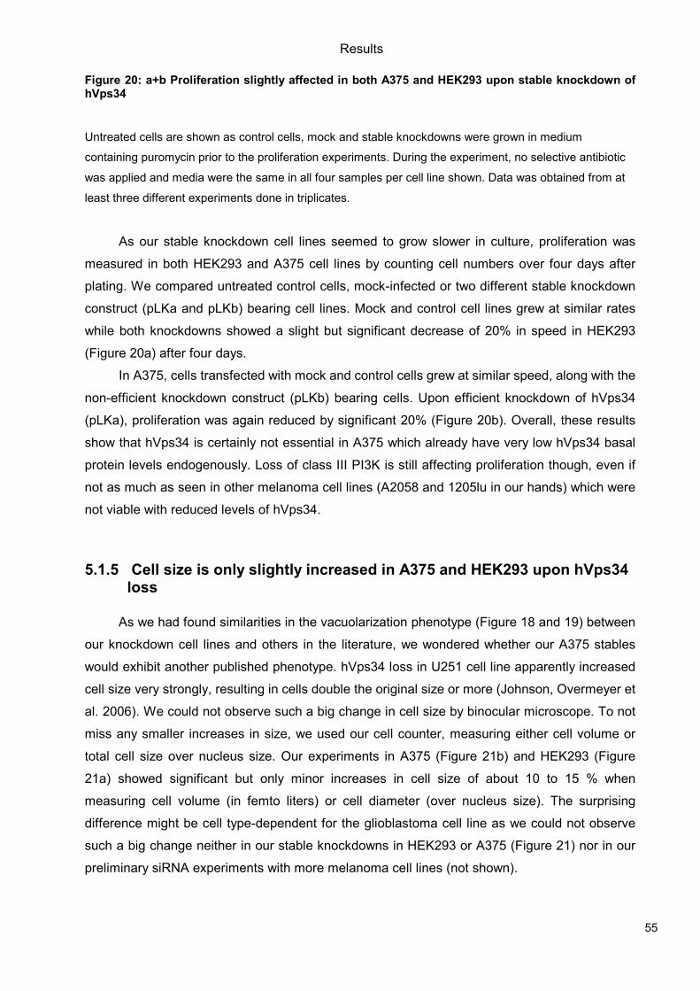
Results
55
Figure 20: a+b Proliferation slightly affected in both A375 and HEK293 upon stable knockdown of hVps34
Untreated cells are shown as control cells, mock and stable knockdowns were grown in medium
containing puromycin prior to the proliferation experiments. During the experiment, no selective antibiotic
was applied and media were the same in all four samples per cell line shown. Data was obtained from at
least three different experiments done in triplicates.
As our stable knockdown cell lines seemed to grow slower in culture, proliferation was
measured in both HEK293 and A375 cell lines by counting cell numbers over four days after
plating. We compared untreated control cells, mock-infected or two different stable knockdown
construct (pLKa and pLKb) bearing cell lines. Mock and control cell lines grew at similar rates
while both knockdowns showed a slight but significant decrease of 20% in speed in HEK293
(Figure 20a) after four days.
In A375, cells transfected with mock and control cells grew at similar speed, along with the
non-efficient knockdown construct (pLKb) bearing cells. Upon efficient knockdown of hVps34
(pLKa), proliferation was again reduced by significant 20% (Figure 20b). Overall, these results
show that hVps34 is certainly not essential in A375 which already have very low hVps34 basal
protein levels endogenously. Loss of class III PI3K is still affecting proliferation though, even if
not as much as seen in other melanoma cell lines (A2058 and 1205lu in our hands) which were
not viable with reduced levels of hVps34.
5.1.5 Cell size is only slightly increased in A375 and HEK293 upon hVps34 loss
As we had found similarities in the vacuolarization phenotype (Figure 18 and 19) between
our knockdown cell lines and others in the literature, we wondered whether our A375 stables
would exhibit another published phenotype. hVps34 loss in U251 cell line apparently increased
cell size very strongly, resulting in cells double the original size or more (Johnson, Overmeyer et
al. 2006). We could not observe such a big change in cell size by binocular microscope. To not
miss any smaller increases in size, we used our cell counter, measuring either cell volume or
total cell size over nucleus size. Our experiments in A375 (Figure 21b) and HEK293 (Figure
21a) showed significant but only minor increases in cell size of about 10 to 15 % when
measuring cell volume (in femto liters) or cell diameter (over nucleus size). The surprising
difference might be cell type-dependent for the glioblastoma cell line as we could not observe
such a big change neither in our stable knockdowns in HEK293 or A375 (Figure 21) nor in our
preliminary siRNA experiments with more melanoma cell lines (not shown).
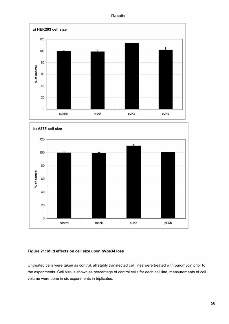
Results
56
a) HEK293 cell size
0
20
40
60
80
100
120
control mock pLKa pLKb
% o
f contr
ol
b) A375 cell size
0
20
40
60
80
100
120
control mock pLKa pLKb
% o
f contr
ol
Figure 21: Mild effects on cell size upon hVps34 loss
Untreated cells were taken as control, all stably transfected cell lines were treated with puromycin prior to
the experiments. Cell size is shown as percentage of control cells for each cell line, measurements of cell
volume were done in six experiments in triplicates.
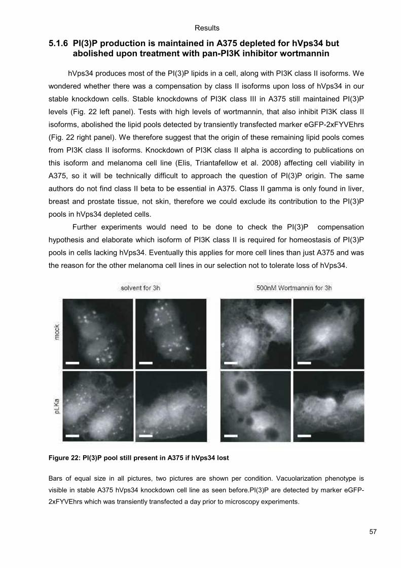
Results
57
5.1.6 PI(3)P production is maintained in A375 depleted for hVps34 but abolished upon treatment with pan-PI3K inhibitor wortmannin
hVps34 produces most of the PI(3)P lipids in a cell, along with PI3K class II isoforms. We
wondered whether there was a compensation by class II isoforms upon loss of hVps34 in our
stable knockdown cells. Stable knockdowns of PI3K class III in A375 still maintained PI(3)P
levels (Fig. 22 left panel). Tests with high levels of wortmannin, that also inhibit PI3K class II
isoforms, abolished the lipid pools detected by transiently transfected marker eGFP-2xFYVEhrs
(Fig. 22 right panel). We therefore suggest that the origin of these remaining lipid pools comes
from PI3K class II isoforms. Knockdown of PI3K class II alpha is according to publications on
this isoform and melanoma cell line (Elis, Triantafellow et al. 2008) affecting cell viability in
A375, so it will be technically difficult to approach the question of PI(3)P origin. The same
authors do not find class II beta to be essential in A375. Class II gamma is only found in liver,
breast and prostate tissue, not skin, therefore we could exclude its contribution to the PI(3)P
pools in hVps34 depleted cells.
Further experiments would need to be done to check the PI(3)P compensation
hypothesis and elaborate which isoform of PI3K class II is required for homeostasis of PI(3)P
pools in cells lacking hVps34. Eventually this applies for more cell lines than just A375 and was
the reason for the other melanoma cell lines in our selection not to tolerate loss of hVps34.
Figure 22: PI(3)P pool still present in A375 if hVps34 lost
Bars of equal size in all pictures, two pictures are shown per condition. Vacuolarization phenotype is
visible in stable A375 hVps34 knockdown cell line as seen before.PI(3)P are detected by marker eGFP-
2xFYVEhrs which was transiently transfected a day prior to microscopy experiments.
Results
58
5.2 Pharmacological Approach: Yeast Screening
USE OF TRANSGENIC YEAST TO DISCOVER
MAMMALIAN PI-3-KINASE ISOFORM INHIBITORS OF NATURAL ORIGIN
(manuscript in preparation)
Ann C. Mertz1, Anna Melone1, Peter Küenzi2, Michael Adams2, Sabine Kiefer2, Matthias
Hamburger2, Matthias P. Wymann1
1Institute of Biochemistry and Genetics, Center for Biomedicine, University of Basel,
Mattenstrasse 28, CH-4058 Basel, Switzerland; 2Institute of Pharmaceutical Biology, University
of Basel, Switzerland
5.2.1 Abstract
Phosphoinositide 3-kinase (PI3K) activity is found in all eukaryotic cells. These lipid
kinases are linked to a diverse set of key cellular functions, including proliferation and motility,
thus contributing to chronic inflammation and metastasis. Different PI3K isoforms seem to be
involved in distinct cellular processes, but it is currently difficult to assess these functions due to
the lack of PI3K isoform-specific inhibitors. We are therefore using Saccharomyces cerevisiae
(Baker’s yeast) to screen PI3K/inhibitor interactions as exploratory genetic tools. Yeast strains
are transformed with plasmids coding for hybrid yeast/mammalian PI3K class III (Vps34). By
nature, Saccharomyces cerevisiae exclusively expresses a single PI3K isoform, Vps34p, a
class III PI3K producing PtdIns 3-P. Here, a hybrid Saccharomyces cerevisiae/Homo sapiens
Vps34p (Sc/Hs_Vps34p) was engineered to complement deletion of vps34 and allow growth at
37°C. When this hybrid protein is inactivated, rescue is lost. Compounds which reduce growth
rates of vps34 yeast expressing Sc/Hs_Vps34p can thus be identified as inhibitors of Vps34p.
As this lipid kinase is involved in autophagy and phago/lysosome fusion processes, it is a
putative drug target in cancer and inflammation.
In our studies we screened various libraries of natural compounds for inhibitory effects
on Vps34. The compounds were mixed extracts of different origins. Upon selection, these
extracts were further fractionated and the fractions retested. In order to confirm the isoform-
specificity, the positive fractions were checked for inhibition of the other PI3K isoforms by
applying them to mammalian cells.
Our data suggests that a fraction of Citrus medica extract had inhibitory effects on the
human but not the yeast Vps34p and did not affect the mammalian PI3K class I isoforms.
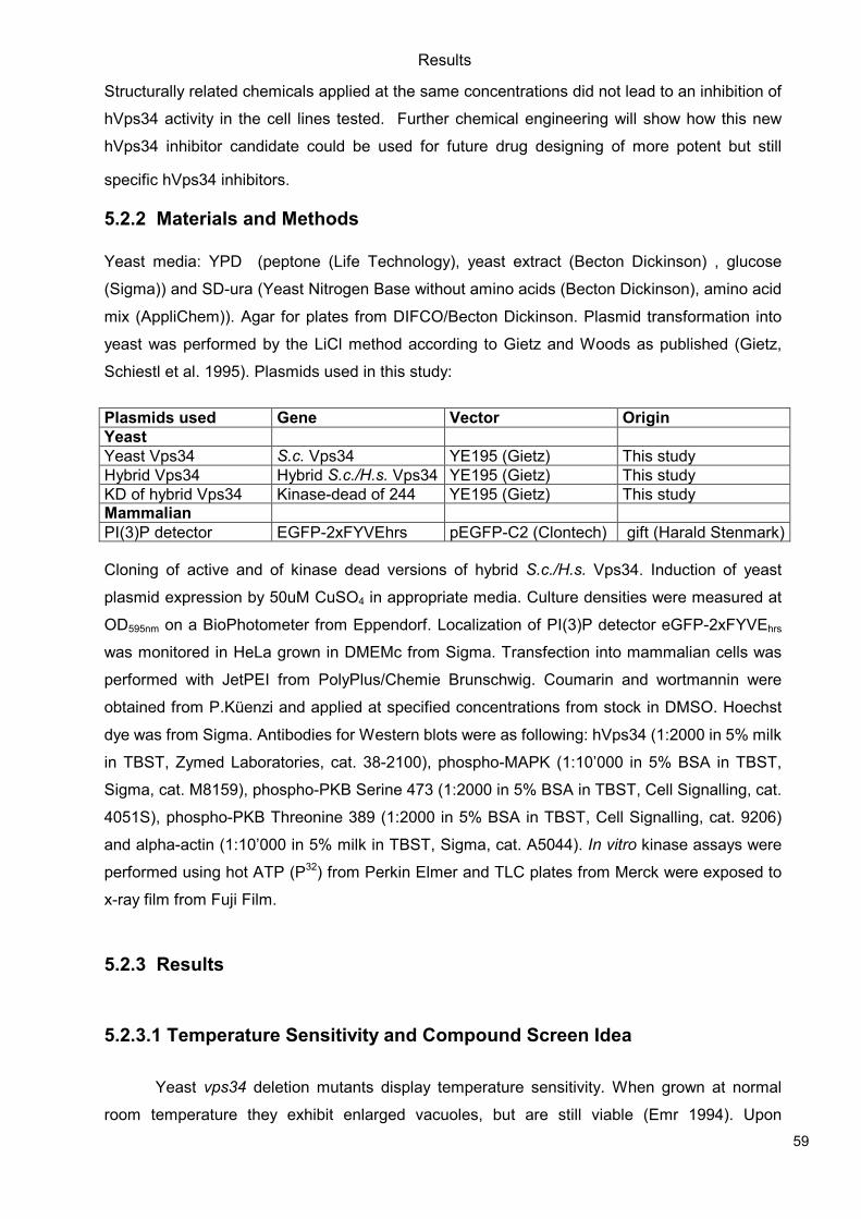
Results
59
Structurally related chemicals applied at the same concentrations did not lead to an inhibition of
hVps34 activity in the cell lines tested. Further chemical engineering will show how this new
hVps34 inhibitor candidate could be used for future drug designing of more potent but still
specific hVps34 inhibitors.
5.2.2 Materials and Methods
Yeast media: YPD (peptone (Life Technology), yeast extract (Becton Dickinson) , glucose
(Sigma)) and SD-ura (Yeast Nitrogen Base without amino acids (Becton Dickinson), amino acid
mix (AppliChem)). Agar for plates from DIFCO/Becton Dickinson. Plasmid transformation into
yeast was performed by the LiCl method according to Gietz and Woods as published (Gietz,
Schiestl et al. 1995). Plasmids used in this study:
Plasmids used Gene Vector Origin
Yeast
Yeast Vps34 S.c. Vps34 YE195 (Gietz) This study
Hybrid Vps34 Hybrid S.c./H.s. Vps34 YE195 (Gietz) This study
KD of hybrid Vps34 Kinase-dead of 244 YE195 (Gietz) This study Mammalian
PI(3)P detector EGFP-2xFYVEhrs pEGFP-C2 (Clontech) gift (Harald Stenmark)
Cloning of active and of kinase dead versions of hybrid S.c./H.s. Vps34. Induction of yeast
plasmid expression by 50uM CuSO4 in appropriate media. Culture densities were measured at
OD595nm on a BioPhotometer from Eppendorf. Localization of PI(3)P detector eGFP-2xFYVEhrs
was monitored in HeLa grown in DMEMc from Sigma. Transfection into mammalian cells was
performed with JetPEI from PolyPlus/Chemie Brunschwig. Coumarin and wortmannin were
obtained from P.Küenzi and applied at specified concentrations from stock in DMSO. Hoechst
dye was from Sigma. Antibodies for Western blots were as following: hVps34 (1:2000 in 5% milk
in TBST, Zymed Laboratories, cat. 38-2100), phospho-MAPK (1:10’000 in 5% BSA in TBST,
Sigma, cat. M8159), phospho-PKB Serine 473 (1:2000 in 5% BSA in TBST, Cell Signalling, cat.
4051S), phospho-PKB Threonine 389 (1:2000 in 5% BSA in TBST, Cell Signalling, cat. 9206)
and alpha-actin (1:10’000 in 5% milk in TBST, Sigma, cat. A5044). In vitro kinase assays were
performed using hot ATP (P32) from Perkin Elmer and TLC plates from Merck were exposed to
x-ray film from Fuji Film.
5.2.3 Results
5.2.3.1 Temperature Sensitivity and Compound Screen Idea
Yeast vps34 deletion mutants display temperature sensitivity. When grown at normal
room temperature they exhibit enlarged vacuoles, but are still viable (Emr 1994). Upon
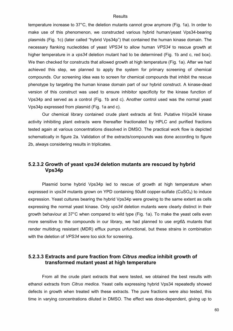
Results
60
temperature increase to 37°C, the deletion mutants cannot grow anymore (Fig. 1a). In order to
make use of this phenomenon, we constructed various hybrid human/yeast Vps34-bearing
plasmids (Fig. 1c) (later called “hybrid Vps34p”) that contained the human kinase domain. The
necessary flanking nucleotides of yeast VPS34 to allow human VPS34 to rescue growth at
higher temperature in a vps34 deletion mutant had to be determined (Fig. 1b and c, red box).
We then checked for constructs that allowed growth at high temperature (Fig. 1a). After we had
achieved this step, we planned to apply the system for primary screening of chemical
compounds. Our screening idea was to screen for chemical compounds that inhibit the rescue
phenotype by targeting the human kinase domain part of our hybrid construct. A kinase-dead
version of this construct was used to ensure inhibitor specificity for the kinase function of
Vps34p and served as a control (Fig. 1b and c). Another control used was the normal yeast
Vps34p expressed from plasmid (Fig. 1a and c).
Our chemical library contained crude plant extracts at first. Putative hVps34 kinase
activity inhibiting plant extracts were thereafter fractionated by HPLC and purified fractions
tested again at various concentrations dissolved in DMSO. The practical work flow is depicted
schematically in figure 2a. Validation of the extracts/compounds was done according to figure
2b, always considering results in triplicates.
5.2.3.2 Growth of yeast vps34 deletion mutants are rescued by hybrid Vps34p
Plasmid borne hybrid Vps34p led to rescue of growth at high temperature when
expressed in vps34 mutants grown on YPD containing 50uM copper-sulfate (CuSO4) to induce
expression. Yeast cultures bearing the hybrid Vps34p were growing to the same extent as cells
expressing the normal yeast kinase. Only vps34 deletion mutants were clearly distinct in their
growth behaviour at 37°C when compared to wild type (Fig. 1a). To make the yeast cells even
more sensitive to the compounds in our library, we had planned to use erg6∆ mutants that
render multidrug resistant (MDR) efflux pumps unfunctional, but these strains in combination
with the deletion of VPS34 were too sick for screening.
5.2.3.3 Extracts and pure fraction from Citrus medica inhibit growth of transformed mutant yeast at high temperature
From all the crude plant extracts that were tested, we obtained the best results with
ethanol extracts from Citrus medica. Yeast cells expressing hybrid Vps34 repeatedly showed
defects in growth when treated with these extracts. The pure fractions were also tested, this
time in varying concentrations diluted in DMSO. The effect was dose-dependent, giving up to
Results
61
60% reduction in growth compared to vehicle-treated cells when in liquid culture (Fig.3a). Best
results were obtained with fraction “MACMS2”, that turned out to be limettin (Fig. 3b). Limettin
(5,7-dimethoxycoumarin) is part of the coumarin-related compounds and has a molecular weight
of 206.19 g/mol. Structurally-related compounds (Fig. 3c) that are commercially available were
checked for similar effects, but none of them showed comparable effects, even at high
concentrations (Fig.3a).
5.2.3.4 Pure fraction of Citrus medica compound does not influence class I PI3K isoforms
In order to check for specificity of our most promising pure fraction of Citrus medica
extracts (MACMS2/limettin), we treated HEK293 cells with different concentrations of the
compound (Fig.4). Lower concentrations than those applied in yeast tests were checked. In
general yeast requires higher dosages of kinase inhibitors than mammalian cells due to their
efflux pumps which often cause multidrug resistance (MDR) in yeast (Prasad, Gaur et al. 2006).
We chose HEK293 since all class I PI3K isoforms are expressed and endogenous hVps34
levels are high when compared to other cell lines available in our lab. Wortmannin is a well-
known pan-PI3K inhibitor (Arcaro and Wymann 1993) and blocks phosphorylation of PKB/Akt at
Serine 473 and phosphorylation of S6K1 at Threonine 389 at the concentration applied, hence
we used it as a positive control. As a negative control we treated the cells with 10uM coumarin,
a compound which is structurally related (Fig. 3c) to our putative hVps34 inhibitor and since it
had not exhibited strong effects in yeast. Wortmannin led to the expected decreases of
phosphorylation, but none of the other compounds had any effects as can be seen in Fig.4.
pMAPK was checked to interpret any further downstream effects, but none were observed.
5.2.3.5 Less PI(3)P detected by microscopy in HeLa treated with pure fraction
A way to confirm the efficiency of our candidate hVps34 inhibitor MACMS2 in
mammalian cells was via microscopy. We checked whether a GFP-labelled 2xFYVE domain
originating from Hrs was still detecting PI(3)P lipids, the product of hVps34 kinase activity.
PI(3)P usually localize at the endosomes in punctuate structures (Raiborg, Bremnes et al.
2001). We used HeLa cells as a model system, as these cells are bigger than HEK293 and
hence more suitable for microscopy. In cells treated with 200uM of putative inhibitor, we
observed less GFP-dots per cell, which we interpreted as equal to less PI(3)P produced due to
inhibition of the kinase. Cells observed by binocular microscope seemed to be healthy after the
treatment, so no cytotoxicity assay was performed.
Results
62
Wortmannin was once more used as positive control and led to total depletion of any
PI(3)P, leaving only cytosolic GFP-signal. 100uM coumarin did not have any obvious effect on
the GFP-signal when compared to vehicle-treatment (Fig.5).
5.2.3.6 In vitro kinase assay confirms reduction of hVps34 activity upon short incubation with pure fraction
As a last confirmation of the inhibitory effect of our compound MACMS2 on hVps34, we
performed in vitro kinase assays with hVps34 immunoprecipitated from HEK293.
Immunoprecipitated kinase was treated with a titration of MACMS2 ranging from 2uM to 200uM
for 10 minutes prior to kinase reactions. As controls, treatment with 1uM wortmannin and
overexpression of kinase-dead hVps34 was used to demonstrate clear inhibition of PI(3)P
production (Fig. 6a). Comparing the quantified PI(3)P production of inhibitor treated hVp34, we
observed a reduction of about 40% versus vehicle-treated kinase (Fig.6b). As seen in the
growth studies in yeast at 37°C (Fig. 3a), we again documented a dose-dependence of the
inhibitor. This recurrent dose-dependence of our putative hVps34 inhibitor suggests a real
specificity for the human Vps34 kinase domain, even if working concentrations are high.
5.2.4 Discussion
hVps34 is involved in many cellular processes such as endocytosis, protein sorting to
the lysosome (Wurmser and Emr 1998) and autophagy (Zeng, Overmeyer et al. 2006), and is
therefore a rapidly expanding field nowadays. Further characterization of the presented
compound-kinase interaction might be able to lead to more potent hVps34 inhibitors, a
challenging task, as there are no specific class III PI3K inhibitors on the market to date. A recent
publication (Miller, Tavshanjian et al.) opens up new possibilities to design class III-specific PI3K
inhibitors. S. Miller et al. discovered differences in the ATP-binding pockets of class III versus
class I PI3K isoforms. Wortmannin, the most popular pan-PI3K inhibitor (Arcaro and Wymann
1993), clearly exhibits a difference in concentration required for inhibition (IC-50), even if it
shows no stronger specificity for one class over the other.
We suggest that MACMS2/limettin be used as a scaffold structure for more optimized
specific hVps34 kinase activity inhibitors of the future. Now that the structure of hVps34’s ATP-
binding pocket is known in this field of research, drug designing can be implemented
subsequently. Specific class III PI3K inhibitors promise to be interesting tools in future medical
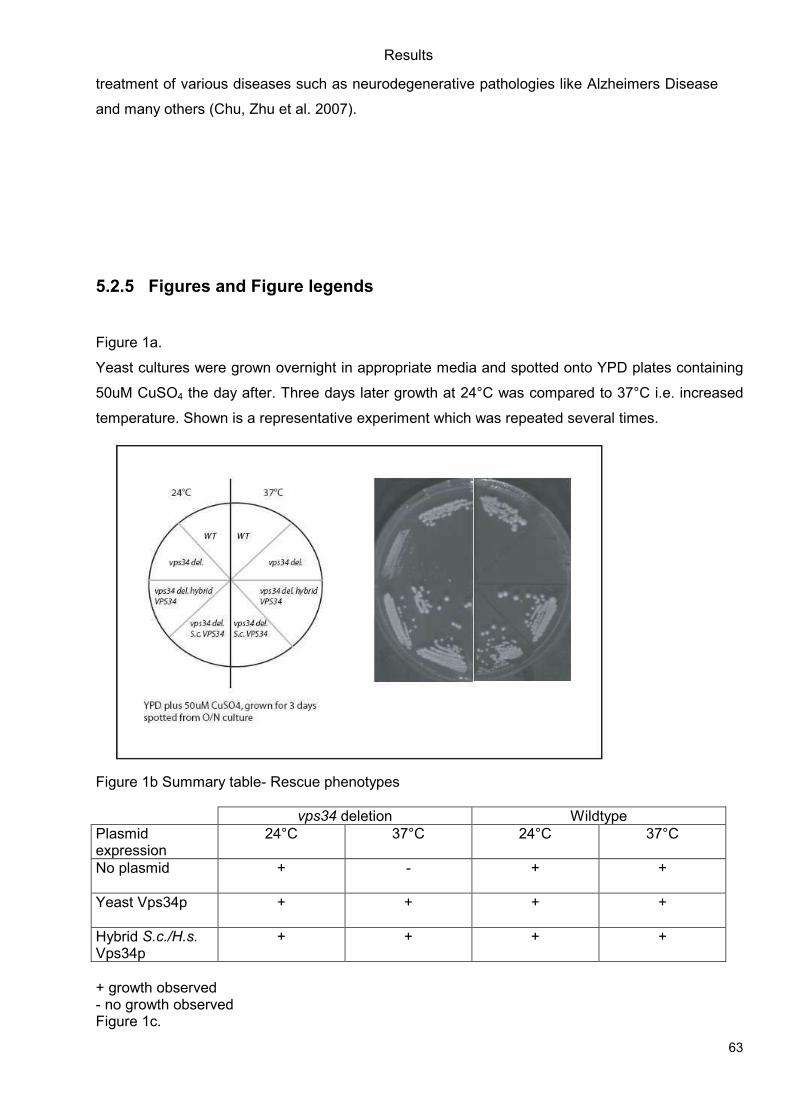
Results
63
treatment of various diseases such as neurodegenerative pathologies like Alzheimers Disease
and many others (Chu, Zhu et al. 2007).
5.2.5 Figures and Figure legends
Figure 1a.
Yeast cultures were grown overnight in appropriate media and spotted onto YPD plates containing
50uM CuSO4 the day after. Three days later growth at 24°C was compared to 37°C i.e. increased
temperature. Shown is a representative experiment which was repeated several times.
Figure 1b Summary table- Rescue phenotypes
vps34 deletion Wildtype
Plasmid expression
24°C 37°C 24°C 37°C
No plasmid
+ - + +
Yeast Vps34p
+ + + +
Hybrid S.c./H.s. Vps34p
+ + + +
+ growth observed - no growth observed Figure 1c.
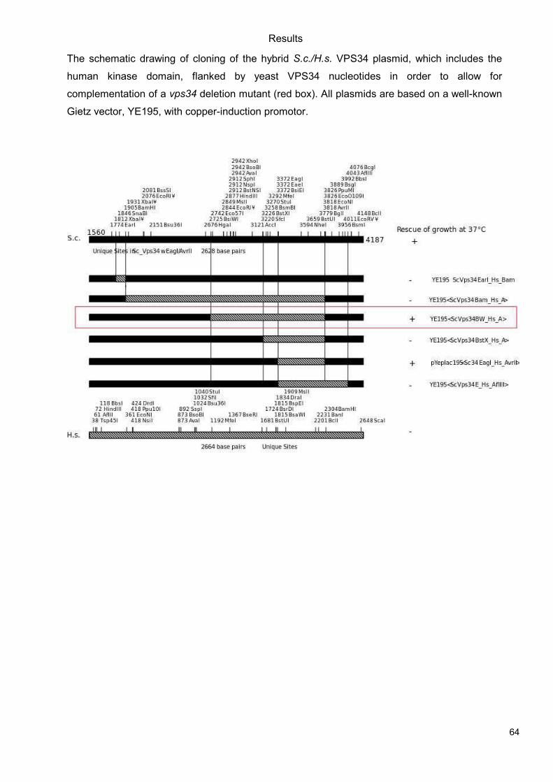
Results
64
The schematic drawing of cloning of the hybrid S.c./H.s. VPS34 plasmid, which includes the
human kinase domain, flanked by yeast VPS34 nucleotides in order to allow for
complementation of a vps34 deletion mutant (red box). All plasmids are based on a well-known
Gietz vector, YE195, with copper-induction promotor.
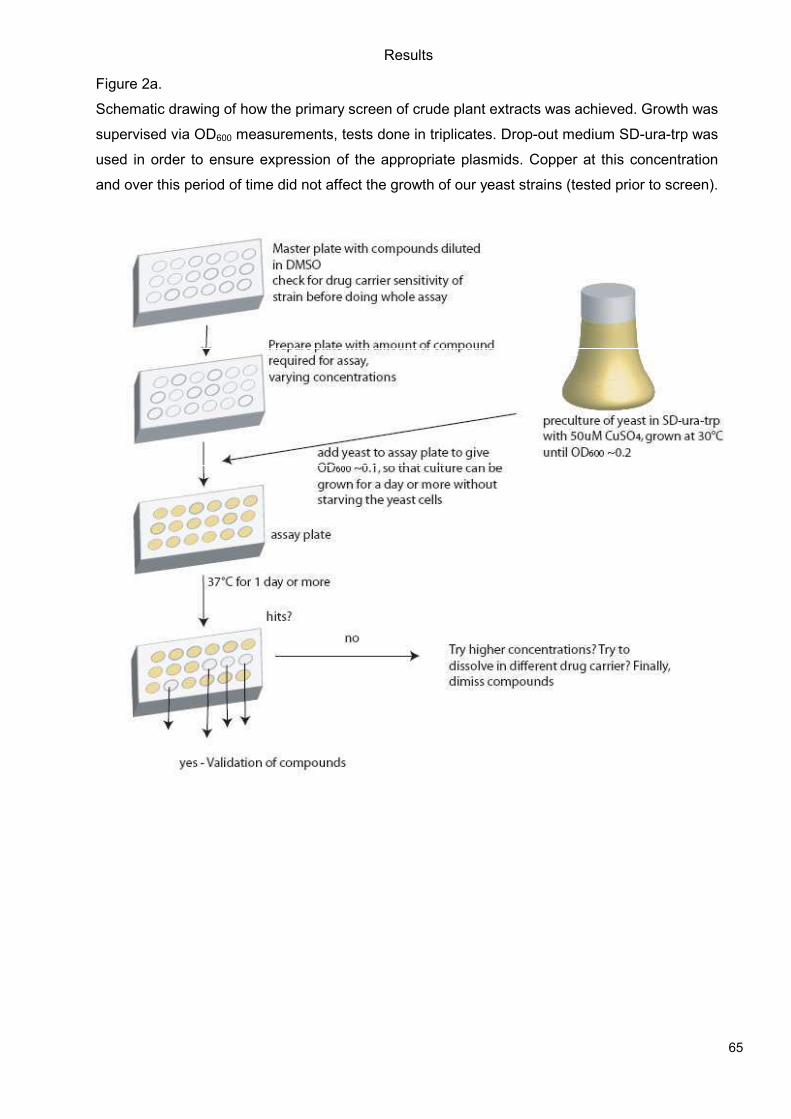
Results
65
Figure 2a.
Schematic drawing of how the primary screen of crude plant extracts was achieved. Growth was
supervised via OD600 measurements, tests done in triplicates. Drop-out medium SD-ura-trp was
used in order to ensure expression of the appropriate plasmids. Copper at this concentration
and over this period of time did not affect the growth of our yeast strains (tested prior to screen).
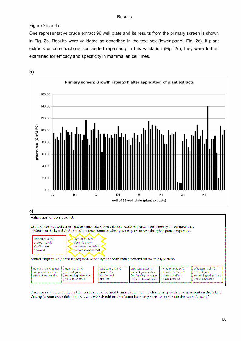
Results
66
Figure 2b and c.
One representative crude extract 96 well plate and its results from the primary screen is shown
in Fig. 2b. Results were validated as described in the text box (lower panel, Fig. 2c). If plant
extracts or pure fractions succeeded repeatedly in this validation (Fig. 2c), they were further
examined for efficacy and specificity in mammalian cell lines.
b)
Primary screen: Growth rates 24h after application of plant extracts
0.00
20.00
40.00
60.00
80.00
100.00
120.00
140.00
160.00
A1 B1 C1 D1 E1 F1 G1 H1
well of 96-well plate (plant extracts)
gro
wth
rate
(%
of 24°C
)
c)
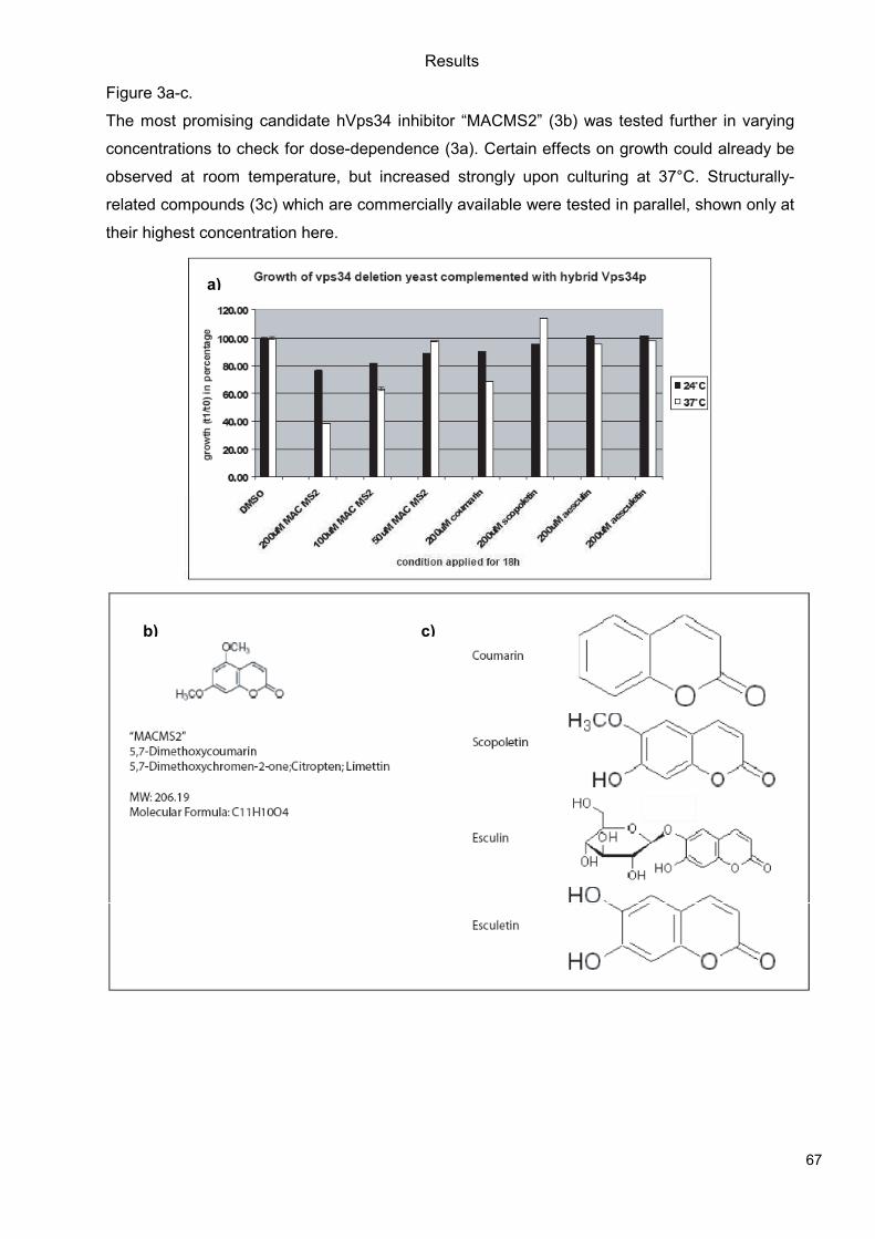
Results
67
Figure 3a-c.
The most promising candidate hVps34 inhibitor “MACMS2” (3b) was tested further in varying
concentrations to check for dose-dependence (3a). Certain effects on growth could already be
observed at room temperature, but increased strongly upon culturing at 37°C. Structurally-
related compounds (3c) which are commercially available were tested in parallel, shown only at
their highest concentration here.
a)
b) c)
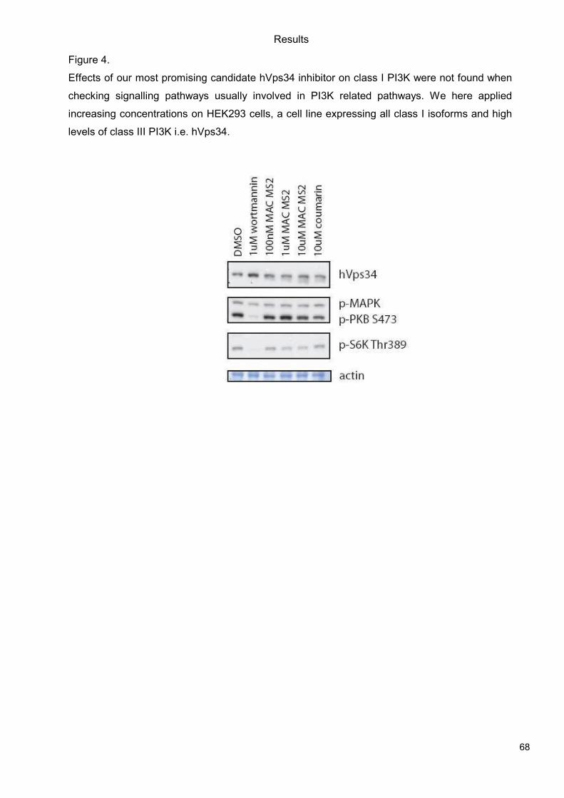
Results
68
Figure 4.
Effects of our most promising candidate hVps34 inhibitor on class I PI3K were not found when
checking signalling pathways usually involved in PI3K related pathways. We here applied
increasing concentrations on HEK293 cells, a cell line expressing all class I isoforms and high
levels of class III PI3K i.e. hVps34.
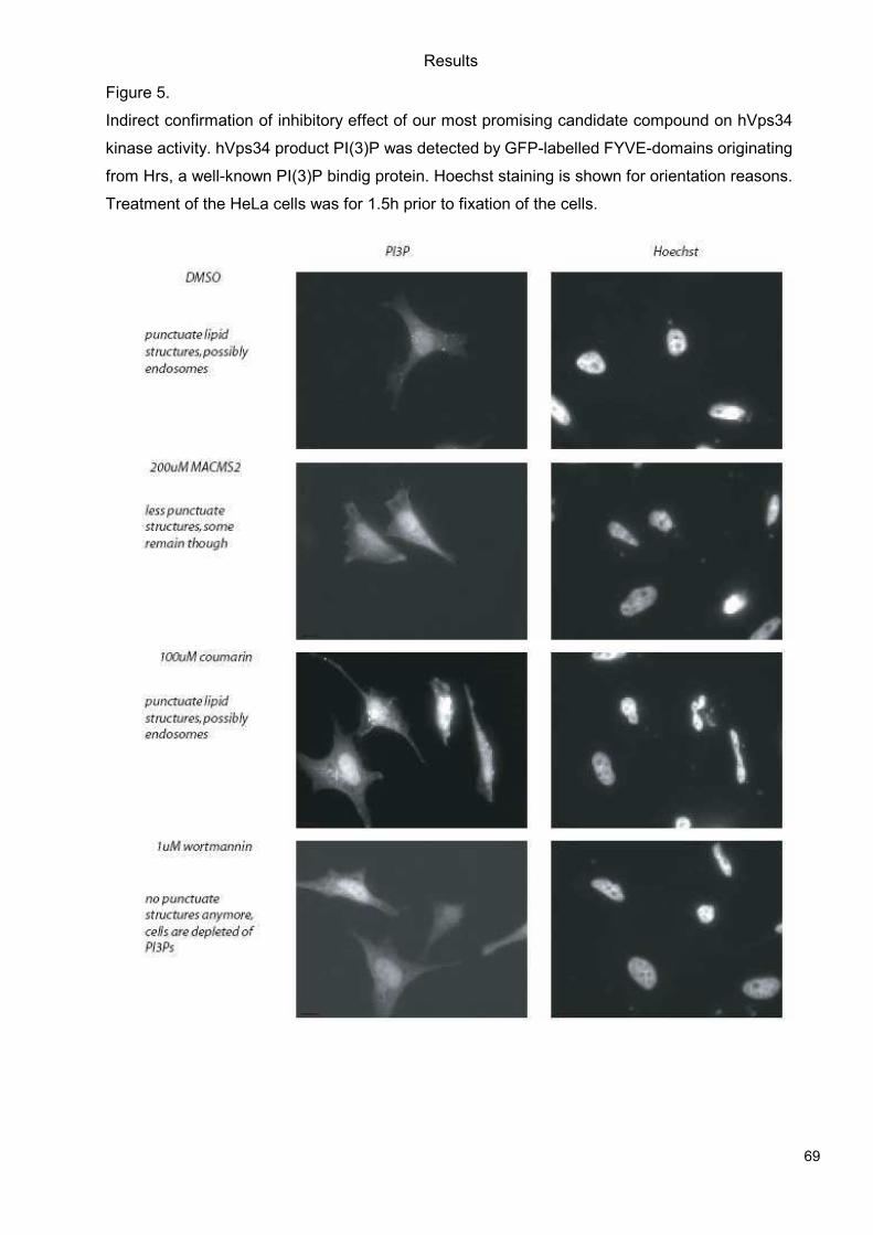
Results
69
Figure 5.
Indirect confirmation of inhibitory effect of our most promising candidate compound on hVps34
kinase activity. hVps34 product PI(3)P was detected by GFP-labelled FYVE-domains originating
from Hrs, a well-known PI(3)P bindig protein. Hoechst staining is shown for orientation reasons.
Treatment of the HeLa cells was for 1.5h prior to fixation of the cells.
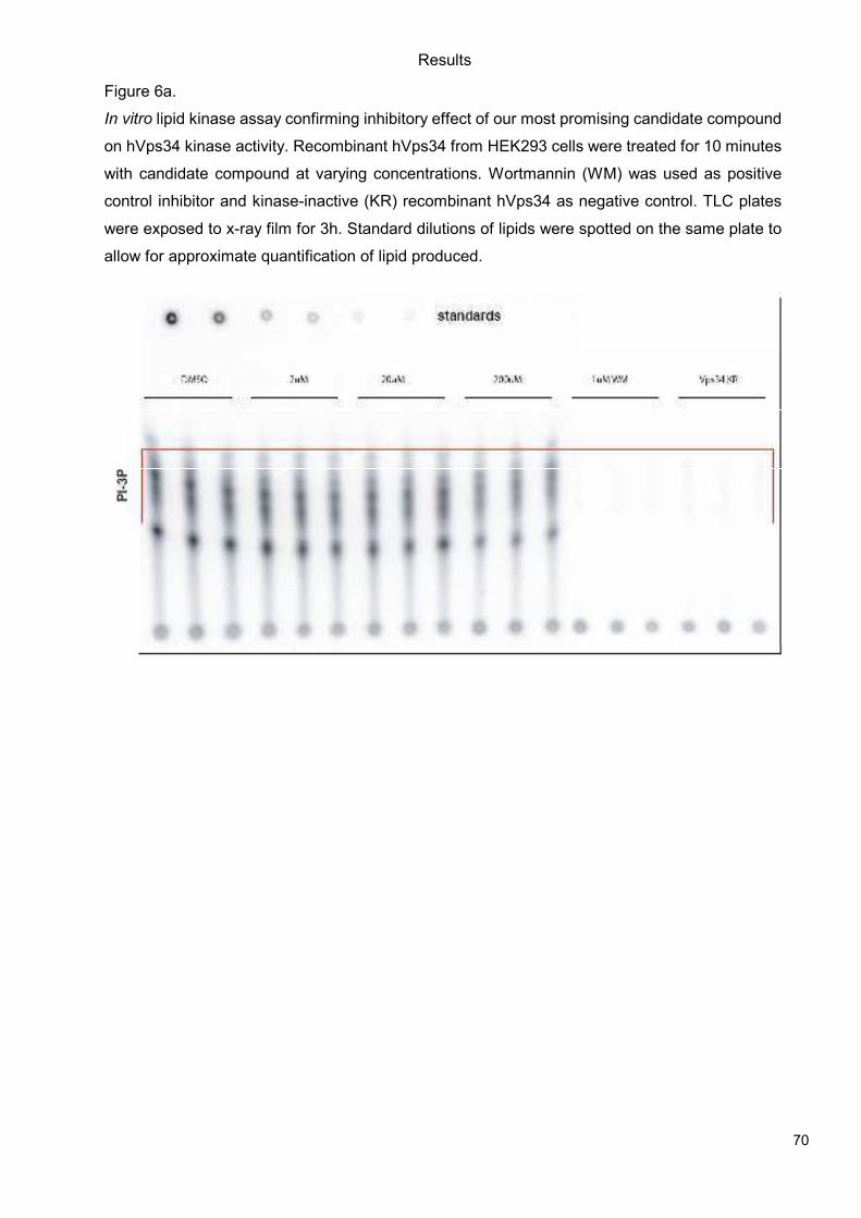
Results
70
Figure 6a.
In vitro lipid kinase assay confirming inhibitory effect of our most promising candidate compound
on hVps34 kinase activity. Recombinant hVps34 from HEK293 cells were treated for 10 minutes
with candidate compound at varying concentrations. Wortmannin (WM) was used as positive
control inhibitor and kinase-inactive (KR) recombinant hVps34 as negative control. TLC plates
were exposed to x-ray film for 3h. Standard dilutions of lipids were spotted on the same plate to
allow for approximate quantification of lipid produced.
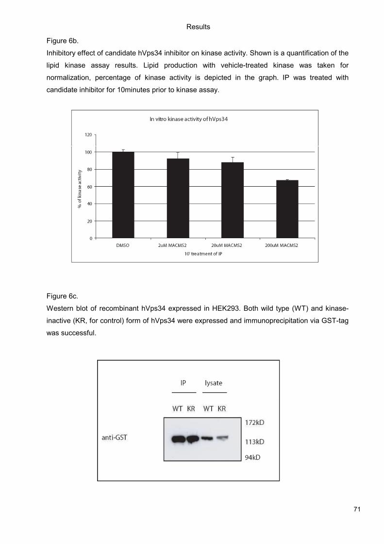
Results
71
Figure 6b.
Inhibitory effect of candidate hVps34 inhibitor on kinase activity. Shown is a quantification of the
lipid kinase assay results. Lipid production with vehicle-treated kinase was taken for
normalization, percentage of kinase activity is depicted in the graph. IP was treated with
candidate inhibitor for 10minutes prior to kinase assay.
Figure 6c.
Western blot of recombinant hVps34 expressed in HEK293. Both wild type (WT) and kinase-
inactive (KR, for control) form of hVps34 were expressed and immunoprecipitation via GST-tag
was successful.
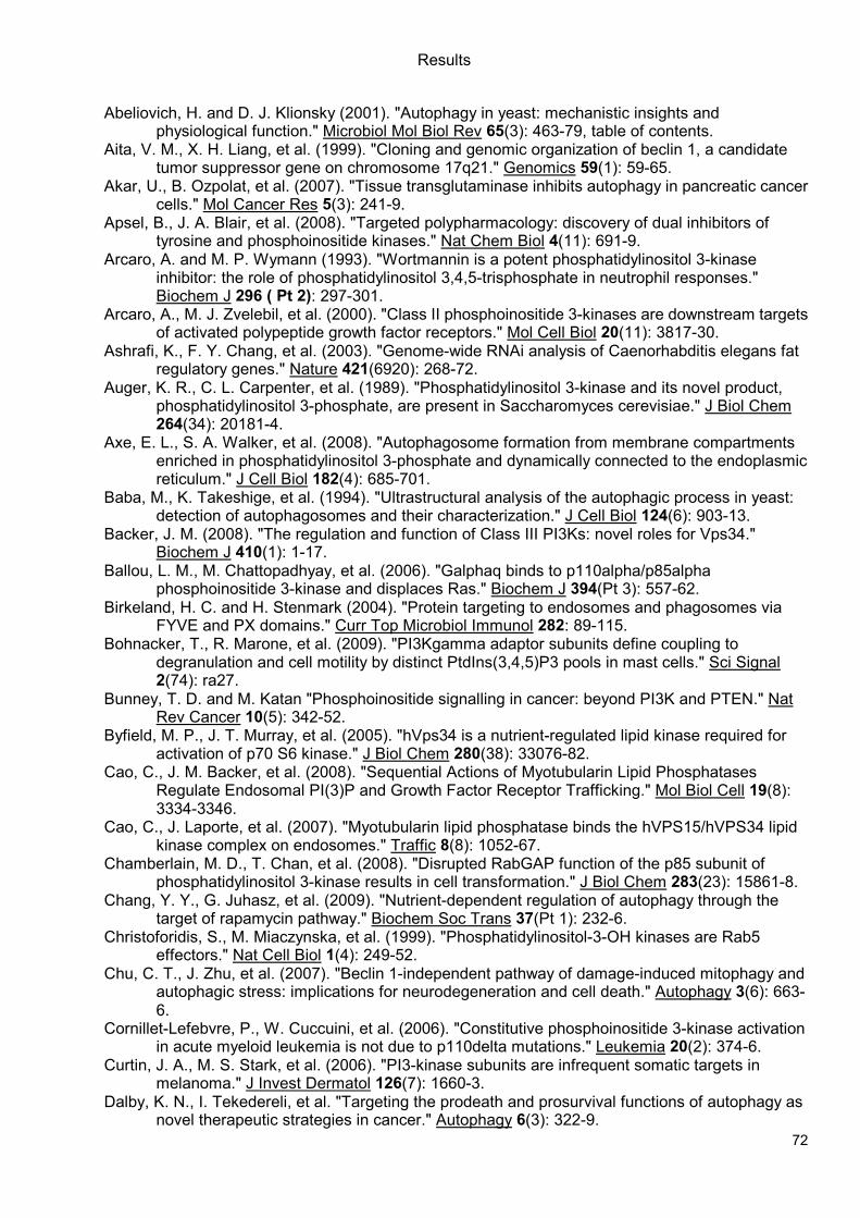
Results
72
Abeliovich, H. and D. J. Klionsky (2001). "Autophagy in yeast: mechanistic insights and
physiological function." Microbiol Mol Biol Rev 65(3): 463-79, table of contents. Aita, V. M., X. H. Liang, et al. (1999). "Cloning and genomic organization of beclin 1, a candidate
tumor suppressor gene on chromosome 17q21." Genomics 59(1): 59-65. Akar, U., B. Ozpolat, et al. (2007). "Tissue transglutaminase inhibits autophagy in pancreatic cancer
cells." Mol Cancer Res 5(3): 241-9. Apsel, B., J. A. Blair, et al. (2008). "Targeted polypharmacology: discovery of dual inhibitors of
tyrosine and phosphoinositide kinases." Nat Chem Biol 4(11): 691-9. Arcaro, A. and M. P. Wymann (1993). "Wortmannin is a potent phosphatidylinositol 3-kinase
inhibitor: the role of phosphatidylinositol 3,4,5-trisphosphate in neutrophil responses." Biochem J 296 ( Pt 2): 297-301.
Arcaro, A., M. J. Zvelebil, et al. (2000). "Class II phosphoinositide 3-kinases are downstream targets of activated polypeptide growth factor receptors." Mol Cell Biol 20(11): 3817-30.
Ashrafi, K., F. Y. Chang, et al. (2003). "Genome-wide RNAi analysis of Caenorhabditis elegans fat regulatory genes." Nature 421(6920): 268-72.
Auger, K. R., C. L. Carpenter, et al. (1989). "Phosphatidylinositol 3-kinase and its novel product, phosphatidylinositol 3-phosphate, are present in Saccharomyces cerevisiae." J Biol Chem 264(34): 20181-4.
Axe, E. L., S. A. Walker, et al. (2008). "Autophagosome formation from membrane compartments enriched in phosphatidylinositol 3-phosphate and dynamically connected to the endoplasmic reticulum." J Cell Biol 182(4): 685-701.
Baba, M., K. Takeshige, et al. (1994). "Ultrastructural analysis of the autophagic process in yeast: detection of autophagosomes and their characterization." J Cell Biol 124(6): 903-13.
Backer, J. M. (2008). "The regulation and function of Class III PI3Ks: novel roles for Vps34." Biochem J 410(1): 1-17.
Ballou, L. M., M. Chattopadhyay, et al. (2006). "Galphaq binds to p110alpha/p85alpha phosphoinositide 3-kinase and displaces Ras." Biochem J 394(Pt 3): 557-62.
Birkeland, H. C. and H. Stenmark (2004). "Protein targeting to endosomes and phagosomes via FYVE and PX domains." Curr Top Microbiol Immunol 282: 89-115.
Bohnacker, T., R. Marone, et al. (2009). "PI3Kgamma adaptor subunits define coupling to degranulation and cell motility by distinct PtdIns(3,4,5)P3 pools in mast cells." Sci Signal 2(74): ra27.
Bunney, T. D. and M. Katan "Phosphoinositide signalling in cancer: beyond PI3K and PTEN." Nat Rev Cancer 10(5): 342-52.
Byfield, M. P., J. T. Murray, et al. (2005). "hVps34 is a nutrient-regulated lipid kinase required for activation of p70 S6 kinase." J Biol Chem 280(38): 33076-82.
Cao, C., J. M. Backer, et al. (2008). "Sequential Actions of Myotubularin Lipid Phosphatases Regulate Endosomal PI(3)P and Growth Factor Receptor Trafficking." Mol Biol Cell 19(8): 3334-3346.
Cao, C., J. Laporte, et al. (2007). "Myotubularin lipid phosphatase binds the hVPS15/hVPS34 lipid kinase complex on endosomes." Traffic 8(8): 1052-67.
Chamberlain, M. D., T. Chan, et al. (2008). "Disrupted RabGAP function of the p85 subunit of phosphatidylinositol 3-kinase results in cell transformation." J Biol Chem 283(23): 15861-8.
Chang, Y. Y., G. Juhasz, et al. (2009). "Nutrient-dependent regulation of autophagy through the target of rapamycin pathway." Biochem Soc Trans 37(Pt 1): 232-6.
Christoforidis, S., M. Miaczynska, et al. (1999). "Phosphatidylinositol-3-OH kinases are Rab5 effectors." Nat Cell Biol 1(4): 249-52.
Chu, C. T., J. Zhu, et al. (2007). "Beclin 1-independent pathway of damage-induced mitophagy and autophagic stress: implications for neurodegeneration and cell death." Autophagy 3(6): 663-6.
Cornillet-Lefebvre, P., W. Cuccuini, et al. (2006). "Constitutive phosphoinositide 3-kinase activation in acute myeloid leukemia is not due to p110delta mutations." Leukemia 20(2): 374-6.
Curtin, J. A., M. S. Stark, et al. (2006). "PI3-kinase subunits are infrequent somatic targets in melanoma." J Invest Dermatol 126(7): 1660-3.
Dalby, K. N., I. Tekedereli, et al. "Targeting the prodeath and prosurvival functions of autophagy as novel therapeutic strategies in cancer." Autophagy 6(3): 322-9.
Results
73
Devarajan, E., J. Chen, et al. (2002). "Human breast cancer MCF-7 cell line contains inherently drug-resistant subclones with distinct genotypic and phenotypic features." Int J Oncol 20(5): 913-20.
Devarajan, E., A. A. Sahin, et al. (2002). "Down-regulation of caspase 3 in breast cancer: a possible mechanism for chemoresistance." Oncogene 21(57): 8843-51.
Di Paolo, G. and P. De Camilli (2006). "Phosphoinositides in cell regulation and membrane dynamics." Nature 443(7112): 651-7.
Domin, J., F. Pages, et al. (1997). "Cloning of a human phosphoinositide 3-kinase with a C2 domain that displays reduced sensitivity to the inhibitor wortmannin." Biochem J 326 ( Pt 1): 139-47.
Dunn, W. A., Jr. (1990). "Studies on the mechanisms of autophagy: formation of the autophagic vacuole." J Cell Biol 110(6): 1923-33.
Eickholt, B. J., A. I. Ahmed, et al. (2007). "Control of axonal growth and regeneration of sensory neurons by the p110delta PI 3-kinase." PLoS One 2(9): e869.
Elis, W., E. Triantafellow, et al. (2008). "Down-regulation of class II phosphoinositide 3-kinase alpha expression below a critical threshold induces apoptotic cell death." Mol Cancer Res 6(4): 614-23.
Ellson, C. D., K. E. Anderson, et al. (2001). "Phosphatidylinositol 3-phosphate is generated in phagosomal membranes." Curr Biol 11(20): 1631-5.
Emr, S. a. (1994). "Vps34p required for yeast vacuolar protein sorting is a multiple specificity kinase that exhibits both protein kinase and phophatidylinositol-specific PI 3-kinase activities." The Journal of Biological Chemistry 269(Dec 16): 31552-31562.
Falasca, M., W. E. Hughes, et al. (2007). "The role of phosphoinositide 3-kinase C2alpha in insulin signaling." J Biol Chem 282(38): 28226-36.
Falasca, M. and T. Maffucci (2007). "Role of class II phosphoinositide 3-kinase in cell signalling." Biochem Soc Trans 35(Pt 2): 211-4.
Fimia, G. M., A. Stoykova, et al. (2007). "Ambra1 regulates autophagy and development of the nervous system." Nature 447(7148): 1121-5.
Findlay, G. M., L. Yan, et al. (2007). "A MAP4 kinase related to Ste20 is a nutrient-sensitive regulator of mTOR signalling." Biochem J 403(1): 13-20.
Folkman, J. (2006). "Angiogenesis." Annu Rev Med 57: 1-18. Frazzetto, M., C. Suphioglu, et al. (2008). "Dissecting isoform selectivity of PI3K inhibitors: the role
of non-conserved residues in the catalytic pocket." Biochem J 414(3): 383-90. Furuya, N., J. Yu, et al. (2005). "The evolutionarily conserved domain of Beclin 1 is required for
Vps34 binding, autophagy and tumor suppressor function." Autophagy 1(1): 46-52. Futter, C. E., L. M. Collinson, et al. (2001). "Human VPS34 is required for internal vesicle formation
within multivesicular endosomes." J Cell Biol 155(7): 1251-64. Gietz, R. D., R. H. Schiestl, et al. (1995). "Studies on the transformation of intact yeast cells by the
LiAc/SS-DNA/PEG procedure." Yeast 11(4): 355-60. Gulati, P., L. D. Gaspers, et al. (2008). "Amino acids activate mTOR complex 1 via Ca2+/CaM
signaling to hVps34." Cell Metab 7(5): 456-65. Gulati, P. and G. Thomas (2007). "Nutrient sensing in the mTOR/S6K1 signalling pathway."
Biochem Soc Trans 35(Pt 2): 236-8. Gupta, S., A. R. Ramjaun, et al. (2007). "Binding of ras to phosphoinositide 3-kinase p110alpha is
required for ras-driven tumorigenesis in mice." Cell 129(5): 957-68. Harada, K., A. B. Truong, et al. (2005). "The class II phosphoinositide 3-kinase C2beta is not
essential for epidermal differentiation." Mol Cell Biol 25(24): 11122-30. Herman, P. K. and S. D. Emr (1990). "Characterization of VPS34, a gene required for vacuolar
protein sorting and vacuole segregation in Saccharomyces cerevisiae." Mol Cell Biol 10(12): 6742-54.
Hirsch, E., L. Braccini, et al. (2009). "Twice upon a time: PI3K's secret double life exposed." Trends Biochem Sci 34(5): 244-8.
Hurley, J. H. (2006). "Membrane binding domains." Biochim Biophys Acta 1761(8): 805-11. Im, Y. J., T. Wollert, et al. (2009). "Structure and function of the ESCRT-II-III interface in
multivesicular body biogenesis." Dev Cell 17(2): 234-43. Jin, S. and E. White (2008). "Tumor suppression by autophagy through the management of
metabolic stress." Autophagy 4(5): 563-6.
Results
74
Johnson, E. E., J. H. Overmeyer, et al. (2006). "Gene silencing reveals a specific function of hVps34 phosphatidylinositol 3-kinase in late versus early endosomes." J Cell Sci 119(Pt 7): 1219-32.
Juhasz, G., J. H. Hill, et al. (2008). "The class III PI(3)K Vps34 promotes autophagy and endocytosis but not TOR signaling in Drosophila." J Cell Biol 181(4): 655-66.
Kihara, A., T. Noda, et al. (2001). "Two distinct Vps34 phosphatidylinositol 3-kinase complexes function in autophagy and carboxypeptidase Y sorting in Saccharomyces cerevisiae." J Cell Biol 152(3): 519-30.
Kim, E., P. Goraksha-Hicks, et al. (2008). "Regulation of TORC1 by Rag GTPases in nutrient response." Nat Cell Biol 10(8): 935-45.
Kinchen, J. M., K. Doukoumetzidis, et al. (2008). "A pathway for phagosome maturation during engulfment of apoptotic cells." Nat Cell Biol 10(5): 556-66.
Knight, Z. A., B. Gonzalez, et al. (2006). "A pharmacological map of the PI3-K family defines a role for p110alpha in insulin signaling." Cell 125(4): 733-47.
Kok, K., B. Geering, et al. (2009). "Regulation of phosphoinositide 3-kinase expression in health and disease." Trends Biochem Sci 34(3): 115-27.
Kurig, B., A. Shymanets, et al. (2009). "Ras is an indispensable coregulator of the class IB phosphoinositide 3-kinase p87/p110gamma." Proc Natl Acad Sci U S A 106(48): 20312-7.
Kurosu, H. and T. Katada (2001). "Association of phosphatidylinositol 3-kinase composed of p110beta-catalytic and p85-regulatory subunits with the small GTPase Rab5." J Biochem 130(1): 73-8.
Kurosu, H., T. Maehama, et al. (1997). "Heterodimeric phosphoinositide 3-kinase consisting of p85 and p110beta is synergistically activated by the betagamma subunits of G proteins and phosphotyrosyl peptide." J Biol Chem 272(39): 24252-6.
Lahtz, C., R. Stranzenbach, et al. (2010). "Methylation of PTEN as a prognostic factor in malignant melanoma of the skin." J Invest Dermatol 130(2): 620-2.
Lehmann, K., J. P. Muller, et al. (2009). "PI3Kgamma controls oxidative bursts in neutrophils via interactions with PKCalpha and p47phox." Biochem J 419(3): 603-10.
Lemmon, M. A. (2008). "Membrane recognition by phospholipid-binding domains." Nat Rev Mol Cell Biol 9(2): 99-111.
Leung, W. H., T. Tarasenko, et al. (2009). "Differential roles for the inositol phosphatase SHIP in the regulation of macrophages and lymphocytes." Immunol Res 43(1-3): 243-51.
Levine, B. (2007). "Cell biology: autophagy and cancer." Nature 446(7137): 745-7. Levine, B. and G. Kroemer (2008). "Autophagy in the pathogenesis of disease." Cell 132(1): 27-42. Liang, C., P. Feng, et al. (2006). "Autophagic and tumour suppressor activity of a novel Beclin1-
binding protein UVRAG." Nat Cell Biol 8(7): 688-99. Liang, C., J. S. Lee, et al. (2008). "Beclin1-binding UVRAG targets the class C Vps complex to
coordinate autophagosome maturation and endocytic trafficking." Nat Cell Biol 10(7): 776-87.
Liang, X. H., S. Jackson, et al. (1999). "Induction of autophagy and inhibition of tumorigenesis by beclin 1." Nature 402(6762): 672-6.
Liu, B., Y. Cheng, et al. (2009). "Polygonatum cyrtonema lectin induces apoptosis and autophagy in human melanoma A375 cells through a mitochondria-mediated ROS-p38-p53 pathway." Cancer Lett 275(1): 54-60.
MacDougall, L. K., M. E. Gagou, et al. (2004). "Targeted expression of the class II phosphoinositide 3-kinase in Drosophila melanogaster reveals lipid kinase-dependent effects on patterning and interactions with receptor signaling pathways." Mol Cell Biol 24(2): 796-808.
Maira, S. M., F. Stauffer, et al. (2008). "Identification and characterization of NVP-BEZ235, a new orally available dual phosphatidylinositol 3-kinase/mammalian target of rapamycin inhibitor with potent in vivo antitumor activity." Mol Cancer Ther 7(7): 1851-63.
Mandelker, D., S. B. Gabelli, et al. (2009). "A frequent kinase domain mutation that changes the interaction between PI3Kalpha and the membrane." Proc Natl Acad Sci U S A 106(40): 16996-7001.
Marone, R., V. Cmiljanovic, et al. (2008). "Targeting phosphoinositide 3-kinase: moving towards therapy." Biochim Biophys Acta 1784(1): 159-85.
Marone, R., D. Erhart, et al. (2009). "Targeting melanoma with dual phosphoinositide 3-kinase/mammalian target of rapamycin inhibitors." Mol Cancer Res 7(4): 601-13.
Results
75
Mauvezin, C., M. Orpinell, et al. "The nuclear cofactor DOR regulates autophagy in mammalian and Drosophila cells." EMBO Rep 11(1): 37-44.
Melendez, A., Z. Talloczy, et al. (2003). "Autophagy genes are essential for dauer development and life-span extension in C. elegans." Science 301(5638): 1387-91.
Miled, N., Y. Yan, et al. (2007). "Mechanism of two classes of cancer mutations in the phosphoinositide 3-kinase catalytic subunit." Science 317(5835): 239-42.
Miller, S., B. Tavshanjian, et al. (2010). "Shaping development of autophagy inhibitors with the structure of the lipid kinase Vps34." Science 327(5973): 1638-42.
Moretti, L., A. Attia, et al. (2007). "Crosstalk between Bak/Bax and mTOR signaling regulates radiation-induced autophagy." Autophagy 3(2): 142-4.
Murray, J. T., C. Panaretou, et al. (2002). "Role of Rab5 in the recruitment of hVps34/p150 to the early endosome." Traffic 3(6): 416-27.
Nicklin, P., P. Bergman, et al. (2009). "Bidirectional transport of amino acids regulates mTOR and autophagy." Cell 136(3): 521-34.
Nobukuni, T., M. Joaquin, et al. (2005). "Amino acids mediate mTOR/raptor signaling through activation of class 3 phosphatidylinositol 3OH-kinase." Proc Natl Acad Sci U S A 102(40): 14238-43.
Novak, I., V. Kirkin, et al. "Nix is a selective autophagy receptor for mitochondrial clearance." EMBO Rep 11(1): 45-51.
Omholt, K., D. Krockel, et al. (2006). "Mutations of PIK3CA are rare in cutaneous melanoma." Melanoma Res 16(2): 197-200.
Orme, M. H., S. Alrubaie, et al. (2006). "Input from Ras is required for maximal PI(3)K signalling in Drosophila." Nat Cell Biol 8(11): 1298-302.
Papakonstanti, E. A., A. J. Ridley, et al. (2007). "The p110delta isoform of PI 3-kinase negatively controls RhoA and PTEN." Embo J 26(13): 3050-61.
Papakonstanti, E. A., O. Zwaenepoel, et al. (2008). "Distinct roles of class IA PI3K isoforms in primary and immortalised macrophages." J Cell Sci 121(Pt 24): 4124-33.
Petiot, A., F. Strappazzon, et al. (2008). "Alix differs from ESCRT proteins in the control of autophagy." Biochem Biophys Res Commun 375(1): 63-8.
Prasad, N. K., M. Tandon, et al. (2008). "Phosphoinositol phosphatase SHIP2 promotes cancer development and metastasis coupled with alterations in EGF receptor turnover." Carcinogenesis 29(1): 25-34.
Prasad, R., N. A. Gaur, et al. (2006). "Efflux pumps in drug resistance of Candida." Infect Disord Drug Targets 6(2): 69-83.
Ragnarsson-Olding, B. K., P. J. Nilsson, et al. (2009). "Primary ano-rectal malignant melanomas within a population-based national patient series in Sweden during 40 years." Acta Oncol 48(1): 125-31.
Raiborg, C., B. Bremnes, et al. (2001). "FYVE and coiled-coil domains determine the specific localisation of Hrs to early endosomes." J Cell Sci 114(Pt 12): 2255-63.
Robinson, J. S., D. J. Klionsky, et al. (1988). "Protein sorting in Saccharomyces cerevisiae: isolation of mutants defective in the delivery and processing of multiple vacuolar hydrolases." Mol Cell Biol 8(11): 4936-48.
Roggo, L., V. Bernard, et al. (2002). "Membrane transport in Caenorhabditis elegans: an essential role for VPS34 at the nuclear membrane." Embo J 21(7): 1673-83.
Rohrschneider, L. R., J. F. Fuller, et al. (2000). "Structure, function, and biology of SHIP proteins." Genes Dev 14(5): 505-20.
Rothman, J. H. and T. H. Stevens (1986). "Protein sorting in yeast: mutants defective in vacuole biogenesis mislocalize vacuolar proteins into the late secretory pathway." Cell 47(6): 1041-51.
Samuels, Y., Z. Wang, et al. (2004). "High frequency of mutations of the PIK3CA gene in human cancers." Science 304(5670): 554.
Sancak, Y., T. R. Peterson, et al. (2008). "The Rag GTPases bind raptor and mediate amino acid signaling to mTORC1." Science 320(5882): 1496-501.
Schu, P. V., K. Takegawa, et al. (1993). "Phosphatidylinositol 3-kinase encoded by yeast VPS34 gene essential for protein sorting." Science 260(5104): 88-91.
Sengupta, S., T. R. Peterson, et al. (2010). "Regulation of the mTOR complex 1 pathway by nutrients, growth factors, and stress." Mol Cell 40(2): 310-22.
Results
76
Shayesteh, L., Y. Lu, et al. (1999). "PIK3CA is implicated as an oncogene in ovarian cancer." Nat Genet 21(1): 99-102.
Shin, H. W., M. Hayashi, et al. (2005). "An enzymatic cascade of Rab5 effectors regulates phosphoinositide turnover in the endocytic pathway." J Cell Biol 170(4): 607-18.
Siddhanta, U., J. McIlroy, et al. (1998). "Distinct roles for the p110alpha and hVPS34 phosphatidylinositol 3'-kinases in vesicular trafficking, regulation of the actin cytoskeleton, and mitogenesis." J Cell Biol 143(6): 1647-59.
Simonsen, A., H. C. Birkeland, et al. (2004). "Alfy, a novel FYVE-domain-containing protein associated with protein granules and autophagic membranes." J Cell Sci 117(Pt 18): 4239-51.
Simonsen, A. and H. Stenmark (2008). "Self-eating from an ER-associated cup." J Cell Biol 182(4): 621-2.
Simonsen, A. and S. A. Tooze (2009). "Coordination of membrane events during autophagy by multiple class III PI3-kinase complexes." J Cell Biol 186(6): 773-82.
Singh, S. B., A. S. Davis, et al. (2006). "Human IRGM induces autophagy to eliminate intracellular mycobacteria." Science 313(5792): 1438-41.
Slessareva, J. E., S. M. Routt, et al. (2006). "Activation of the phosphatidylinositol 3-kinase Vps34 by a G protein alpha subunit at the endosome." Cell 126(1): 191-203.
Song, X., W. Xu, et al. (2001). "Phox homology domains specifically bind phosphatidylinositol phosphates." Biochemistry 40(30): 8940-4.
Spowart, J. and J. J. Lum "Opening a new DOR to autophagy." EMBO Rep 11(1): 4-5. Stahelin, R. V., D. Karathanassis, et al. (2006). "Structural and membrane binding analysis of the
Phox homology domain of phosphoinositide 3-kinase-C2alpha." J Biol Chem 281(51): 39396-406.
Stark, M. and N. Hayward (2007). "Genome-wide loss of heterozygosity and copy number analysis in melanoma using high-density single-nucleotide polymorphism arrays." Cancer Res 67(6): 2632-42.
Stein, M. P., Y. Feng, et al. (2003). "Human VPS34 and p150 are Rab7 interacting partners." Traffic 4(11): 754-71.
Taboubi, S., J. Milanini, et al. (2007). "G alpha(q/11)-coupled P2Y2 nucleotide receptor inhibits human keratinocyte spreading and migration." Faseb J 21(14): 4047-58.
Takahashi, Y., D. Coppola, et al. (2007). "Bif-1 interacts with Beclin 1 through UVRAG and regulates autophagy and tumorigenesis." Nat Cell Biol 9(10): 1142-51.
Takahashi, Y., D. Coppola, et al. (2007). "Bif-1 interacts with Beclin 1 through UVRAG and regulates autophagy and tumorigenesis." Nat Cell Biol 9(10): 1142-51.
Takahashi, Y., C. L. Meyerkord, et al. (2008). "BARgaining membranes for autophagosome formation: Regulation of autophagy and tumorigenesis by Bif-1/Endophilin B1." Autophagy 4(1): 121-4.
Takeshige, K., M. Baba, et al. (1992). "Autophagy in yeast demonstrated with proteinase-deficient mutants and conditions for its induction." J Cell Biol 119(2): 301-11.
Tang, R., X. Zhao, et al. (2008). "Investigation of variants in the promoter region of PIK3C3 in schizophrenia." Neurosci Lett 437(1): 42-4.
Tormo, D., A. Checinska, et al. (2009). "Targeted activation of innate immunity for therapeutic induction of autophagy and apoptosis in melanoma cells." Cancer Cell 16(2): 103-14.
Tsukada, M. and Y. Ohsumi (1993). "Isolation and characterization of autophagy-defective mutants of Saccharomyces cerevisiae." FEBS Lett 333(1-2): 169-74.
Vanhaesebroeck, B., K. Ali, et al. (2005). "Signalling by PI3K isoforms: insights from gene-targeted mice." Trends Biochem Sci 30(4): 194-204.
Vanhaesebroeck, B., J. Guillermet-Guibert, et al. (2010). "The emerging mechanisms of isoform-specific PI3K signalling." Nat Rev Mol Cell Biol 11(5): 329-41.
Vanhaesebroeck, B., S. J. Leevers, et al. (2001). "Synthesis and function of 3-phosphorylated inositol lipids." Annu Rev Biochem 70: 535-602.
Vanhaesebroeck, B., S. J. Leevers, et al. (1997). "Phosphoinositide 3-kinases: a conserved family of signal transducers." Trends Biochem Sci 22(7): 267-72.
Vergne, I., E. Roberts, et al. (2009). "Control of autophagy initiation by phosphoinositide 3-phosphatase Jumpy." Embo J 28(15): 2244-58.
Results
77
Virbasius, J. V., A. Guilherme, et al. (1996). "Mouse p170 is a novel phosphatidylinositol 3-kinase containing a C2 domain." J Biol Chem 271(23): 13304-7.
Vlahos, C. J., W. F. Matter, et al. (1994). "A specific inhibitor of phosphatidylinositol 3-kinase, 2-(4-morpholinyl)-8-phenyl-4H-1-benzopyran-4-one (LY294002)." J Biol Chem 269(7): 5241-8.
Vogt, P. K., S. Kang, et al. (2007). "Cancer-specific mutations in phosphatidylinositol 3-kinase." Trends Biochem Sci 32(7): 342-9.
Volinia, S., R. Dhand, et al. (1995). "A human phosphatidylinositol 3-kinase complex related to the yeast Vps34p-Vps15p protein sorting system." Embo J 14(14): 3339-48.
Walker, E. H., M. E. Pacold, et al. (2000). "Structural determinants of phosphoinositide 3-kinase inhibition by wortmannin, LY294002, quercetin, myricetin, and staurosporine." Mol Cell 6(4): 909-19.
Wang, X. and C. G. Proud (2009). "Nutrient control of TORC1, a cell-cycle regulator." Trends Cell Biol 19(6): 260-7.
Wang, Z., W. A. Wilson, et al. (2001). "Antagonistic controls of autophagy and glycogen accumulation by Snf1p, the yeast homolog of AMP-activated protein kinase, and the cyclin-dependent kinase Pho85p." Mol Cell Biol 21(17): 5742-52.
Whitman, M., D. Kaplan, et al. (1987). "Evidence for two distinct phosphatidylinositol kinases in fibroblasts. Implications for cellular regulation." Biochem J 247(1): 165-74.
Wollert, T., D. Yang, et al. (2009). "The ESCRT machinery at a glance." J Cell Sci 122(Pt 13): 2163-6.
Wu, H., V. Goel, et al. (2003). "PTEN signaling pathways in melanoma." Oncogene 22(20): 3113-22.
Wullschleger, S., R. Loewith, et al. (2006). "TOR signaling in growth and metabolism." Cell 124(3): 471-84.
Wurmser, A. E. and S. D. Emr (1998). "Phosphoinositide signaling and turnover: PtdIns(3)P, a regulator of membrane traffic, is transported to the vacuole and degraded by a process that requires lumenal vacuolar hydrolase activities." Embo J 17(17): 4930-42.
Wymann, M. P. and R. Marone (2005). "Phosphoinositide 3-kinase in disease: timing, location, and scaffolding." Curr Opin Cell Biol 17(2): 141-9.
Wymann, M. P. and L. Pirola (1998). "Structure and function of phosphoinositide 3-kinases." Biochim Biophys Acta 1436(1-2): 127-50.
Xue, Y., H. Fares, et al. (2003). "Genetic analysis of the myotubularin family of phosphatases in Caenorhabditis elegans." J Biol Chem 278(36): 34380-6.
Yaguchi, S., Y. Fukui, et al. (2006). "Antitumor activity of ZSTK474, a new phosphatidylinositol 3-kinase inhibitor." J Natl Cancer Inst 98(8): 545-56.
Yan, Y., R. J. Flinn, et al. (2009). "hVps15, but not Ca2+/CaM, is required for the activity and regulation of hVps34 in mammalian cells." Biochem J 417(3): 747-55.
Zeng, X., J. H. Overmeyer, et al. (2006). "Functional specificity of the mammalian Beclin-Vps34 PI 3-kinase complex in macroautophagy versus endocytosis and lysosomal enzyme trafficking." J Cell Sci 119(Pt 2): 259-70.
Zhang, H., J. P. Stallock, et al. (2000). "Regulation of cellular growth by the Drosophila target of rapamycin dTOR." Genes Dev 14(21): 2712-24.
Zhao, L. and P. K. Vogt (2008). "Helical domain and kinase domain mutations in p110alpha of phosphatidylinositol 3-kinase induce gain of function by different mechanisms." Proc Natl Acad Sci U S A 105(7): 2652-7.
Zhong, Y., Q. J. Wang, et al. (2009). "Distinct regulation of autophagic activity by Atg14L and Rubicon associated with Beclin 1-phosphatidylinositol-3-kinase complex." Nat Cell Biol 11(4): 468-76.
Zoncu, R., A. Efeyan, et al. (2010). "mTOR: from growth signal integration to cancer, diabetes and ageing." Nat Rev Mol Cell Biol 12(1): 21-35.
Publications
78
6 Peer-reviewed publications
6.1 “Targeting Melanoma with Dual PI3K/mTOR Inhibitors”
Romina Marone, Dominik Erhart, Ann C Mertz, Thomas Bohnacker, Christian Schnell, Vladimir Cmiljanovic, Frédéric Stauffer, Carlos Garcia-Echeverria, Bernd Giese, Sauveur-Michel Maira, Matthias P. Wymann. “Targeting Melanoma with Dual PI3K/mTOR Inhibitors”, published in Molecular Cancer Research, 2009
6.2 “(E,Z)-3-(3’,5’-dimethoxy-4’-hydroxy-benzylidene)-2-indolinone (Indolinone) blocks mast cell degranulation”
Sabine Kiefer, Ann C Mertz, A. Koryakina, M. Hamburger, P. Küenzi. „(E,Z)-3-(3’,5’-dimethoxy-4’-hydroxy-benzylidene)-2-indolinone (Indolinone) blocks mast cell degranulation“, published in European Journal of Pharmaceutical Sciences, 2010
6.3 “Separation and detection of all phosphoinositide isomers by ESI-MS”
Sabine Kiefer, J. Rogger, Anna Melone, Ann C Mertz, A. Koryakina, P. Küenzi. “Separation and detection of all phosphoinositide isomers by ESI-MS”, published in Journal of Pharmaceutical and Biomedical Analysis, 2010
Fulltext of each publication at end of thesis

Publications
79
Discussion
80
7 Discussion
7.1 Vps34 in disease model systems There are not many publications on hVps34 in a disease context yet, most researchers
focus on its role in endosomal transport or autophagy. How class III PI3K is indirectly involved in
various diseases, even if it has never been found to be the cause thereof, starts to get the
attention of the field. A recent article on hVps34 in an Alzheimer’s Disease (AD) model system
links regulation of hVps34 kinase activity with cell-cycle control and prognosis of AD. During
mitosis, Vps34 is phosphorylated at Thr159 of the C2 domain by Cdk1, negatively regulating its
interaction with Beclin1 and hence inhibiting autophagy. This happens simultaneously to an
increase in cyclin B levels during mitosis. Furuya et al. claim to have discovered the key
mechanism by which autophagy is downregulated during mitosis, resulting from a decrease of
PI(3)P production originally coming from inhibition of Vps34 kinase activity through Cdk1
phosphorylation. Cdk5/p25, which is known to act on certain substrates of Cdk1, can
phosphorylate the same position in the class III PI3K isoform. The authors suggest that
phosphorylation of Thr159 in Vps34 is a key regulatory mechanism by which class III PI3K
activity in cell-cycle control, development and human diseases such as Alzheimer’s Disease is
controlled. (Furuya, Yu et al.)
In our mammalian cell study, we used a cancer model system, i.e. a selection of melanoma
cell lines with genetically different backgrounds. Our main focus was on the importance of
hVps34 in these cells: Could the cells survive without hVps34 on short or long term? Is hVps34
a putative target for drugs in melanoma therapy? Can we find a specific inhibitor for this isoform
which is not affecting the other PI3K classes? Which function of hVps34 is essential, is its
depletion balanced through production of PI(3)P by class II isoforms?
7.1.1 Vps34 in melanoma
hVps34, as an early regulator of autophagy, has been linked to a protective role in
cellular and animal models of Alzheimer’s Disease (AD) and other neuropathies. (Furuya, Yu et
al.) (Cao, Backer et al. 2008; Tang, Zhao et al. 2008) According to our knowledge, no articles
have so far been published on functions of the class III PI3K isoform in the melanoma subset
described in our study. The aim of our study was to describe the effects of genetic
downregulation or pharmacological inhibition of hVps34 in a melanoma model system. Overall
we found similarities between the characteristics of our subset of melanoma cell lines and
published data on neuronal cell lines upon reduction or loss of hVps34. (Johnson, Overmeyer et
Discussion
81
al. 2006) To our surprise, tolerance to loss of hVps34 was very diverse between individual
melanoma cell lines tested. Short term and slow reduction of the kinase by siRNA technique
was mildly affecting proliferation in many melanoma cell lines. On the other hand, stable
knockdown via lentiviral expression of shRNA targeting hVps34 led to cell death in several
melanoma cell lines (e.g. A2058 and 1205lu repeatedly in our hands) and was only tolerated in
A375 melanoma cell line in our studies. The same genetic modulations did not abrogate viability
in other cell types such as the standard lab cell line HEK293 and other cancer cell lines, e.g.
breast cancer cell line MCF-7. Up to now we cannot explain which characteristics allowed loss
of hVps34 in A375 while other melanoma cell lines would not tolerate a longterm reduction.
Our results suggest that targeting class III PI3K isoform in certain melanoma types might
be beneficial for patients by slowing down or even abolishing growth of the cancer cells while in
other genetic contexts it would require additional inhibition of other effectors of the same
pathway. Among the selection of melanoma we analyzed, only A375 with wildtype PTEN
survived longterm knockdown of hVps34. We hypothesize that the PTEN status of the
melanoma cell lines and hence its normal or upregulated PI(3,4,5)P3 signalling levels led to
tolerance or loss of viability in the cell lines tested. This hypothesis requires more investigations
on a wider selection of cell lines similar to A375 but with varying PTEN stati.
7.2 Autophagy in melanoma
Tormo et al. described the therapeutic induction of autophagy in melanoma cells in a
recent publication in Cancer Cell (Tormo, Checinska et al. 2009). They used mouse models to
show that melanoma cells retain an innate immune system response to recognize cytosolic
double-stranded RNA (dsRNA) and start stress response programs to block tumor growth. The
dsRNA was identified to be an inducer of autophagy, requiring Atg5 for the cell death process.
Knockdown of Beclin1 led to cellular senescence or cell death in many melanoma cell lines
tested. As Atg5 knockdown was lethal, too, they used Atg5-/- MEFs which showed a similar
reaction to dsRNA as observed earlier in their melanoma cell lines. The authors conclude from
their results that their artificial dsRNA induced an early but persistent autophagy and a late
apoptotic program, hence killing the melanoma cells (Tormo, Checinska et al. 2009).
Another publication describing apoptosis and autophagy induction in melanoma cell lines
was written by Liu et al. They conclude that in A375 melanoma cell line, application of
polygonatum cyrtonema lectin induces cell death by autophagy followed by apoptosis through
mitochondria-mediated ROS-p38-p53 pathways. Beclin1 levels were increased after treatment
with PC lectin and mature forms of LC-3 were observed. ROS (reactive oxygen species) were
generated and p38 (MAPK) and p53 (tumor suppressor involved in cell cycle control) activated
as well (Liu, Cheng et al. 2009).
Discussion
82
In our studies we did not focus on autophagy in melanoma. It would be interesting to see
though how increased or reduced autophagy levels influence the prognosis of melanoma
patients. Genetic targeting of hVps34 turned out to be lethal for at least some melanoma cell
types we had tested, but not for non-cancer cell line HEK293. As for the cell lines (melanoma
A375 and standard HEK293) we could use for our long term knockdown study, we can predict
milder effects on their proliferation and very little change in cell size, as no drastic changes
were observed. Up to now we do not know enough about the possible side effects of loss of
class III PI3K in in vivo systems i.e. whole organisms. So far there is no mouse model for
hVps34 that we are aware of, which could give clues about systemic effects of this enzyme’s
inhibition. From our mammalian cell line studies, we can suggest that at least in some
melanoma types, chemical targeting of hVps34 as a regulator of autophagy would probably be
beneficial for the patients.
7.3 Phospholipids in melanoma
Wu et al. published a review on the role of PTEN in melanoma. Loss of the tumor
suppressor PTEN results in both aberrant cell growth and cell spreading/migration. So far, its
role in melanoma has always been seen as a late event. (Wu, Goel et al. 2003) A more recent
article by Lahtz et al. described the methylation status of PTEN as determining prognosis factor.
Patients with methylated PTEN showed a decreased survival rate. (Lahtz, Stranzenbach et al.
2010)
In a so far unpublished study (collaboration with Anna Melone, University of Basel,
Switzerland; data not shown) we investigated the role of phospholipids, i.e. especially the PI(3)P
production, in a melanoma cell line and HEK293. Upon stable knockdown of hVps34, production
of PI(3)P did not seem to be abrogated but somehow thought to be balanced by class II PI3K
isoforms. PI(3)P levels were therefore assumed to be crucial for cell survival. They might locate
mTOR and its complexes properly within the cell, be it under normal or nutrient deprived
conditions. Or they serve as absolutely essential substrates for further phospho-modifications by
class I PI3K isoforms and reduction of PI(3)P would hence lead to fatal effects in this signalling
context. Today we know that an overload of PI(3,4,5)P3 (upon PTEN loss as found in many
tumors) is causing increased signalling and generation of tumor cell characteristics. How a cell
would react to the absolute loss of PI(3)P is not known. As these lipids are involved in
endosomal sorting and autophagosome formation, we guess that transport from the endosomes
would be severely affected which might be lethal for the cell. If not under nutrient rich conditions,
a change in nutrient availability might certainly do so. We could assume that the melanoma cell
lines we had stably transfected with knockdown constructs against hVps34, had diverse PI(3)P
levels from the beginning and hence relied more or less strongly on these lipids. Reduction of

Discussion
83
the lipids upon knockdown might therefore have been too critical for the cell lines that usually
produce high levels of PI(3)P, as they had high hVps34 levels. A375 on the other hand, which
exhibit low levels of hVps34 endogenously, might have had an advantage in turning on class II
PI3K to compensate for the reduction. We would need to measure and compare between cell
lines the lipid levels in all the melanoma cell lines in our selection and in addition test HEK293
and melanocytes as well.
Detailed mechanisms underlying the importance of balanced phospholipid levels (here,
PI(3)P) are content of current investigations.
Materials and Methods
84
8 Materials and Methods
8.1 Protocols
8.1.1 Yeast Cell Culture
Yeast strains were grown in standard yeast extract-peptone-dextrose (YPD) medium (1% yeast extract, 2%
bacto peptone, 2% glucose, yeast extract from Gibco BRL or DIFCO laboratories, bacto peptone from Becton
Dickinson), or in SD (synthetic growth medium with 2% glucose, lacking essential amino acids, yeast nitrogen
base from Becton Dickinson) minimal medium with ammonium sulfate or without, supplemented with essential
amino acids as required for maintenance of plasmids.
Transformation of yeast was performed according to Gietz et al., 1995. Yeast cells were harvested at an
OD600 0.1-0.8 and washed once in 1ml sterile water. About 109 yeast cells were incubated in 100 µl Li Acetate
(pH 7.5) for 30 minutes at 25°C. 10 µl single stranded salmon sperm carrier DNA (20mg/ml) and 0.1-10µg of
plasmid DNA were added and cells were incubated for 30 minutes at 25°C. Sterile 40% PEG 4000 solution
was added to a final concentration of 35% and cells were incubated for 30 minutes at 25°C. Heat shock was
performed at 42°C in a waterbath for 15 minutes and cells were incubated on ice for two minutes afterwards.
500µl TE (10mM Tris-HCl, 1mM EDTA, pH 7.5) were added and cells were collected by centrifugation. The
pellet was resuspended in TE and plated onto appropriate medium.
8.1.2 Mammalian Cell Culture
Cell lines were grown at 37°C in 5% CO2 incubators. Cell media for A375, 1205lu and WM115: RPMI
complete. Cell media for HEK293, HEK293T, A2058, SKMel23, SKMel28 and PG-02 and SchM99: DMEM
complete. Supplementation for complete media: 10% heat-inactivated FCS, 1% glutamine and 1% penicillin-
streptomycin (all Sigma-Aldrich).
8.1.3 Molecular Biology
RNA isolation
Materials:
- Various cells
- Trizol (Invitrogen, 15596-026) or Tri-Reagent (Sigma, T9424)
- Chloroform (Fluka, 25690)
- Isopropanol
- 75% (v/v) Ethanol
- RNAse free water
- Filter tips
Materials and Methods
85
Procedure:
- Clean bench and pipettes carefully, wear gloves and lab coat to avoid contamination with RNAse.
- RNA isolation was performed according to the manufacturer’s instruction,
details see: http://tools.invitrogen.com/content/sfs/manuals/15596018%20pps%20Tri
zol%20Reagent%20061207.pdf
- Measure RNA concentration in a spectrophotometer.
- Use 2 µg total RNA for cDNA synthesis
cDNA synthesis:
Materials:
- RNA
- Oligo-(dT)15 primer (0.5 µg/µl; Microsynth)
- DTT (supplied with M-MLV RTase)
- 5x RT buffer (supplied with M-MLV RTase)
- 10 mM dNTPs (Invitrogen, 10297-018)
- RNAsin (Promega, N2111)
- M-MLV RTase (Invitrogen, 28025-013)
Procedure (one reaction):
- For primer annealing mix 2 µg RNA and 8 µl oligo(dT)15
- Fill up to 23µl final volume
- Incubate for 10 min. at 70°C
- In the meanwhile mix 4 µl 0.1M DTT, 8 µl 5xRT buffer, 2 µl 10 mM dNTPs, 1 µl RNAsin and 2 µl M-
MLV RTase.
- Place annealing reaction on ice (2 min.)
- Add 4 µl 0.1M DTT, 8 µl 5xRT buffer, 2 µl 10 mM dNTPs, 1 µl RNAsin and 2 µl M- MLV RTase to the
annealing reaction.
- Incubate at 37°C, 90 min.
- Heat to 95°C for 10 min.
- store at -20°C until used for PCR.
Plasmid DNA amplification and isolation:
Materials:
- LB-media (5 g NaCl (LB Miller = 10 g NaCl), 5 g yeast extract; 10 g bacto-tryptone, 5 ml of NaOH 1M,
add 1 liter with H2O (autoclave)
- LB-agar (LB media + 12.5 g Agar [DIFCO, 281230]), autoclave.
- For antibiotic selection add 100 µg/ml Ampicilline or 25µg/ml Kanamycin to the LB media or LB-agar
- Chemically competent bacteria (usually E.coli XL-1 Blue strain)
- Template plasmid DNA or ligation reaction
- TE- buffer (10 mM Tris-HCl, pH 7,6; 1mM EDTA, pH 8.0)
Materials and Methods
86
1. Preparation of chemically competent cells (CaCl2 method)
Procedure:
- Take bacteria from -80°C stock by scratching the frozen tube content with a sterile tip and
plate on LB agar solid media overnight at 37°C.
- Pick single colony and inoculate a 100 ml LB-Miller, grow overnight (shaking in 37oC, 300
rpm)
- Inoculate 1 liter LB-Miller with the overnight culture, inoculation 1:100.
- Grow in shaker (37oC, ca. 300 rpm) until OD600= 0.6. Then put the flask on ice. Wait 20 min until
culture is really cold. Pre-cool CaCl2 solutions on ice!
- From now keep at 4oC (coldroom)
- Transfer bacteria to the centrifuge bottles and spin in the pre-cooled centrifuge about 20 min.
3000 rpm at 4oC.
- Discard the supernatant, resuspend the pellet in 500 ml ice-cold CaCl2 (1/2 of initial volume). Mix
strongly initially, then gently.
- Leave the bacteria for few hours at 4oC, mix gently occasionally. Cool down solution of CaCl2/10-15%
glycerol on ice.
- Spin 15 min at 4oC, 3000 rpm, discard the supernatant and resuspend very gently in 100 ml of pre-
cooled CaCl2/10-15% glycerol.
- Immediately aliquot the bacteria in 500 µl portions to Eppendorf tubes and freeze in liquid N2
immediately.
- Store tubes at -80oC.
2. Transformation of competent bacteria
Procedure
- Thaw tube with bacterial suspension on ice.
- Aliquot bacterial suspension to 100 µl portions in pre-cooled tubes. Add DNA (plasmid [100 ng] or
ligation [up to15 µl]).
- Mix and incubate for 30 min. on ice
- Heat shock for 45- 90s at 42°C. Place tube on ice.
- Add 900 µl of SOC media and shake for (1 hour, 37°C)
- Collect cells by centrifugation (1 min. 3000g)
- Remove 800 µl supernatant, resuspend cells in the remaining 200 µl.
- Plate suspension on LB agar plate with the respective antibiotic (for plasmid selection). When the fluid
is absorbed, incubate plate up side down overnight at 37°C.
3. Plasmid amplification and purification:
Material:
- Transformed E. coli
- LB media, LB agar, supplemented with antibiotics
- Endofree plasmid maxi kit (Qiagen, 12362) or Genelute high performance endotoxin-free (Sigma, NA
0410) or Genelute Plasmid Miniprep kit (Sigma, PLN-350)
- TE-buffer
Materials and Methods
87
- Isopropanol
- 70% Ethanol
Procedure:
- Pick colonies form the transformed E.coli plates, streak backup on LB agar and inoculate LB media,
both supplemented with the selective antibiotic.
- Growth conditions and plasmid isolation were performed according to the manufacturer’s instructions.
- Plasmid DNA was eluted with 1xTE buffer
- The concentration and purity of the plasmid was measured in a spectrophotometer
- (Optional: ethanol precipitation, according to the manufacturer’s instructions, to increase plasmid
concentration and purity)
- Adjust plasmid DNA to 0.5 or 1.0 µg/µl in 1x TE
Agarose gel-electrophoresis
Material:
- Agarose, standard (Eurobio, 018054)
- 1x TAE buffer (40 mM Tris-acetate pH 8.0, 1mM EDTA)
- ethidium bromide
- Loading buffer (0.4% bromphenol, 0.4% xylene cyanol FF, 50% glycerol)
- Lambda marker, l HindIII/EcoRI (Labforce, 1695/1),
- pBR322 DNA/AluI Marker (Fermentas, SM0121)
- GenElute Gel Extraction Kit (Sigma, NA1111)
Procedure:
- Prepare 1-2% agarose gels.
- Melt agarose in TAE (40 mM Tris-acetate pH 8.0, 1mM EDTA)
- Add 0.3 µg/ml ethidium bromide.
- Pour liquid agarose on glass plates
- Place the combs and wait until agarose has solidified.
- Mix DNA samples (PCR reactions, restriction digests) with 0.1 volume loading buffer and load on the
gel together with a DNA marker
- Separate DNA by electrophoresis (constant current 70 V)
- Visualize DNA under UV-light.
- For preparative electrophoresis excise DNA fragments of interest and purify with GenElute
Gel Extraction Kit according to the instructions
- Run another gel for purified fragments to approximate the DNA amount, if used for ligation
Restriction endonuclease digests
Materials:
- PCR product or plasmid DNA 1-5µl
- Reaction buffers (2µl, NEB, type depends on restriction enzymes)
Materials and Methods
88
- NEB restriction enzymes (0.5µl)
- (Optional add 1:100 BSA, supplied with NEB enzymes)
- ddH2O, up to 20µl final volume.
Procedure:
- Mix all required components (see material)
- Incubate 1h at 37°C, if required incubate additionally at 65°C afterwards
- Analyse and isolate digest products by agarose gel electrophoresis.
Ligation:
Material:
- Purified restriction endonuclease digest products (approx. equimolar concentrations, with compatible
ends, 0.5- 2µl)
- 10x T4 Ligase buffer (1.5µl, NEB)
- T4 DNA Ligase (0.5µl, NEB, 102378)
- ddH2O, fill up to 15µl final volume
Procedure:
- Thaw T4 Ligase buffer on ice.
- Mix all required components (see material)
- Incubate for 5 min. on ice, then for 16 h at 16°C or for 1h at room temperature
- Transform competent E. coli with ligation products.
Polymerase Chain Reaction (PCR)
Material:
- Template DNA (plasmid DNA [10-100 ng/ml, 1 µl], cDNA [1µl])
- 10x PCR reaction buffer (5 µl)
- 50 mM MgCl2 stock solution (1.5 µl)
- Taq-DNA Polymerase (NEB, 102373) or Pwo-DNA Polymerase (Roche, 1164495500; 0.5 µl)
- 10 mM dNTPs (form dNTP set PCR grade, Invitrogen, 10297-018 [2.5 mM of dATP, dCTP, dTTP,
dGTP; 1µl])
- Forward primer (10µM, 1µl)
- Reverse primer (10µM, 1µl)
- ddH2O (fill up to final volume of 50 µl)
- 0.5 ml PCR tubes (Molecular BioProducts, 3430)
- T3 Thermocycler (Biometra, Göppingen)
Procedure:
- Thaw all frozen components on ice, except for DNA-Polymerase
- Combine all components on ice, except for the DNA Polymerase in 0.5 ml PCR tubes, volumes are
given in material for one PCR reaction)
- Example of a PCR protocol:
- Heated lid (105°C)
- 95°C for 5 min, add DNA-Polymerase after 4 min (hot start)
Materials and Methods
89
a) Denaturing: 95°C, 45 s
b) Annealing: 62-65°C, 40 s
c) Elongation: 72°C, time dependent on size of the PCR product
d) Repeat step a)-c) 30 times
e) 72°C, 10 min.
f) 4°C
- Prepare PCR samples for agarose gel electrophoresis.
8.1.4 Protein Methods
SDS-PAGE Electrophoresis
Solutions
- Ultra pure Accu Gel 29:1 (40%), National Diagnostic, EC852
- Tris-HCl, pH8.8 (1.875 M)
- Tris-HCl, pH 6.8 (1.25M)
- 10% SDS (Sodium Dodecyl Sulfate solution):
- 10% Ammonium Persulfate solution
- 10x Electrode Buffer (Tris-Glycine):
Glycine (144.2 g), Tris (30.3 g), SDS (10 g). Dissolve in 800 ml H2O.
1x: Dilute 100 ml of 10xElectrode Buffer with 900 ml distilled water.
- Cell lysis buffer (20 mM Tris/HCl, pH 8.0, 138 mM NaCl, 2.7 mM KCl, 5% glycerol), store at -20°C,
prior usage finish lysis buffer by addition of (final conc.):
- 1% NP 40; Protease and phosphatase inhibitors (20 µM Leupeptin,18 µM Pepstatin, 20mmol/L NaF, 1
mM PMSF, 20 mM Sodiumfluoride, 1 mM Sodium-ortho-vanadate)
- 5x Loading Buffer: 1.25 M Tris-HCl pH 6.8 (2.5 ml), SDS (1g), 2-Mercaptoethanol (2.5 ml) Glycerol
87% (5.8 ml), Bromophenol blue (5 mg), H2O distilled (35 ml)
- Staining solution: Coomassie Brillant Blue G250 (0,1%), 50%Methanol, 7%Acetic acid, 43% ddH2O -
- Destaining solution: Methanol (20%), Acetic acid (5%), Distilled water (75%)
- PBS
- Cell lifter (Corning, F21222F)
Sample preparation:
For adherent cells: place cell culture dishes on ice
- Aspirate medium, wash 2x with cold PBS
- Add 100-200 µl cell lysis buffer and incubate on ice
- Scrape cells from the dishes and transfer cell lysates to eppendorf tubes
- Incubate 15 min. on ice
- Spin (16000g, 10 min., 4°C)
- Transfer supernatant to new eppendorf tubes, measure protein concentration with BioRad Protein
Assay (BioRad, 500-0006), store at -20°C or -80°C, or proceed by addition 1/5th of 5x loading buffer.
Heat to 95°C for 7 min.
- Store at -20°C until usage for SDS-PAGE

Materials and Methods
90
Gel preparation:
- Clean alumina and glass plates (Hoefer, SE202N-10 and SE202P-10), the spacers and the combs
- Assemble 10 gel units in the multicasting cassette with a plastic plate in between the single units (do
not place the combs at this time)
- Prepare the resolving gel mixture (see table next page) and fill the multicasting cassette
- Overlay each gel unit with 1ml isopropanol. Polymerization takes about 30 min.
- Discard the isopropanol, wash gel surface with water and remove residual water with whatman paper
- Prepare the stacking gel solution and overlay the resolving gel with the stacking gel solution. Insert
combs.
- After polymerization disassemble the cassette and store each gel in a plastic bag with 1 ml 1xTris, pH
8.8 at 4°C until usage.
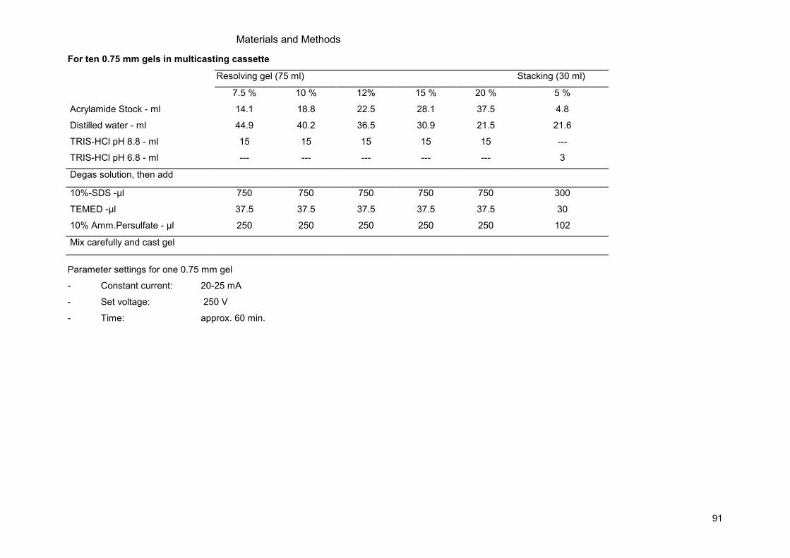
Materials and Methods
91
For ten 0.75 mm gels in multicasting cassette
Parameter settings for one 0.75 mm gel
- Constant current: 20-25 mA
- Set voltage: 250 V
- Time: approx. 60 min.
Resolving gel (75 ml) Stacking (30 ml)
7.5 % 10 % 12% 15 % 20 % 5 %
Acrylamide Stock - ml 14.1 18.8 22.5 28.1 37.5 4.8
Distilled water - ml 44.9 40.2 36.5 30.9 21.5 21.6
TRIS-HCl pH 8.8 - ml 15 15 15 15 15 ---
TRIS-HCl pH 6.8 - ml --- --- --- --- --- 3
Degas solution, then add
10%-SDS -µl 750 750 750 750 750 300
TEMED -µl 37.5 37.5 37.5 37.5 37.5 30
10% Amm.Persulfate - µl 250 250 250 250 250 102
Mix carefully and cast gel
Materials and Methods
92
Electrophoretic Transfer / Semi-dry blotting
Material and solution:
- Transfer buffer (25 mM Tris, 192 mM Glycine, 20% methanol, pH 8.3)
- Immobilon FL (Millipore, IPFL00010), Immobilon P PVDF (Millipore, IPVH00010) cutted to gel
size (9x6.5 cm)
- For one gel 6x whatman paper (9x6.5 cm)
- Semidry blotting device
- Methanol
Procedure:
- Label and activate membrane in methanol for 1 min.
- Equilibrate in Transfer buffer.
- Mount SDS-PAGE/membrane sandwich.
- Blot proteins to the membrane with constant current of approx. 10 mA per cm2 for 75 min
(0.75mm gel) or 120 min. (1.5 mm gel).
- Disassemble sandwich, incubate in blocking buffer for 30 min.
- Add 1st antidbody (see antibody table below for their dilution, blocking buffer, time).
- Wash 3x 5 min. with TBS, 0.1%Tween
- Add 2nd
antibody (HRP) for 1 hour
- Wash 3x 5 min. with TBS, 0.1%Tween
- Detection: either enhanced chemoluminiscence (ECL)
8.1.5 Microscopy
Preparation of mounting solution:
Material:
- 50 mM Tris-HCl, pH7.5-8.0
- Mowiol (Plüss-Staufer, Oftringen, Hoechst 4-88/cmB339161)
- Glycerol (Fluka, 49770)
- n-Propylgallat (Sigma, P3130)
Procedure:
- Stir 5 g Mowiol in 20 ml Tris-HCl buffer (0.05M, pH7.5-8.0) on a magnet stirrer (30-40°C, 16h).
- Add 10 ml 100% Glycerol and stir 16 h.
- Spin 10 min. at 1500 rpm at room temperature.
- Add 10 mg/ ml n-propylgallat as anti-fading agent.
- Aliquot the Mowiol solution
- Spin aliquots to remove air bubbles, store at -20°C.
- Thaw the aliquot at least 30 min. before usage at 37°C
Materials and Methods
93
Preparation of 10% para-formaldehyde in PBS (10% PFA/PBS):
Material:
- Weigh 100 g PFA to 400 ml water.
- Boil in a waterbath under the extractor hood
- Shake from time to time.
- Add cautiously drops of 5N NaOH to completely dissolve the PFA (PFA is not soluble at that pH
without NaOH).
- After complete solution of the PFA add 100 ml 10x PBS.
- Fill up to 1 liter with water.
- Solution can be further diluted to 4% PFA/PBS. Store at 4°C
Preparation of adherent cells:
Material
- PBS
- 4% para-formaldehyde in PBS pH = 8.0
- Mounting solution, Mowiol, see above
- Hoechst 33342 (Molecular Probes, H-1399), cell permeable
- 12- well, or 24- well plate
- 18 mm or 12 mm diameter coverslips
- Microscopy slides
- ddH2O
Procedure:
- Grow cells on sterile coverslips in a 12 well or 24-well plate
- Transfect according to experiment
- Perform your experiment of interest
- Wash cells twice with PBS, aspirate PBS, add ice cold 4% PFA in PBS.
- Incubate on ice for 30 min.
- Wash with PBS.
- Optional nuclear staining: add PBS with 1:1000 Hoechst 33342 (stock solution 1 mM in ddH2O),
incubate (15 min., 4°C, in the dark).
Preparation of the microscopy slides:
- Put one drop of mounting solution onto a microscopy slide
- Take the coverslip with fine forceps and wash with H2O, quickly.
- Soak residual liquid from the edge of the coverslip with a fine paper towel.
- Place coverslip upside down onto the drop of mounting solution on the microscopy slide.
- Dry for min. 30 min. at room temperature in the dark.
- Then store at 4°C in the dark.
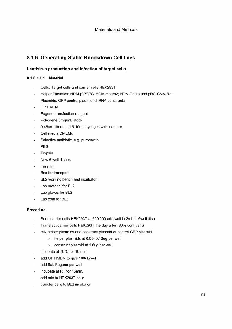
Materials and Methods
94
8.1.6 Generating Stable Knockdown Cell lines
Lentivirus production and infection of target cells 8.1.6.1.1.1 Material
- Cells: Target cells and carrier cells HEK293T
- Helper Plasmids: HDM-pVSV/G; HDM-Hpgm2; HDM-Tat1b and pRC-CMV-RaII
- Plasmids: GFP control plasmid; shRNA constructs
- OPTIMEM
- Fugene transfection reagent
- Polybrene 3mg/mL stock
- 0.45um filters and 5-10mL syringes with luer lock
- Cell media DMEMc
- Selective antibiotic, e.g. puromycin
- PBS
- Trypsin
- New 6 well dishes
- Parafilm
- Box for transport
- BL2 working bench and incubator
- Lab material for BL2
- Lab gloves for BL2
- Lab coat for BL2
Procedure
- Seed carrier cells HEK293T at 600’000cells/well in 2mL in 6well dish
- Transfect carrier cells HEK293T the day after (80% confluent)
- mix helper plasmids and construct plasmid or control GFP plasmid
o helper plasmids at 0.08- 0.16ug per well
o construct plasmid at 1.6ug per well
- incubate at 70°C for 10 min.
- add OPTIMEM to give 100uL/well
- add 8uL Fugene per well
- incubate at RT for 15min.
- add mix to HEK293T cells
- transfer cells to BL2 incubator
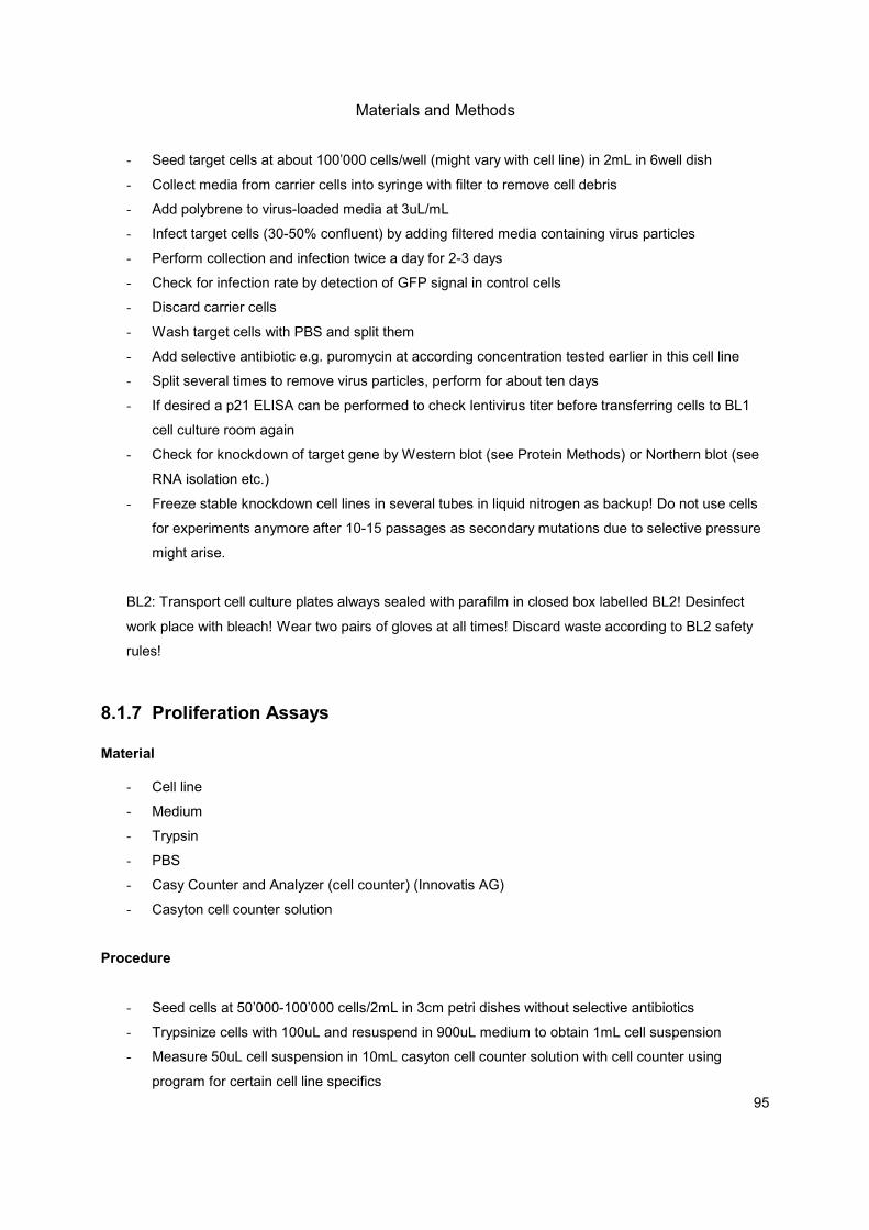
Materials and Methods
95
- Seed target cells at about 100’000 cells/well (might vary with cell line) in 2mL in 6well dish
- Collect media from carrier cells into syringe with filter to remove cell debris
- Add polybrene to virus-loaded media at 3uL/mL
- Infect target cells (30-50% confluent) by adding filtered media containing virus particles
- Perform collection and infection twice a day for 2-3 days
- Check for infection rate by detection of GFP signal in control cells
- Discard carrier cells
- Wash target cells with PBS and split them
- Add selective antibiotic e.g. puromycin at according concentration tested earlier in this cell line
- Split several times to remove virus particles, perform for about ten days
- If desired a p21 ELISA can be performed to check lentivirus titer before transferring cells to BL1
cell culture room again
- Check for knockdown of target gene by Western blot (see Protein Methods) or Northern blot (see
RNA isolation etc.)
- Freeze stable knockdown cell lines in several tubes in liquid nitrogen as backup! Do not use cells
for experiments anymore after 10-15 passages as secondary mutations due to selective pressure
might arise.
BL2: Transport cell culture plates always sealed with parafilm in closed box labelled BL2! Desinfect
work place with bleach! Wear two pairs of gloves at all times! Discard waste according to BL2 safety
rules!
8.1.7 Proliferation Assays
Material
- Cell line
- Medium
- Trypsin
- PBS
- Casy Counter and Analyzer (cell counter) (Innovatis AG)
- Casyton cell counter solution
Procedure
- Seed cells at 50’000-100’000 cells/2mL in 3cm petri dishes without selective antibiotics
- Trypsinize cells with 100uL and resuspend in 900uL medium to obtain 1mL cell suspension
- Measure 50uL cell suspension in 10mL casyton cell counter solution with cell counter using
program for certain cell line specifics

Materials and Methods
96
- Do measurements in triplicates, repeat experiment several times
- Check confluency and health of cell cultures with binocular microscope, take pictures daily
- Measure cell number daily over 3-7 days
8.1.8 Cell Size Assays
- perform similar to proliferation assays
- measure cell size, eventually normalize to nuclear volume
- normalization to nuclear volume:
o To a second batch, resuspend cells in casyton solution containing 0.5% triton X-100 to
obtain nuclear volume for normalization if necessary
- measure cell size/volume daily over 3 days
8.2 Consumables Restriction enzymes were purchased from New England Biolabs. Chemicals were from Sigma-Aldrich if not stated differently.
Materials and Methods
97
Product Supplier Cat. No.
2- Mercaptoethanol Sigma M 7522
AccuGel 40% National Diagnostics EC-852
Agar Technical DIFCO/BD 281230
Agarose, standard Eurobio 018054
Ammonium persulfate (APS) Bio-RAD 161-0700
Ampicillin trihydrate Sigma 10045
Chloroform Fluka 25690
Dimethyl sulfoxide Sigma 154938
dNTP set PCR grade Invitrogen 10297-018
Dulbecco’s MEM medium Bioconcept 1-26F01
ECL Western Blotting Detection Amersham RPN2106
Endofree plasmid maxi kit Qiagen 12362
GenElute Gel Extraction Kit Sigma NA1111
Genelute high performance endotoxin-free Sigma NA 0410
GenElute Plasmid Miniprep Sigma PLN-350
Glucose D(+) Monohydrate Sigma 49159
HEPES Sigma H3375
HOECHST 33342 Juro supply H-1399
Hyperfilm ECL Amersham RPN3103
Immobilon Western substrate reagents Millipore WBKLS0500
Immobilon-P PVDF membrane Millipore IPVH00010
Jet PEI transfection reagent Polyplus Transfection 101-40
Lambda marker, l HindIII/EcoRI Labforce 1695/1
Leupeptin Alexis 260-009-M025
L-Glutamine (200mM) Sigma G7513
Luria Broth Base life technologies 127-95-027
Magnesium chloride Fluka 63068
M-MLV RTase Invitrogen 28025-013
N,N,N,N'-Tetramethyl-ethylenediamine, TEMED Bio-Rad 161-0800
Oligo(dT)15 primer Microsynth non commercial
Penicillin-Streptomycin 100x Sigma P0781
Pepstatin A Fluka 77170
Potassium chloride Fluka 60128
Pwo DNA Polymerase Roche 11644955001
RNasin Promega N2111
RPMI 1640 medium Gibco/Invitrogen 31870-025
Sodium chloride Fluka 71376
Sodium dodecyl sulfate Fluka 71729
Sodium orthovanadate Sigma S6508
Super RX film Fuji F57164052
T4 DNA ligase NEB 102378
Taq DNA Polymerase NEB 102373
Taq DNA Polymerase recombinant Invitrogen 10342-020
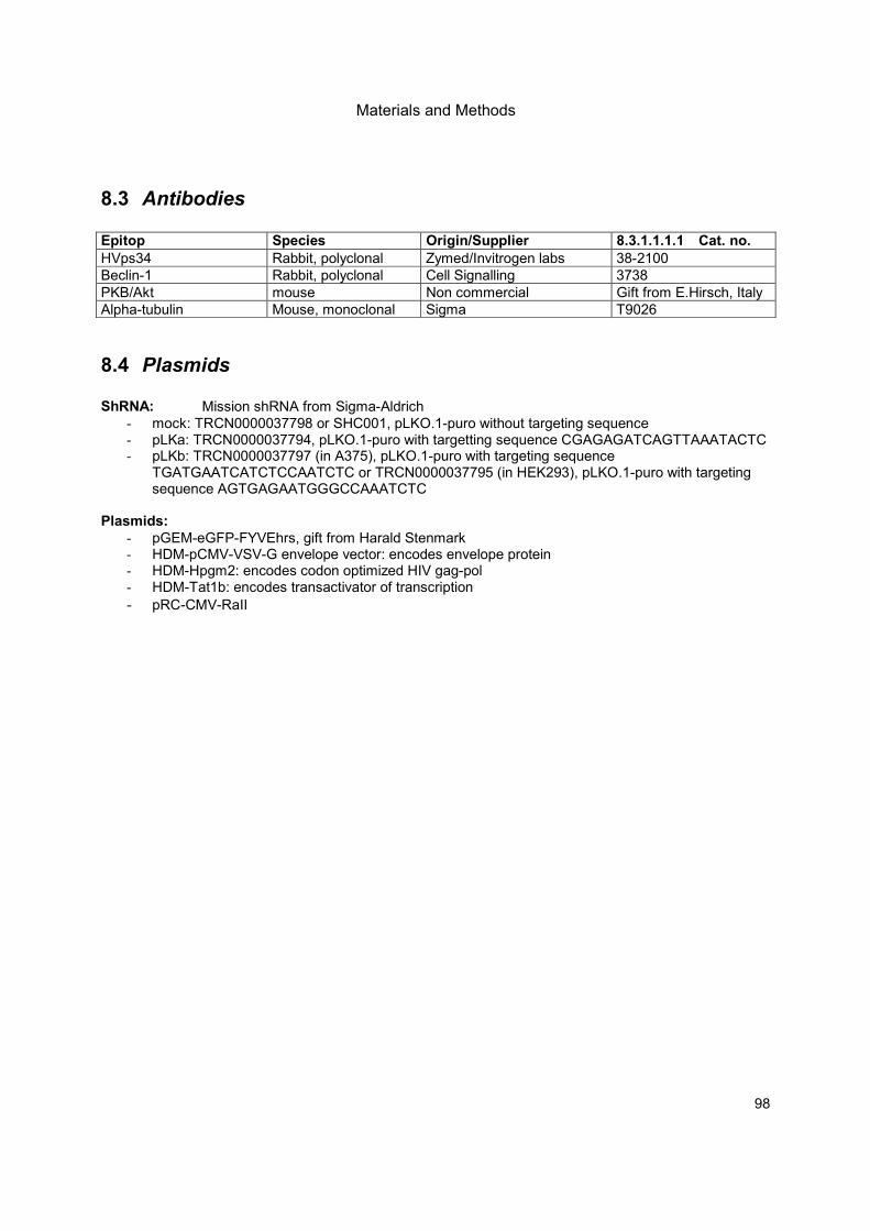
Materials and Methods
98
8.3 Antibodies Epitop Species Origin/Supplier 8.3.1.1.1.1 Cat. no.
HVps34 Rabbit, polyclonal Zymed/Invitrogen labs 38-2100
Beclin-1 Rabbit, polyclonal Cell Signalling 3738
PKB/Akt mouse Non commercial Gift from E.Hirsch, Italy
Alpha-tubulin Mouse, monoclonal Sigma T9026
8.4 Plasmids ShRNA: Mission shRNA from Sigma-Aldrich
- mock: TRCN0000037798 or SHC001, pLKO.1-puro without targeting sequence - pLKa: TRCN0000037794, pLKO.1-puro with targetting sequence CGAGAGATCAGTTAAATACTC - pLKb: TRCN0000037797 (in A375), pLKO.1-puro with targeting sequence
TGATGAATCATCTCCAATCTC or TRCN0000037795 (in HEK293), pLKO.1-puro with targeting sequence AGTGAGAATGGGCCAAATCTC
Plasmids:
- pGEM-eGFP-FYVEhrs, gift from Harald Stenmark - HDM-pCMV-VSV-G envelope vector: encodes envelope protein - HDM-Hpgm2: encodes codon optimized HIV gag-pol - HDM-Tat1b: encodes transactivator of transcription
- pRC-CMV-RaII
Abbreviations
99
9 Abbreviations
3-MA 3-methyladenine
AA amino acids
AD Alzheimer's Disease
Akt/PKB protein kinase B
Arg arginine
BCR homology domain breakpoint cluster region homology domain
Ca calcium
CaM calmodulin
COP 1 coat protein complex 1
CORVET class C core vacuole/endosome tethering
CPY carboxypeptidase Y
C-term carboxy-terminus
Cvt cytoplasm-to-vacuole-transport
DsRNA double-stranded RNA
EEA essential amino acids
EGF epidermal growth factor
EGTA calcium chelator
ER endoplasmatic reticulum
ESCRT endosomal sorting complex required for transport
FYVE domain PI(3)P binding domain
GAP GTPase activating protein
GDP guanosine diphosphate
GEF guanine nucleotide exchange factor
GFP green fluorescent protein
Glu glutamine
GPCR G-protein coupled receptor
GTP guanosine triphosphate
GTPase molecular switch either bound by GDP or GTP
HEK293 human embryonic kidney cell line
His histidine
HOPS homotypic vacuole fusion and protein sorting complex
IFN-gamma interferon gamma
IRG immunity related GTPase
KD kinase dead
kDa kilo Dalden
Leu leucine
LiAc lithium acetate
LPA lysophosphatidic acid
Lys lysine
MDC mono-dansyl cadaverine
Abbreviations
100
MEF mouse embryonic fibroblasts
mL milliliters
mM millimolar
mut mutated
MVB multivesicular body
nM nanomolar
N-term amino terminus
PDGF platelet-derived growth factor
PH domain pleckstrin homology domain
PI phosphoinositides
PI(3,4,5)P3 " " with three phosphate groups added
PI3K phosphoinositide-3 kinases
PI(3)P PI with one phosphate group added
PI-4,5P2 PI with two phosphate groups added
PI-4P PI with one phosphate group added
PKC protein kinase C
PM plasma membrane
PTEN phosphatase and tensin homolog
PX domain phox domain
RBD Ras binding domain
RGP radial growth phase
Rheb Ras homolog enriched in brain
ROS reactive oxygen species
RT room temperature
S6K RPS-6 kinase
SD synthetic yeast medium, selective
Ser serine
SH2 domain src homology domain 2
SH3 domain src homology domain 3
ShRNA short hairpin RNA
SiRNA small interfering RNA
Tg Thapsigargine
TGN trans-Golgi network
Thr threonine
TNF-alpha tumor-necrosis factor alpha
Tor target of rapamycin
TORC1 " " complex 1
TORC2 " " complex 2
TSC1/2 tuberous sclerosis complex 1/2
uL microliters
uM micromolars
UPR unfolded protein response
VEGF vascular endothelial growth factor

Abbreviations
101
VGP vertical growth phase
Vps vacuolar protein sorting
WD repeats beta-transducin repeats, usually WD 40
Wm wortmannin
WT wild type
YPD yeast full medium

Abbreviations
102

Acknowledgments
103
10 Acknowledgments First I would like to thank… … Matthias for giving me the chance to perform my PhD studies in his lab. When I had to abruptly stop the bench work, you still gave me the opportunity to write up my thesis at home and have the defense a bit later than originally planned. Merci for your understanding! Many thanks go to… … the present and former members of the institute who helped me along my way. Lab work was tough and frustrating at times, but your scientific advice and collegiality always brought me back my motivation and smiles! … my parents who always believe in me. Moral support is most important for graduate students! My deepest gratitude… … I would like to express towards my husband, Akos. Your love and support is priceless! Last but not least… … I would like to thank my little daughter, Eva. Your strength and will to live amazed me from the start, you are the biggest source of motivation. I can no longer imagine my world without your smiles! “Scio me nihil scire.” Ich weiss, dass ich nichts weiss. (Socrate, 469-399 BC)

Acknowledgments
104

Curriculum Vitae
105
11 Curriculum Vitae
Ann Christine Mertz Biro Date of Birth: 07.10.1982 Place of Birth: Basel, Switzerland Marital Status: married, 1 child, born 23.07.2010 Nationality: Swiss Address:
Waldenburgerstrasse 16 CH-4052 Basel Tel. +41 61 312 99 81 (Private) E-Mail: [email protected]
Education 06/2006 - today PhD studies
in the laboratory of Prof. Matthias P. Wymann at Center for Biomedicine, Department of Biomedicine, University of Basel, Switzerland “Class III PI3K in Melanoma”
09/2004 – 11/2005 University of Basel (Biozentrum) Masters degree of Science (MSc) in Molecular Biology, Major in Biochemistry (Lab of Prof. Michael N. Hall) Degree: cum laude “Pib2p, a novel vacuolar membrane and endosome localized FYVE protein, mediates exocytosis of the general amino acid permease in yeast, Gap1p” under supervision of Dr. Stephen B. Helliwell,
Biochemistry Department, Biozentrum 10/2001 – 09/2004 University of Basel (Biozentrum)
Bachelor degree of Science (BSc) in Molecular Biology Disciplines: Biochemistry, Biophysics and Structural Biology, Microbiology, Immunology, Cell Biology, Neurobiology and Medical Biology
12/2000-12/2005 Student job at the English Bookstore “Bider and Tanner”, Basel 08/1996 – 06/2000 Gymnasium BZB to obtain Latin-Matura (Eidg. Matura)
Scholarships 08/1996 – 06/2000 Scholarship for highly gifted children of the Kanton Baselland

Curriculum Vitae
106
Research Experience 06/2006 to present Research Associate
in the laboratory of Prof. Matthias P. Wymann at Center for Biomedicine, Department of Biomedicine, University of Basel, Switzerland
2008 to present Assist. Clinical Study Reports Evaluation (Pharmacology and Toxicology) SDPS Pharma Solutions, Victoria, Canada
01/2006 - 05/2006 Scientific Associate, Internship, Molecular Biology and Microbiology techniques, Novartis Institutes of Biomedical Research (NIBR), Basel
07/2001- 10/2001 Validation and introduction of new database (Reference Manager V.10) at Novartis Ophthalmics, Basel
Teaching Experience
2006 to present Tutor in “Basic Biology” for students at the University of Basel Assistant in “Enzyme kinetics course” for Medical students at University of Basel
1/2009 – 6/2009 Supervisor of Master student in Pharmacology at the University of Basel
2005 Tutor in „Applied Ethics“ course for Biology students at the University of Basel
Methodological Training 2/2007 Practical and Theoretical Laboratory animal course (LTK1), Roche,
Switzerland 2002 Practical course in Medical Biology, University of Basel
Methodological Skills Yeast and bacteria:
• microscopy (fluorescence, light, indirect immunofluorescence)
• yeast genetics (incl. practical work such as mating and dissecting of yeast)
• Tandem-Affinity-Purification
• GST-fusion-protein purification
• liquid LacZ assays
• Yeast-Two-Hybrid screening
• synthetic lethality screening (in yeast)
• standard molecular biology techniques (PCR (polymerase chain reaction), Westernblotting, Co-immunoprecipitations, Cloning etc.)
• radioactively (C14
) labelled amino acids uptakes in yeast Mammalian cell lines and mice:
• mammalian cell transfections
• siRNA and shRNA (generation of stable cell lines) application in various cell lines
• lab work at BL2 level (lentiviral shRNA)
• in vitro lipid kinase assays (P32
)
• various in vitro recombinant kinase assay (KinaseGlo, Adapta etc.)
• inhibitor screening of natural compounds in mammalian and yeast cell systems, assay design
• TR-FRET (Time-resolved Fluorescence-Fluorescence Resonance Energy Transfer)
• genotyping of mice strains (by PCR)
IT Skills MS Windows, MS Office (Word, Excel, Power Point), basic MacOS, Reference Manager V.10 or EndNotes, Adobe Photoshop/Illustrator, PubMed
Language Skills German mother tongue English Proficient user, understanding, reading, oral and written production C2
Level within the European Framework of Reference 2006: TOEFL score within top 4 % (653 of 677 points) Travelling through Canada, USA, New Zealand
Curriculum Vitae
107
1996: English Course in Cambridge, UK 1985-1986: Preschool in New Jersey, USA
French Independent user, understanding and reading at B2 Level within the European Framework of Reference, oral and written production B1 Level
Latin Latin-Matura
Memberships Schweizerische Gesellschaft für Zellbiologie, Molekularbiologie und Genetik (ZMG, Swiss Society for Cell Biology, Molecular Biology and Genetics, member of USGEB)
References Supervisor during PhD studies: Matthias P. Wymann PhD Institute of Biochemistry and Genetics Department of Biomedicine University of Basel Mattenstrasse 28 CH-4058 Basel Switzerland phone +41 61 695 30 46 [email protected]
Supervisor during Masters studies: Stephen B. Helliwell PhD Novartis Institutes for BioMedical Research CH-4057 Basel Switzerland phone +41 61 696 31 67 fax +41 61 696 31 70 [email protected]
Supervisor during Internship at Novartis: Dominic Hoepfner PhD Novartis Institutes for BioMedical Research CH-4057 Basel Switzerland phone +41 61 696 31 65 fax +41 61 696 31 70 [email protected]
Supervisor at SDPS Pharma Solutions Beat P. Mertz PhD 89 Howe St V8V 4K2 Victoria BC Canada phone +1 250 384 51 40 [email protected]
Peer-Reviewed Articles Sabine Kiefer, Ann C Mertz, A. Koryakina, M. Hamburger, P. Küenzi. „(E,Z)-3-(3’,5’-dimethoxy-4’-
hydroxy-benzylidene)-2-indolinone (Indolinone) blocks mast cell degranulation“, published in European Journal of Pharmaceutical Sciences, 2010
Sabine Kiefer, J. Rogger, Anna Melone, Ann C Mertz, A. Koryakina, P. Küenzi. “Separation and detection
of all phosphoinositide isomers by ESI-MS”, published in Journal of Pharmaceutical and Biomedical Analysis, 2010
Romina Marone, Dominik Erhart, Ann C Mertz, Thomas Bohnacker, Christian Schnell, Vladimir
Cmiljanovic, Frédéric Stauffer, Carlos Garcia-Echeverria, Bernd Giese, Sauveur-Michel Maira, Matthias P. Wymann. “Targeting Melanoma with Dual PI3K/mTOR Inhibitors”, published in Molecular Cancer Research, 2009
Articles in Preparation Anna Melone, Ann C Mertz, Matthias P. Wymann. “Regulation of nutrient sensing by cellular PI(3)P and
endomembrane localization of mTORC1”, in preparation Ann C Mertz, Anna Melone, Peter Küenzi, Sabine Kiefer, Michael Adams, M. Hamburger, Matthias P.
Wymann. “Use of transgenic yeast to discover mammalian PI-3-kinase isoform inhibitors of natural origin”, in preparation
Ann C Mertz, Danilo Ritz, Matthias Peter, Mike Tyers, Stephen B. Helliwell. „Pib2p, a novel endosome
and vacuolar membrane localized FYVE protein, mediates exocytosis of the general amino acid permease, Gap1p” submitted Dec 2010
Presentation
Ann C Mertz, Peter Küenzi, Sabine Kiefer, Matthias P. Wymann. „Using transgenic yeast to uncover functions of mammalian PI-3-kinase isoforms”, at MAIN students’ meeting, 5/2007
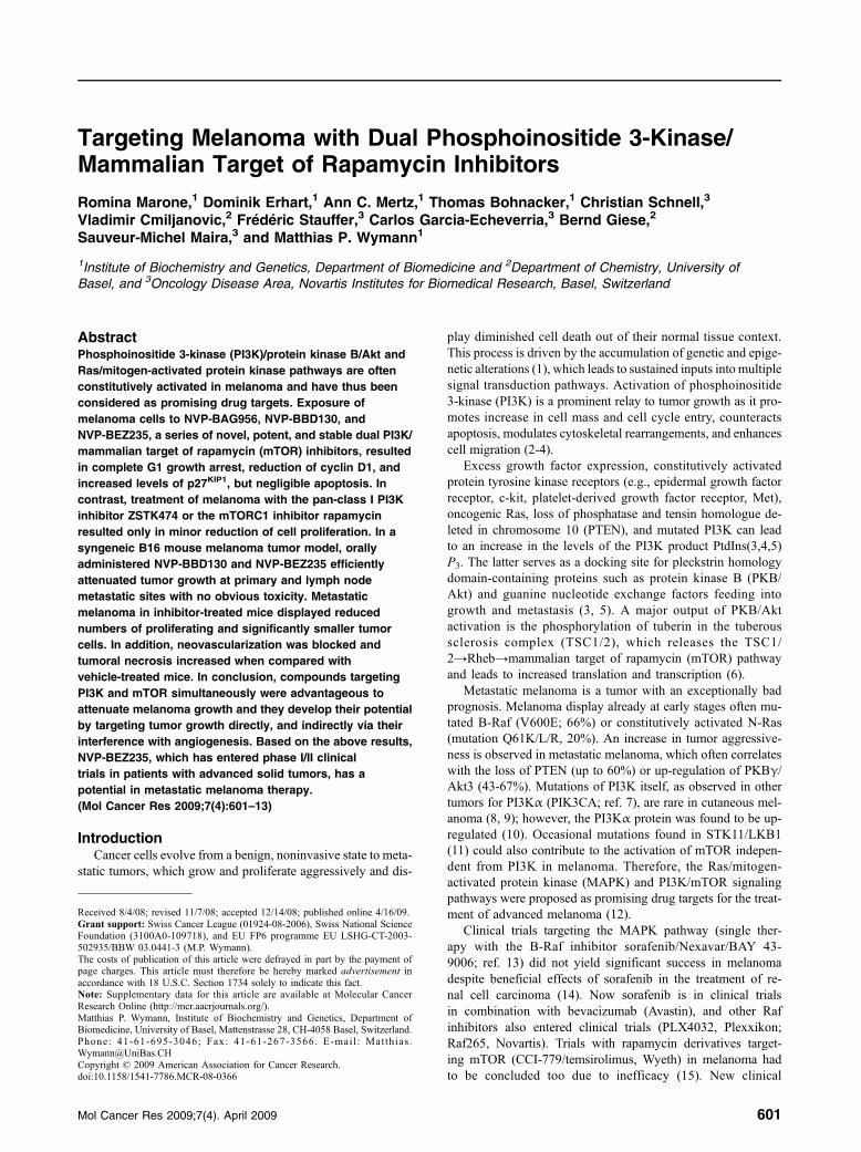
Targeting Melanoma with Dual Phosphoinositide 3-Kinase/Mammalian Target of Rapamycin Inhibitors
Romina Marone,1 Dominik Erhart,1 Ann C. Mertz,1 Thomas Bohnacker,1 Christian Schnell,3
Vladimir Cmiljanovic,2 Frédéric Stauffer,3 Carlos Garcia-Echeverria,3 Bernd Giese,2
Sauveur-Michel Maira,3 and Matthias P. Wymann1
1Institute of Biochemistry and Genetics, Department of Biomedicine and 2Department of Chemistry, University of
Basel, and 3Oncology Disease Area, Novartis Institutes for Biomedical Research, Basel, Switzerland
AbstractPhosphoinositide 3-kinase (PI3K)/protein kinase B/Akt and
Ras/mitogen-activated protein kinase pathways are often
constitutively activated in melanoma and have thus been
considered as promising drug targets. Exposure of
melanoma cells to NVP-BAG956, NVP-BBD130, and
NVP-BEZ235, a series of novel, potent, and stable dual PI3K/
mammalian target of rapamycin (mTOR) inhibitors, resulted
in complete G1 growth arrest, reduction of cyclin D1, and
increased levels of p27KIP1, but negligible apoptosis. In
contrast, treatment of melanoma with the pan-class I PI3K
inhibitor ZSTK474 or the mTORC1 inhibitor rapamycin
resulted only in minor reduction of cell proliferation. In a
syngeneic B16 mouse melanoma tumor model, orally
administered NVP-BBD130 and NVP-BEZ235 efficiently
attenuated tumor growth at primary and lymph node
metastatic sites with no obvious toxicity. Metastatic
melanoma in inhibitor-treated mice displayed reduced
numbers of proliferating and significantly smaller tumor
cells. In addition, neovascularization was blocked and
tumoral necrosis increased when compared with
vehicle-treated mice. In conclusion, compounds targeting
PI3K and mTOR simultaneously were advantageous to
attenuate melanoma growth and they develop their potential
by targeting tumor growth directly, and indirectly via their
interference with angiogenesis. Based on the above results,
NVP-BEZ235, which has entered phase I/II clinical
trials in patients with advanced solid tumors, has a
potential in metastatic melanoma therapy.
(Mol Cancer Res 2009;7(4):601–13)
IntroductionCancer cells evolve from a benign, noninvasive state to meta-
static tumors, which grow and proliferate aggressively and dis-
play diminished cell death out of their normal tissue context.
This process is driven by the accumulation of genetic and epige-
netic alterations (1), which leads to sustained inputs into multiple
signal transduction pathways. Activation of phosphoinositide
3-kinase (PI3K) is a prominent relay to tumor growth as it pro-
motes increase in cell mass and cell cycle entry, counteracts
apoptosis, modulates cytoskeletal rearrangements, and enhances
cell migration (2-4).
Excess growth factor expression, constitutively activated
protein tyrosine kinase receptors (e.g., epidermal growth factor
receptor, c-kit, platelet-derived growth factor receptor, Met),
oncogenic Ras, loss of phosphatase and tensin homologue de-
leted in chromosome 10 (PTEN), and mutated PI3K can lead
to an increase in the levels of the PI3K product PtdIns(3,4,5)
P3. The latter serves as a docking site for pleckstrin homology
domain-containing proteins such as protein kinase B (PKB/
Akt) and guanine nucleotide exchange factors feeding into
growth and metastasis (3, 5). A major output of PKB/Akt
activation is the phosphorylation of tuberin in the tuberous
sclerosis complex (TSC1/2), which releases the TSC1/
2→Rheb→mammalian target of rapamycin (mTOR) pathway
and leads to increased translation and transcription (6).
Metastatic melanoma is a tumor with an exceptionally bad
prognosis. Melanoma display already at early stages often mu-
tated B-Raf (V600E; 66%) or constitutively activated N-Ras
(mutation Q61K/L/R, 20%). An increase in tumor aggressive-
ness is observed in metastatic melanoma, which often correlates
with the loss of PTEN (up to 60%) or up-regulation of PKBγ/
Akt3 (43-67%). Mutations of PI3K itself, as observed in other
tumors for PI3Kα (PIK3CA; ref. 7), are rare in cutaneous mel-
anoma (8, 9); however, the PI3Kα protein was found to be up-
regulated (10). Occasional mutations found in STK11/LKB1
(11) could also contribute to the activation of mTOR indepen-
dent from PI3K in melanoma. Therefore, the Ras/mitogen-
activated protein kinase (MAPK) and PI3K/mTOR signaling
pathways were proposed as promising drug targets for the treat-
ment of advanced melanoma (12).
Clinical trials targeting the MAPK pathway (single ther-
apy with the B-Raf inhibitor sorafenib/Nexavar/BAY 43-
9006; ref. 13) did not yield significant success in melanoma
despite beneficial effects of sorafenib in the treatment of re-
nal cell carcinoma (14). Now sorafenib is in clinical trials
in combination with bevacizumab (Avastin), and other Raf
inhibitors also entered clinical trials (PLX4032, Plexxikon;
Raf265, Novartis). Trials with rapamycin derivatives target-
ing mTOR (CCI-779/temsirolimus, Wyeth) in melanoma had
to be concluded too due to inefficacy (15). New clinical
Received 8/4/08; revised 11/7/08; accepted 12/14/08; published online 4/16/09.
Grant support: Swiss Cancer League (01924-08-2006), Swiss National ScienceFoundation (3100A0-109718), and EU FP6 programme EU LSHG-CT-2003-
502935/BBW 03.0441-3 (M.P. Wymann).
The costs of publication of this article were defrayed in part by the payment ofpage charges. This article must therefore be hereby marked advertisement in
accordance with 18 U.S.C. Section 1734 solely to indicate this fact.Note: Supplementary data for this article are available at Molecular Cancer
Research Online (http://mcr.aacrjournals.org/).
Matthias P. Wymann, Institute of Biochemistry and Genetics, Department ofBiomedicine, University of Basel, Mattenstrasse 28, CH-4058 Basel, Switzerland.
Phone: 41-61-695-3046; Fax: 41-61-267-3566. E-mai l : Matthias [email protected]
Copyright © 2009 American Association for Cancer Research.doi:10.1158/1541-7786.MCR-08-0366
Mol Cancer Res 2009;7(4). April 2009 601
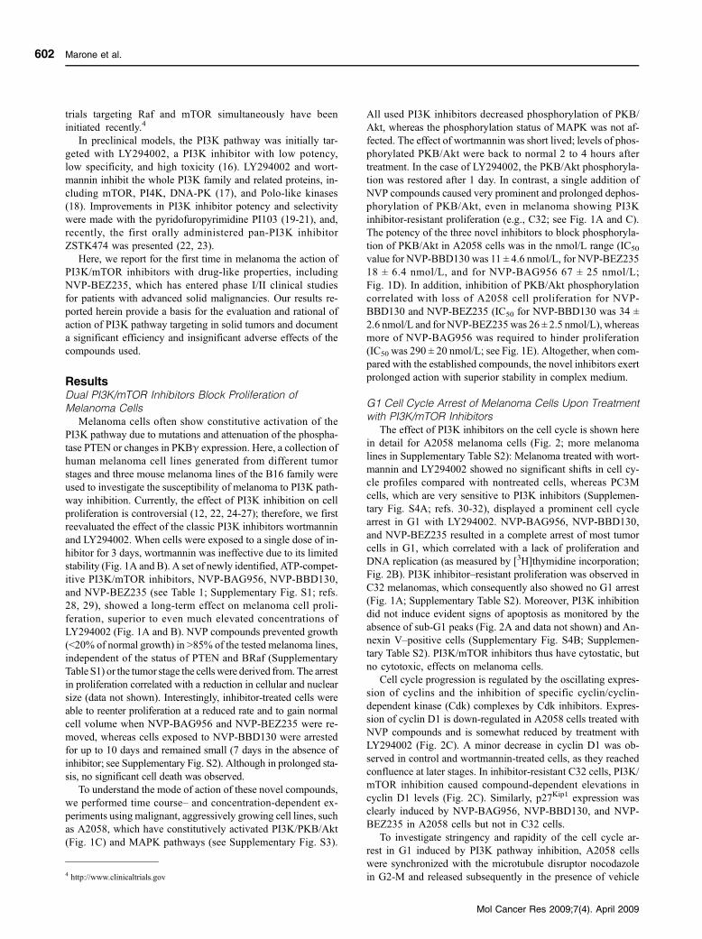
trials targeting Raf and mTOR simultaneously have been
initiated recently.4
In preclinical models, the PI3K pathway was initially tar-
geted with LY294002, a PI3K inhibitor with low potency,
low specificity, and high toxicity (16). LY294002 and wort-
mannin inhibit the whole PI3K family and related proteins, in-
cluding mTOR, PI4K, DNA-PK (17), and Polo-like kinases
(18). Improvements in PI3K inhibitor potency and selectivity
were made with the pyridofuropyrimidine PI103 (19-21), and,
recently, the first orally administered pan-PI3K inhibitor
ZSTK474 was presented (22, 23).
Here, we report for the first time in melanoma the action of
PI3K/mTOR inhibitors with drug-like properties, including
NVP-BEZ235, which has entered phase I/II clinical studies
for patients with advanced solid malignancies. Our results re-
ported herein provide a basis for the evaluation and rational of
action of PI3K pathway targeting in solid tumors and document
a significant efficiency and insignificant adverse effects of the
compounds used.
ResultsDual PI3K/mTOR Inhibitors Block Proliferation of
Melanoma Cells
Melanoma cells often show constitutive activation of the
PI3K pathway due to mutations and attenuation of the phospha-
tase PTEN or changes in PKBγ expression. Here, a collection of
human melanoma cell lines generated from different tumor
stages and three mouse melanoma lines of the B16 family were
used to investigate the susceptibility of melanoma to PI3K path-
way inhibition. Currently, the effect of PI3K inhibition on cell
proliferation is controversial (12, 22, 24-27); therefore, we first
reevaluated the effect of the classic PI3K inhibitors wortmannin
and LY294002. When cells were exposed to a single dose of in-
hibitor for 3 days, wortmannin was ineffective due to its limited
stability (Fig. 1A and B). A set of newly identified, ATP-compet-
itive PI3K/mTOR inhibitors, NVP-BAG956, NVP-BBD130,
and NVP-BEZ235 (see Table 1; Supplementary Fig. S1; refs.
28, 29), showed a long-term effect on melanoma cell proli-
feration, superior to even much elevated concentrations of
LY294002 (Fig. 1A and B). NVP compounds prevented growth
(<20% of normal growth) in >85% of the tested melanoma lines,
independent of the status of PTEN and BRaf (Supplementary
Table S1) or the tumor stage the cells were derived from. The arrest
in proliferation correlated with a reduction in cellular and nuclear
size (data not shown). Interestingly, inhibitor-treated cells were
able to reenter proliferation at a reduced rate and to gain normal
cell volume when NVP-BAG956 and NVP-BEZ235 were re-
moved, whereas cells exposed to NVP-BBD130 were arrested
for up to 10 days and remained small (7 days in the absence of
inhibitor; see Supplementary Fig. S2). Although in prolonged sta-
sis, no significant cell death was observed.
To understand the mode of action of these novel compounds,
we performed time course– and concentration-dependent ex-
periments using malignant, aggressively growing cell lines, such
as A2058, which have constitutively activated PI3K/PKB/Akt
(Fig. 1C) and MAPK pathways (see Supplementary Fig. S3).
All used PI3K inhibitors decreased phosphorylation of PKB/
Akt, whereas the phosphorylation status of MAPK was not af-
fected. The effect of wortmannin was short lived; levels of phos-
phorylated PKB/Akt were back to normal 2 to 4 hours after
treatment. In the case of LY294002, the PKB/Akt phosphoryla-
tion was restored after 1 day. In contrast, a single addition of
NVP compounds caused very prominent and prolonged dephos-
phorylation of PKB/Akt, even in melanoma showing PI3K
inhibitor-resistant proliferation (e.g., C32; see Fig. 1A and C).
The potency of the three novel inhibitors to block phosphoryla-
tion of PKB/Akt in A2058 cells was in the nmol/L range (IC50
value for NVP-BBD130 was 11 ± 4.6 nmol/L, for NVP-BEZ235
18 ± 6.4 nmol/L, and for NVP-BAG956 67 ± 25 nmol/L;
Fig. 1D). In addition, inhibition of PKB/Akt phosphorylation
correlated with loss of A2058 cell proliferation for NVP-
BBD130 and NVP-BEZ235 (IC50 for NVP-BBD130 was 34 ±
2.6 nmol/L and for NVP-BEZ235was 26 ± 2.5 nmol/L), whereas
more of NVP-BAG956 was required to hinder proliferation
(IC50was 290 ± 20 nmol/L; see Fig. 1E). Altogether, when com-
pared with the established compounds, the novel inhibitors exert
prolonged action with superior stability in complex medium.
G1 Cell Cycle Arrest of Melanoma Cells Upon Treatment
with PI3K/mTOR Inhibitors
The effect of PI3K inhibitors on the cell cycle is shown here
in detail for A2058 melanoma cells (Fig. 2; more melanoma
lines in Supplementary Table S2): Melanoma treated with wort-
mannin and LY294002 showed no significant shifts in cell cy-
cle profiles compared with nontreated cells, whereas PC3M
cells, which are very sensitive to PI3K inhibitors (Supplemen-
tary Fig. S4A; refs. 30-32), displayed a prominent cell cycle
arrest in G1 with LY294002. NVP-BAG956, NVP-BBD130,
and NVP-BEZ235 resulted in a complete arrest of most tumor
cells in G1, which correlated with a lack of proliferation and
DNA replication (as measured by [3H]thymidine incorporation;
Fig. 2B). PI3K inhibitor–resistant proliferation was observed in
C32 melanomas, which consequently also showed no G1 arrest
(Fig. 1A; Supplementary Table S2). Moreover, PI3K inhibition
did not induce evident signs of apoptosis as monitored by the
absence of sub-G1 peaks (Fig. 2A and data not shown) and An-
nexin V–positive cells (Supplementary Fig. S4B; Supplemen-
tary Table S2). PI3K/mTOR inhibitors thus have cytostatic, but
no cytotoxic, effects on melanoma cells.
Cell cycle progression is regulated by the oscillating expres-
sion of cyclins and the inhibition of specific cyclin/cyclin-
dependent kinase (Cdk) complexes by Cdk inhibitors. Expres-
sion of cyclin D1 is down-regulated in A2058 cells treated with
NVP compounds and is somewhat reduced by treatment with
LY294002 (Fig. 2C). A minor decrease in cyclin D1 was ob-
served in control and wortmannin-treated cells, as they reached
confluence at later stages. In inhibitor-resistant C32 cells, PI3K/
mTOR inhibition caused compound-dependent elevations in
cyclin D1 levels (Fig. 2C). Similarly, p27Kip1 expression was
clearly induced by NVP-BAG956, NVP-BBD130, and NVP-
BEZ235 in A2058 cells but not in C32 cells.
To investigate stringency and rapidity of the cell cycle ar-
rest in G1 induced by PI3K pathway inhibition, A2058 cells
were synchronized with the microtubule disruptor nocodazole
in G2-M and released subsequently in the presence of vehicle4 http://www.clinicaltrials.gov
Marone et al.
Mol Cancer Res 2009;7(4). April 2009
602
or PI3K inhibitors: (a) the initial treatment with nocodazole
arrested cells in G2-M (from 30% to 60% of cells in G2-M;
Fig. 2D); (b) after nocodazole removal, control and wortman-
nin-treated cells showed a profile typical of proliferating cells
after 24 h, whereas cells exposed to LY294002 required
48 h to restart proliferation. In the presence of NVP-BAG956,
NVP-BBD130, or NVP-BEZ235, A2058 cells were only able
to exit G2-M and then remained in G1 (Fig. 2D).
Primary and Metastatic Tumors Require the PI3K Pathway
for Growth In vivo
To evaluate the efficacy of different PI3K/mTOR inhibitors
in vivo, we used a syngeneic B16BL6 mouse melanoma model,
in which cells, injected intradermally in both ears, rapidly prog-
ress to primary tumors and cervical lymph node metastasis.
Treatment with vehicle control, PI3K/mTOR inhibitors, and a
vascular endothelial growth factor receptor (VEGFR) protein
tyrosine kinase inhibitor (PTK787) were started 7 days follow-
ing tumor inoculation. A ∼60% reduction in the primary tumor
size was achieved with different doses and regimens of NVP-
BBD130, NVP-BEZ235 and PI103 (Fig. 3A), and PTK787
(data not shown). Moreover, mice treated with NVP-BBD130,
NVP-BEZ235, or PTK787 showed a significant reduction in
the size of the cervical lymph node metastasis (Fig. 3B;
PTK787: data not shown). For NVP-BBD130, tumor and me-
tastasis size reduction correlated with a stringent reduction in
the amount of phosphorylated PKB/Akt and p70S6K 2 hours
after the last treatment (Fig. 4A). A partial recovery to basal
level was seen after 16 hours. In addition, reduced expression
of cyclin D1 and increased levels of p27Kip1 were detected in
metastatic tissue 2 hours after dosing (Fig. 4B). Intriguingly,
treatment with PI103 at the concentration used here did not
significantly reduce the mass of lymph node metastasis
(Fig. 3B), which was in agreement with overshooting signals
for phosphorylated PKB/Akt and p70S6K in primary and meta-
static tumor tissue (Fig. 4A). As PI103 is cleared rapidly from
the tumor (half life <2 hours; see ref. 20), these elevated and
dose-dependent signals could represent a release of feedback
loops in the PI3K/mTOR signaling system.
A closer analysis of metastatic sections revealed that cell
size was decreased in tumor tissue from inhibitor-treated ani-
mals (Fig. 5A), but not in hepatocytes of the same mice. This
is encouraging, as liver cell size dynamically responds to star-
vation and the current PI3K/mTOR treatment regimen seems
not to fully mimic nutrient deprivation. Plasma levels of liver
marker enzymes and proteins were also not significantly affect-
ed by the treatment with PI3K/mTOR inhibitors (except alanine
aminotransferase; see Supplementary Table S3). Interestingly,
PI3K inhibitor–mediated reduction in cell size went along with
a decreased mitotic index (Fig. 5B). The few remaining mitotic
cells were usually located around preexisting, large blood ves-
sels. In addition to reduced cell proliferation, metastatic tissue
from mice treated with NVP-BEZ235 displayed significantly
increased necrosis levels compared with tumors from vehicle
control–treated mice (Fig. 5C).
To survive and grow, tumors with a diameter larger than
1 mm require blood vessels to be fully supplied with nutrients
and oxygen. Staining of metastatic sections for the endothelial
cell marker CD31 revealed that NVP-BBD130 and NVP-
BEZ235 completely abrogated neoangiogenesis in metastatic
tissue, whereas vehicle control–, PI103-, or PTK787-treated tu-
mors displayed a well-developed microvasculature (Fig. 6A;
quantification in Fig. 6B). Large and preexisting blood vessels
at the tumor border remained unaffected by all treatments (data
not shown). Altogether, the above results show that the novel
NVP compounds have a direct effect on tumor cell growth and
proliferation, and exert, in addition, antiangiogenic effects sup-
porting tumor necrosis.
PI3K/mTOR Inhibitors Are Well Tolerated
Tumor-bearing mice showed a disease-related drop in
body weight by 5% to 10% and had >2-fold enlarged
spleens when compared with healthy controls (Table 2).
PI3K inhibitor–treated animals showed a less dramatic in-
crease in spleen size and an overall tendency to regain body
weight, reaching significance for PTK787 and NVP-BEZ235
(data not shown). The collected data illustrate that the tumor
per se causes dramatic systemic changes, which were partial-
ly reversed by PI3K/mTOR inhibition. Interference with in-
sulin-mediated glucose uptake also seemed negligible, as
blood glucose levels did not significantly increase over con-
centrations determined in healthy and vehicle control–treated
animals (Table 2).
PI3K and mTOR have been reported to play important roles
in the immune system. Spleen and bone marrow of NVP-
BBD130– and PI103-treated mice were therefore analyzed
for adverse effects on immune cells. Tumor-bearing, vehicle
control–treated mice displayed a reduction in the number of
CD3+ T cells, B cells, and natural killer cells, compared with
healthy animals (Supplementary Table S4), and NVP-BBD130
treatment decreased lymphocyte counts in a statistically insig-
nificant manner. Interestingly, treatment with PI103 resulted in
an increase in the number of T cells, B cells, and natural killer
cells back to levels similar to healthy control animals. Granu-
locytes, macrophages, and erythroid cell numbers were normal
in the bone marrow of all tumor-bearing mice, and there was no
indication that the applied PI3K/mTOR inhibitors interfered
with hematopoiesis.
Cooperation of PI3K and mTOR in Melanoma Cell
Proliferation
To discriminate between the importance of PI3K and
mTOR, we ectopically expressed a constitutively active PKB/
Akt (myristoylated PKB, Myr-PKB; ref. 33) to partially bypass
the inhibitor-mediated block in PI3K signaling. To monitor
PKB/Akt action, we used the mTOR-independent nuclear to
cytoplasmic translocation of forkhead transcription factors
(FOXO), which feed negatively into cell cycle progression
and antiapoptotic events (34, 35). Nonphosphorylated FOXOs
are localized to the nucleus but are retained in the cytoplasm
when phosphorylated by PKB/Akt. In A2058 cells, PKB/Akt
is maintained activated in a PI3K-dependent manner even in
serum-free conditions, mainly due to the lack of the lipid phos-
phatase PTEN. As a consequence, ectopically expressed
FOXO1 is quantitatively localized in the cytoplasm. Treatment
of A2058 cells with NVP compounds caused a rapid inactiva-
tion of PKB/Akt and a subsequent translocation of FOXO1 to
the nucleus (Fig. 7A). Stable expression of Myr-PKB in A2058
PI3K/mTOR in Melanoma
Mol Cancer Res 2009;7(4). April 2009
603
was sufficient to prevent nuclear translocation of exogenous
FOXO1 in the presence of PI3K inhibitors, but did not rescue
proliferation in the presence of, e.g., NVP-BEZ235 (Fig. 7B).
In serum-deprived HEK293 cells, Myr-PKB reconstituted
phosphorylation of FOXO and GSK3β and also fed into mTOR
signaling with phosphorylated p70S6K, S6, and 4E-BP1 as read-
outs (Supplementary Fig. S5). Whereas FOXO and GSK-3β
phosphorylation was to a large extent PI3K inhibitor resistant,
signals downstream of mTOR were completely abrogated by
NVP-BAG956, NVP-BBD130, NVP-BEZ235, and PI103, as
well as by the mTORC1 inhibitor rapamycin. This corroborates
mTOR inhibition by NVP compounds observed in TSC1 knock-
out mouse embryonic fibroblasts (Table 1; see ref. 28).
Recently, a selective pan-PI3K inhibitor without activity
against mTOR (ZSTK474) was shown to block tumor cell
growth with an overall 50% sensitivity rate (22, 23). To evalu-
ate the requirement of a dual PI3K and mTOR inhibition to
target melanoma, cells were comparatively exposed to NVP-
BEZ235, ZSTK474, and/or rapamycin. The obtained results
clearly illustrate that targeting either PI3K or mTOR in isola-
tion can attenuate growth and proliferation of particularly sen-
sitive cells (e.g., A375), whereas more resistant melanoma are
only affected by the simultaneous targeting of PI3K and mTOR
(Fig. 8A). Treatment of melanoma cells with a combina-
tion of ZSTK474 and rapamycin results in a more pronounced
proliferation arrest compared with single compounds. The
FIGURE 1. PI3K inhibitors affect proliferation and downstream signaling in melanoma cells. A. Antiproliferative effect of PI3K inhibitors. Melanoma cellswere exposed to the indicated inhibitors (LY294002 at 25 μmol/L; wortmannin at 500 nmol/L; NVP-BAG956, NVP-BBD130, and NVP-BEZ235 at 1 μmol/L) for3 d before cell numbers were determined. Cell numbers are depicted after the subtraction of initially seeded cells in relation to nontreated controls (ratio oftreated over nontreated cultures). Columns, mean of triplicates; bars, SE. B. Time-dependent A2058 proliferation in the presence of PI3K inhibitors. A2058cells were exposed to PI3K inhibitors at day 0 before cells were counted at the indicated time intervals. Points, mean of triplicates; bars, SE. C. Prolongedchanges in phosphorylation of PKB/Akt upon PI3K inhibition. Phosphorylated PKB as detected in total cell lysates of A2058 and C32 cells treated with PI3Kinhibitors for the indicated intervals are shown. An extended version of the figure is presented in Supplementary Fig. S3. D. Concentration-dependent inhi-bition of PKB phosphorylation. A2058 cells were exposed for 3 h to increasing concentrations of NVP-BAG956, NVP-BBD130, and NVP-BEZ235. Total celllysates were subjected to immunoblotting for total and phosphorylated PKB or MAPK. Emerging signals were quantified using fluorescent secondary anti-bodies, and ratios of phosphosphorylated over nonphosphorylated kinases are displayed. Points, mean (n > 3); bars, SE. E. Inhibitory activity of PI3Kinhibitors against melanoma cell proliferation. A2058 cells were treated at day 0 with increasing concentrations of NVP-BAG956, NVP-BBD130, andNVP-BEZ235. Cell proliferation was evaluated 3 d later. Points, mean of triplicates; bars, SE.
Marone et al.
Mol Cancer Res 2009;7(4). April 2009
604
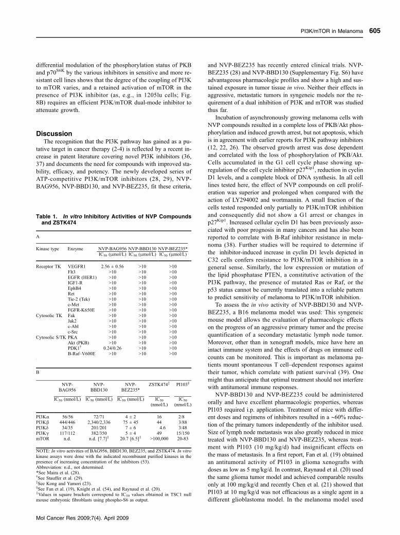
differential modulation of the phosphorylation status of PKB
and p70S6K by the various inhibitors in sensitive and more re-
sistant cell lines shows that the degree of the coupling of PI3K
to mTOR varies, and a retained activation of mTOR in the
presence of PI3K inhibitor (as, e.g., in 1205lu cells; Fig.
8B) requires an efficient PI3K/mTOR dual-mode inhibitor to
attenuate growth.
DiscussionThe recognition that the PI3K pathway has gained as a pu-
tative target in cancer therapy (2-4) is reflected by a recent in-
crease in patent literature covering novel PI3K inhibitors (36,
37) and documents the need for compounds with improved sta-
bility, efficacy, and potency. The newly developed series of
ATP-competitive PI3K/mTOR inhibitors (28, 29), NVP-
BAG956, NVP-BBD130, and NVP-BEZ235, fit these criteria,
and NVP-BEZ235 has recently entered clinical trials. NVP-
BEZ235 (28) and NVP-BBD130 (Supplementary Fig. S6) have
advantageous pharmacologic profiles and show a high and sus-
tained exposure in tumor tissue in vivo. Neither their effects in
aggressive, metastatic tumors in syngeneic models nor the re-
quirement of a dual inhibition of PI3K and mTOR was studied
thus far.
Incubation of asynchronously growing melanoma cells with
NVP compounds resulted in a complete loss of PKB/Akt phos-
phorylation and induced growth arrest, but not apoptosis, which
is in agreement with earlier reports for PI3K pathway inhibitors
(12, 22, 26). The observed growth arrest was dose dependent
and correlated with the loss of phosphorylation of PKB/Akt.
Cells accumulated in the G1 cell cycle phase showing up-
regulation of the cell cycle inhibitor p27Kip1, reduction in cyclin
D1 levels, and a complete block of DNA synthesis. In all cell
lines tested here, the effect of NVP compounds on cell prolif-
eration was superior and prolonged when compared with the
action of LY294002 and wortmannin. A small fraction of the
cells tested responded only partially to PI3K/mTOR inhibition
and consequently did not show a G1 arrest or changes in
p27Kip1. Increased cellular cyclin D1 has been previously asso-
ciated with poor prognosis in many cancers and has also been
reported to correlate with B-Raf inhibitor resistance in mela-
noma (38). Further studies will be required to determine if
the inhibitor-induced increase in cyclin D1 levels depicted in
C32 cells confers resistance to PI3K/mTOR inhibition in a
general sense. Similarly, the low expression or mutation of
the lipid phosphatase PTEN, a constitutive activation of the
PI3K pathway, the presence of mutated Ras or Raf, or the
p53 status cannot be currently translated into a reliable pattern
to predict sensitivity of melanoma to PI3K/mTOR inhibition.
To assess the in vivo activity of NVP-BBD130 and NVP-
BEZ235, a B16 melanoma model was used: This syngeneic
mouse model allows the evaluation of pharmacologic effects
on the progress of an aggressive primary tumor and the precise
quantification of a secondary metastatic lymph node tumor.
Moreover, other than in xenograft models, mice have here an
intact immune system and the effects of drugs on immune cell
counts can be monitored. This is important as melanoma pa-
tients mount spontaneous T cell–dependent responses against
their tumor, which correlate with patient survival (39). One
might thus anticipate that optimal treatment should not interfere
with antitumoral immune responses.
NVP-BBD130 and NVP-BEZ235 could be administered
orally and have excellent pharmacologic properties, whereas
PI103 required i.p. application. Treatment of mice with differ-
ent doses and regimens of inhibitors resulted in a ∼60% reduc-
tion of the primary tumors independently of the inhibitor used.
Size of lymph node metastasis was also greatly reduced in mice
treated with NVP-BBD130 and NVP-BEZ235, whereas treat-
ment with PI103 (10 mg/kg/d) had insignificant effects on
the mass of metastasis. In a first report, Fan et al. (19) obtained
an antitumoral activity of PI103 in glioma xenografts with
doses as low as 5 mg/kg/d. In contrast, Raynaud et al. (20) used
the same glioma tumor model and achieved comparable results
only at 100 mg/kg/d and recently Chen et al. (21) showed that
PI103 at 10 mg/kg/d was not efficacious as a single agent in a
different glioblastoma model. In the melanoma model used
Table 1. In vitro Inhibitory Activities of NVP Compoundsand ZSTK474
A
Kinase type Enzyme NVP-BAG956 NVP-BBD130 NVP-BEZ235*
IC50 (μmol/L) IC50 (μmol/L) IC50 (μmol/L)
Receptor TK VEGFR1 2.56 ± 0.56 >10 >10
Flt3 >10 >10 >10
EGFR (HER1) >10 >10 >10IGF1-R >10 >10 >10
EphB4 >10 >10 >10Ret >10 >10 >10
Tie-2 (Tek) >10 >10 >10c-Met >10 >10 >10
FGFR-K650E >10 >10 >10
Cytosolic TK Fak >10 >10 >10Jak2 >10 >10 >10
c-Abl >10 >10 >10c-Src >10 >10 >10
Cytosolic S/TK PKA >10 >10 >10
Akt (PKB) >10 >10 >10PDK1† 0.24/0.26 >10 >10
B-Raf–V600E >10 >10 >10CDK1 >10 >10 >10
B
NVP-BAG956
NVP-BBD130
NVP-BEZ235*
ZSTK474‡ PI103§
IC50 (nmol/L) IC50 (nmol/L) IC50 (nmol/L) IC50
(nmol/L)
IC50
(nmol/L)
PI3Kα 56/56 72/71 4 ± 2 16 2/8
PI3Kβ 444/446 2,340/2,336 75 ± 45 44 3/88PI3Kδ 34/35 201/201 7 ± 6 4.6 3/48
PI3Kγ 117/112 382/350 5 ± 4 49 15/150
mTOR n.d. n.d. [7.7]∥ 20.7 [6.5]∥ >100,000 20-83
NOTE: In vitro activities of BAG956, BBD130, BEZ235, and ZSTK474. In vitro
kinase assays were done with the indicated recombinant purified kinases in thepresence of increasing concentration of the inhibitors (53).
Abbreviation: n.d., not determined.*See Maira et al. (28).†See Stauffer et al. (29).‡See Kong and Yamori (23).§See Fan et al. (19), Knight et al. (54), and Raynaud et al. (20).∥Values in square brackets correspond to IC50 values obtained in TSC1 nullmouse embryonic fibroblasts using phospho-S6 as output.
PI3K/mTOR in Melanoma
Mol Cancer Res 2009;7(4). April 2009
605
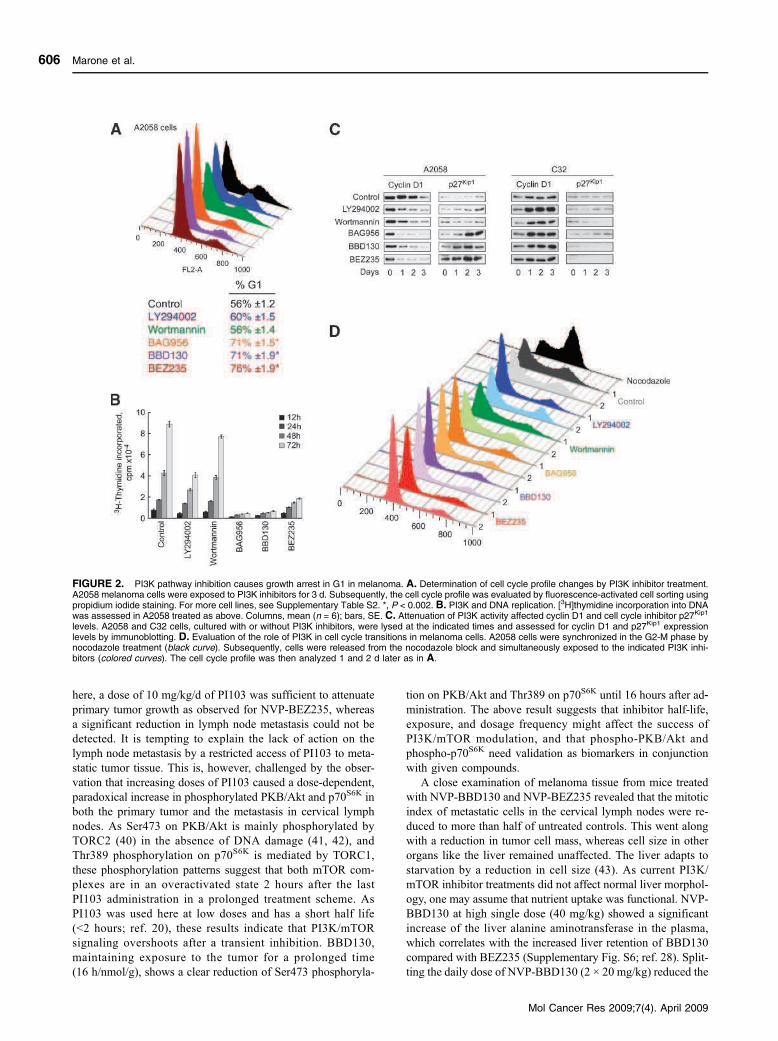
here, a dose of 10 mg/kg/d of PI103 was sufficient to attenuate
primary tumor growth as observed for NVP-BEZ235, whereas
a significant reduction in lymph node metastasis could not be
detected. It is tempting to explain the lack of action on the
lymph node metastasis by a restricted access of PI103 to meta-
static tumor tissue. This is, however, challenged by the obser-
vation that increasing doses of PI103 caused a dose-dependent,
paradoxical increase in phosphorylated PKB/Akt and p70S6K in
both the primary tumor and the metastasis in cervical lymph
nodes. As Ser473 on PKB/Akt is mainly phosphorylated by
TORC2 (40) in the absence of DNA damage (41, 42), and
Thr389 phosphorylation on p70S6K is mediated by TORC1,
these phosphorylation patterns suggest that both mTOR com-
plexes are in an overactivated state 2 hours after the last
PI103 administration in a prolonged treatment scheme. As
PI103 was used here at low doses and has a short half life
(<2 hours; ref. 20), these results indicate that PI3K/mTOR
signaling overshoots after a transient inhibition. BBD130,
maintaining exposure to the tumor for a prolonged time
(16 h/nmol/g), shows a clear reduction of Ser473 phosphoryla-
tion on PKB/Akt and Thr389 on p70S6K until 16 hours after ad‐
ministration. The above result suggests that inhibitor half-life,
exposure, and dosage frequency might affect the success of
PI3K/mTOR modulation, and that phospho-PKB/Akt and
phospho-p70S6K need validation as biomarkers in conjunction
with given compounds.
A close examination of melanoma tissue from mice treated
with NVP-BBD130 and NVP-BEZ235 revealed that the mitotic
index of metastatic cells in the cervical lymph nodes were re-
duced to more than half of untreated controls. This went along
with a reduction in tumor cell mass, whereas cell size in other
organs like the liver remained unaffected. The liver adapts to
starvation by a reduction in cell size (43). As current PI3K/
mTOR inhibitor treatments did not affect normal liver morphol-
ogy, one may assume that nutrient uptake was functional. NVP-
BBD130 at high single dose (40 mg/kg) showed a significant
increase of the liver alanine aminotransferase in the plasma,
which correlates with the increased liver retention of BBD130
compared with BEZ235 (Supplementary Fig. S6; ref. 28). Split-
ting the daily dose of NVP-BBD130 (2 × 20 mg/kg) reduced the
FIGURE 2. PI3K pathway inhibition causes growth arrest in G1 in melanoma. A. Determination of cell cycle profile changes by PI3K inhibitor treatment.A2058 melanoma cells were exposed to PI3K inhibitors for 3 d. Subsequently, the cell cycle profile was evaluated by fluorescence-activated cell sorting usingpropidium iodide staining. For more cell lines, see Supplementary Table S2. *, P < 0.002. B. PI3K and DNA replication. [3H]thymidine incorporation into DNAwas assessed in A2058 treated as above. Columns, mean (n = 6); bars, SE. C. Attenuation of PI3K activity affected cyclin D1 and cell cycle inhibitor p27Kip1
levels. A2058 and C32 cells, cultured with or without PI3K inhibitors, were lysed at the indicated times and assessed for cyclin D1 and p27Kip1 expressionlevels by immunoblotting. D. Evaluation of the role of PI3K in cell cycle transitions in melanoma cells. A2058 cells were synchronized in the G2-M phase bynocodazole treatment (black curve). Subsequently, cells were released from the nocodazole block and simultaneously exposed to the indicated PI3K inhi-bitors (colored curves). The cell cycle profile was then analyzed 1 and 2 d later as in A.
Marone et al.
Mol Cancer Res 2009;7(4). April 2009
606
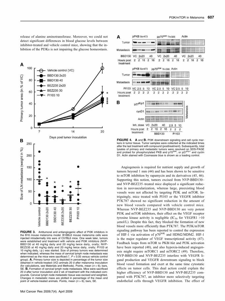
release of alanine aminotransferase. Moreover, we could not
detect significant differences in blood glucose levels between
inhibitor-treated and vehicle control mice, showing that the in-
hibition of the PI3Ks is not impairing the glucose homeostasis.
Angiogenesis is required for nutrient supply and growth of
tumors beyond 1 mm (44) and has been shown to be sensitive
to mTOR inhibition by rapamycin and its derivatives (45, 46).
Supporting this notion, tumors excised from NVP-BBD130–
and NVP-BEZ235–treated mice displayed a significant reduc-
tion in neovascularization, whereas large, preexisting blood
vessels were not affected by targeting PI3K and mTOR. In-
triguingly, mice treated with PI103 or the VEGFR inhibitor
PTK787 showed no significant reduction in the amount of
new blood vessels compared with vehicle control mice.
Whereas NVP-BEZ235 and NVP-BBD130 are very potent
PI3K and mTOR inhibitors, their effect on the VEGF receptor
tyrosine kinase activity is negligible (IC50 for VEGFR1 >10
μmol/L). Despite this fact, they blocked the formation of new
blood vessels more efficiently than PTK787. The PI3K/mTOR
signaling pathway has been reported to control the expression
of HIF-1 via activation of p70S6K and HDM2/MDM2. HIF-1
is the major regulator of VEGF transcriptional activity (47).
Feedback loops from mTOR to PKB/Akt and PI3K activation
have been reported (48), and also hypoxia-induced angiogen-
esis might require mTORC1 and mTORC2 (49). Therefore,
NVP-BBD130 and NVP-BEZ235 interfere with VEGFR li-
gand production and VEGFR downstream signaling to block
blood vessel formation and exert at the same time cytostatic
effects on tumor cells. This dual action could explain the
higher efficiency of NVP-BBD130 and NVP-BEZ235 com-
pared with PTK787, an inhibitor more selectively targeting
endothelial cells through VEGFR inhibition. The effect of
FIGURE 3. Antitumoral and antiangiogenic effect of PI3K inhibitors inthe B16 mouse melanoma model. B16BL6 mouse melanoma cells wereinjected intradermally into ears of C57BL6 mice. One week later, tumorswere established and treatment with vehicle and PI3K inhibitors (NVP-BBD130 at 40 mg/kg daily and 20 mg/kg twice daily, orally; NVP-BEZ235 at 40 mg/kg daily and 20 mg/kg twice daily, orally; PI103 at10 mg/kg daily, i.p.) was started. Size of primary tumors was determinedwhen indicated, whereas the mass of cervical lymph node metastasis wasdetermined as the mice were sacrificed (*, P < 0.05 versus vehicle controlgroup). A. Primary tumor size is depicted in percentage of the tumor sizeobtained in vehicle-treated (VC) animals 20 d after melanoma inoculation(for calculations, see Materials and Methods). Points, mean (n > 6); bars,SE. B. Formation of cervical lymph node metastasis. Mice were sacrificed20 d after tumor inoculation and 2 wk of treatment with the indicated com-pounds. Cervical lymph node metastatic tissue was excised and weighted.Changes in metastatic mass are plotted in percentage of the mean endpoint of vehicle-treated animals. Points, mean (n > 6); bars, SE.
FIGURE 4. A and B. PI3K downstream signaling and cell cycle mar-kers in tumor tissue. Tumor samples were collected at the indicated timesafter the last treatment with compound (posttreatment). Subsequently, totallysates of primary and metastatic tumors were resolved on SDS-PAGEand probed for phosphorylated PKB and p70S6K, or p27Kip1 and cyclinD1. Actin stained with Coomassie blue is shown as a loading control.
PI3K/mTOR in Melanoma
Mol Cancer Res 2009;7(4). April 2009
607
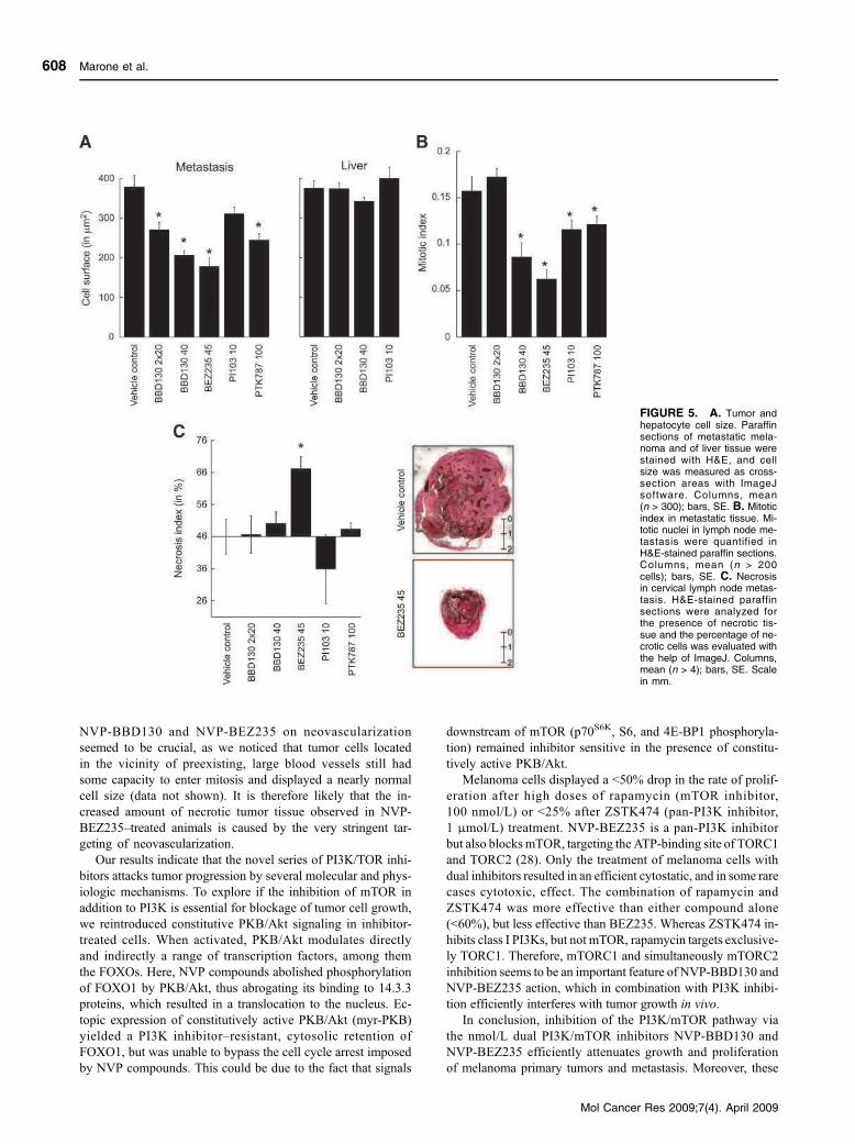
NVP-BBD130 and NVP-BEZ235 on neovascularization
seemed to be crucial, as we noticed that tumor cells located
in the vicinity of preexisting, large blood vessels still had
some capacity to enter mitosis and displayed a nearly normal
cell size (data not shown). It is therefore likely that the in-
creased amount of necrotic tumor tissue observed in NVP-
BEZ235–treated animals is caused by the very stringent tar-
geting of neovascularization.
Our results indicate that the novel series of PI3K/TOR inhi-
bitors attacks tumor progression by several molecular and phys-
iologic mechanisms. To explore if the inhibition of mTOR in
addition to PI3K is essential for blockage of tumor cell growth,
we reintroduced constitutive PKB/Akt signaling in inhibitor-
treated cells. When activated, PKB/Akt modulates directly
and indirectly a range of transcription factors, among them
the FOXOs. Here, NVP compounds abolished phosphorylation
of FOXO1 by PKB/Akt, thus abrogating its binding to 14.3.3
proteins, which resulted in a translocation to the nucleus. Ec-
topic expression of constitutively active PKB/Akt (myr-PKB)
yielded a PI3K inhibitor–resistant, cytosolic retention of
FOXO1, but was unable to bypass the cell cycle arrest imposed
by NVP compounds. This could be due to the fact that signals
downstream of mTOR (p70S6K, S6, and 4E-BP1 phosphoryla-
tion) remained inhibitor sensitive in the presence of constitu-
tively active PKB/Akt.
Melanoma cells displayed a <50% drop in the rate of prolif-
eration after high doses of rapamycin (mTOR inhibitor,
100 nmol/L) or <25% after ZSTK474 (pan-PI3K inhibitor,
1 μmol/L) treatment. NVP-BEZ235 is a pan-PI3K inhibitor
but also blocks mTOR, targeting the ATP-binding site of TORC1
and TORC2 (28). Only the treatment of melanoma cells with
dual inhibitors resulted in an efficient cytostatic, and in some rare
cases cytotoxic, effect. The combination of rapamycin and
ZSTK474 was more effective than either compound alone
(<60%), but less effective than BEZ235. Whereas ZSTK474 in-
hibits class I PI3Ks, but not mTOR, rapamycin targets exclusive-
ly TORC1. Therefore, mTORC1 and simultaneously mTORC2
inhibition seems to be an important feature of NVP-BBD130 and
NVP-BEZ235 action, which in combination with PI3K inhibi-
tion efficiently interferes with tumor growth in vivo.
In conclusion, inhibition of the PI3K/mTOR pathway via
the nmol/L dual PI3K/mTOR inhibitors NVP-BBD130 and
NVP-BEZ235 efficiently attenuates growth and proliferation
of melanoma primary tumors and metastasis. Moreover, these
FIGURE 5. A. Tumor andhepatocyte cell size. Paraffinsections of metastatic mela-noma and of liver tissue werestained with H&E, and cellsize was measured as cross-section areas with ImageJsoftware. Columns, mean(n > 300); bars, SE. B. Mitoticindex in metastatic tissue. Mi-totic nuclei in lymph node me-tastasis were quantified inH&E-stained paraffin sections.Columns, mean (n > 200cells); bars, SE. C. Necrosisin cervical lymph node metas-tasis. H&E-stained paraffinsections were analyzed forthe presence of necrotic tis-sue and the percentage of ne-crotic cells was evaluated withthe help of ImageJ. Columns,mean (n > 4); bars, SE. Scalein mm.
Marone et al.
Mol Cancer Res 2009;7(4). April 2009
608
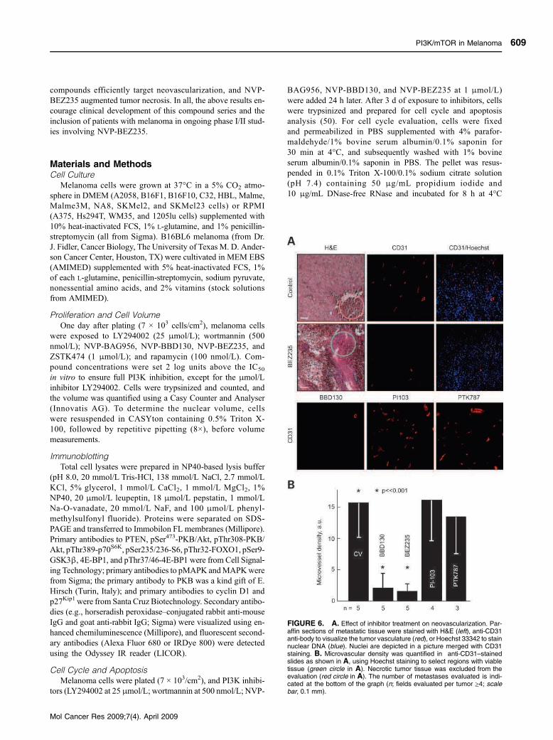
compounds efficiently target neovascularization, and NVP-
BEZ235 augmented tumor necrosis. In all, the above results en-
courage clinical development of this compound series and the
inclusion of patients with melanoma in ongoing phase I/II stud-
ies involving NVP-BEZ235.
Materials and MethodsCell Culture
Melanoma cells were grown at 37°C in a 5% CO2 atmo-
sphere in DMEM (A2058, B16F1, B16F10, C32, HBL, Malme,
Malme3M, NA8, SKMel2, and SKMel23 cells) or RPMI
(A375, Hs294T, WM35, and 1205lu cells) supplemented with
10% heat-inactivated FCS, 1% L-glutamine, and 1% penicillin-
streptomycin (all from Sigma). B16BL6 melanoma (from Dr.
J. Fidler, Cancer Biology, The University of Texas M. D. Ander-
son Cancer Center, Houston, TX) were cultivated in MEM EBS
(AMIMED) supplemented with 5% heat-inactivated FCS, 1%
of each L-glutamine, penicillin-streptomycin, sodium pyruvate,
nonessential amino acids, and 2% vitamins (stock solutions
from AMIMED).
Proliferation and Cell Volume
One day after plating (7 × 10³ cells/cm2), melanoma cells
were exposed to LY294002 (25 μmol/L); wortmannin (500
nmol/L); NVP-BAG956, NVP-BBD130, NVP-BEZ235, and
ZSTK474 (1 μmol/L); and rapamycin (100 nmol/L). Com-
pound concentrations were set 2 log units above the IC50
in vitro to ensure full PI3K inhibition, except for the μmol/L
inhibitor LY294002. Cells were trypsinized and counted, and
the volume was quantified using a Casy Counter and Analyser
(Innovatis AG). To determine the nuclear volume, cells
were resuspended in CASYton containing 0.5% Triton X-
100, followed by repetitive pipetting (8×), before volume
measurements.
Immunoblotting
Total cell lysates were prepared in NP40-based lysis buffer
(pH 8.0, 20 mmol/L Tris-HCl, 138 mmol/L NaCl, 2.7 mmol/L
KCl, 5% glycerol, 1 mmol/L CaCl2, 1 mmol/L MgCl2, 1%
NP40, 20 μmol/L leupeptin, 18 μmol/L pepstatin, 1 mmol/L
Na-O-vanadate, 20 mmol/L NaF, and 100 μmol/L phenyl-
methylsulfonyl fluoride). Proteins were separated on SDS-
PAGE and transferred to Immobilon FL membranes (Millipore).
Primary antibodies to PTEN, pSer473-PKB/Akt, pThr308-PKB/
Akt, pThr389-p70S6K, pSer235/236-S6, pThr32-FOXO1, pSer9-
GSK3β, 4E-BP1, and pThr37/46-4E-BP1 were from Cell Signal‐
ing Technology; primary antibodies to pMAPK andMAPKwere
from Sigma; the primary antibody to PKB was a kind gift of E.
Hirsch (Turin, Italy); and primary antibodies to cyclin D1 and
p27Kip1were from Santa Cruz Biotechnology. Secondary antibo-
dies (e.g., horseradish peroxidase–conjugated rabbit anti-mouse
IgG and goat anti-rabbit IgG; Sigma) were visualized using en-
hanced chemiluminescence (Millipore), and fluorescent second-
ary antibodies (Alexa Fluor 680 or IRDye 800) were detected
using the Odyssey IR reader (LICOR).
Cell Cycle and Apoptosis
Melanoma cells were plated (7 × 10³/cm2), and PI3K inhibi-
tors (LY294002 at 25 μmol/L; wortmannin at 500 nmol/L; NVP-
BAG956, NVP-BBD130, and NVP-BEZ235 at 1 μmol/L)
were added 24 h later. After 3 d of exposure to inhibitors, cells
were trypsinized and prepared for cell cycle and apoptosis
analysis (50). For cell cycle evaluation, cells were fixed
and permeabilized in PBS supplemented with 4% parafor-
maldehyde/1% bovine serum albumin/0.1% saponin for
30 min at 4°C, and subsequently washed with 1% bovine
serum albumin/0.1% saponin in PBS. The pellet was resus-
pended in 0.1% Triton X-100/0.1% sodium citrate solution
(pH 7.4) containing 50 μg/mL propidium iodide and
10 μg/mL DNase-free RNase and incubated for 8 h at 4°C
FIGURE 6. A. Effect of inhibitor treatment on neovascularization. Par-affin sections of metastatic tissue were stained with H&E (left), anti-CD31anti‐body to visualize the tumor vasculature (red), or Hoechst 33342 to stainnuclear DNA (blue). Nuclei are depicted in a picture merged with CD31staining. B. Microvascular density was quantified in anti-CD31–stainedslides as shown in A, using Hoechst staining to select regions with viabletissue (green circle in A). Necrotic tumor tissue was excluded from theevaluation (red circle in A). The number of metastases evaluated is indi-cated at the bottom of the graph (n; fields evaluated per tumor ≥4; scalebar, 0.1 mm).
PI3K/mTOR in Melanoma
Mol Cancer Res 2009;7(4). April 2009
609
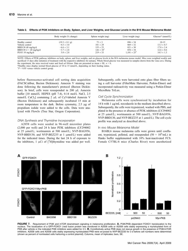
before fluorescence-activated cell sorting data acquisition
(FACSCalibur, Becton Dickinson). Annexin V staining was
done following the manufacturer's protocol (Becton Dickin-
son). In brief, cells were resuspended in 200 μL Annexin
buffer [10 mmol/L HEPES (pH 7.4), 0.14 mol/L NaCl, 2.5
mmol/L CaCl2] containing 2 μL of Cy5-labeled Annexin V
(Becton Dickinson) and subsequently incubated 15 min at
room temperature in the dark. Before cytometry, 2.5 μg of
propidium iodide were added to the cells. Data were ana-
lyzed with FlowJo (Tree Star, Oregon Corporation).
DNA Synthesis and Thymidine Incorporation
A2058 cells were seeded in 96-well microtiter plates
(2,000 per well) and 24 h later PI3K inhibitors (LY294002
at 25 μmol/L; wortmannin at 500 nmol/L; NVP-BAG956,
NVP-BBD130, and NVP-BEZ235 at 1 μmol/L) were added
for the indicated times. During the last 24 h of exposure to
the inhibitors, 1 μCi of [3H]thymidine was added per well.
Subsequently, cells were harvested onto glass fiber filters us-
ing a cell harvester (FilterMate Harvester, Perkin-Elmer) and
incorporated radioactivity was measured using a Perkin-Elmer
MicroBeta TriLux.
Cell Cycle Synchronization
Melanoma cells were synchronized by incubation for
14 h with 1 μg/mL nocodazole in the medium described above.
Subsequently, the cells were trypsinized, washed with PBS, and
plated in the presence or absence of PI3K inhibitors (LY294002
at 25 μmol/L; wortmannin at 500 nmol/L; NVP-BAG956,
NVP-BBD130, and NVP-BEZ235 at 1 μmol/L). The cell cycle
profile was analyzed as described above.
In vivo Mouse Melanoma Model
B16BL6 mouse melanoma cells were grown until conflu-
ent, trypsinized, pelleted, and resuspended (50 × 106/mL) in
Hanks buffer supplemented with 10% heat-inactivated FCS.
Female C57BL/6 mice (Charles River) were anesthetized
Table 2. Effects of PI3K Inhibitors on Body, Spleen, and Liver Weights, and Glucose Levels in the B16 Mouse Melanoma Model
Body weight (% change) Spleen weight (mg) Liver weight (mg) Glucose* (mmol/L)
Healthy control (19.3 ± 0.1 g) 73 ± 3 896 ± 34 10-15
Vehicle control −4.5 ± 3.0 181 ± 29 869 ± 32 13.3 ± 0.7
BBD130 40 mg/kg/d −6.6 ± 3.1 155 ± 23 821 ± 30 17.8 ± 2.4BBD130 2 × 20 mg/kg/d −3.7 ± 2.8 126 ± 10† 854 ± 34 12.6 ± 0.6
PI103 10 mg/kg/d 5.0 ± 2.8 219 ± 33 1,101 ± 25† 14.5 ± 1.3
NOTE: Effects of PI3K pathway inhibitors on body, spleen, and liver weights, and on glucose levels in the B16 melanoma mouse model. Mice were weighted weekly and
sacrificed 13 days after initiation of treatment with the respective inhibitors for autopsy. Whole blood glucose was measured in samples drawn from the vena cava. Duringthe experiment, the mice received water and food ad libitum. Data are presented as mean ± SE, n > 6.
*Healthy mice display normal blood glucose of 10 to 15 mmol/L, depending on their feeding status.†P < 0.05 versus vehicle control group.
FIGURE 7. Requirement of PI3K and mTOR downstream signaling in melanoma proliferation. A. PI3K/PKB–dependent FOXO1 translocation to thenucleus. The localization of a GFP-FOXO1 (red) fusion protein was monitored in A2058 cells or A2058 cells stably expressing a myristoylated form ofPKB after vehicle or the indicated PI3K inhibitors were added for 3 h. B. Constitutively active PKB does not rescue growth in the presence of PI3K/mTORinhibitors. A2058 cells and A2058 cells stably expressing myristoylated PKB were ex‐posed to NVP-BEZ235 for 3 d before cell numbers were determined(shown as percent of nontreated cells harboring a control plasmid). Columns, mean of triplicates; bars, SE.
Marone et al.
Mol Cancer Res 2009;7(4). April 2009
610

with 3% isofluorane in O2 (v/v) and placed on an operation
table maintained at 37°C. Mouse ears were fixed with a
double-sided tape over a steel cone and 1 μL of the cell suspen-
sion was injected intradermally using a microliter syringe with
a 30-G needle. Primary tumor size was recorded every 7 d in
mice anesthetized with isofluorane. One week after cell injec-
tion, oral treatment with vehicle control [10% NMP/PEG
300 (1-methyl-2-pyrrolidone/polyethylene glycol 300);
10:90, v/v], NVP-BBD130 (40 mg/kg daily and twice daily
20 mg/kg), NVP-BEZ235 (30 mg/kg daily and twice daily
20 mg/kg), PTK787 (100 mg/kg/d) and i.p. treatment with
PI103 (2.5, 5, and 10 mg/kg/d) was started. NVP-BBD130,
NVP-BEZ235, and PTK787 were dissolved by sonication in
NMP and then the corresponding volume of PEG 300 was
added. PI103 was instead dissolved in KZI (Cremophor EL/
ethanol absolute 65:35, v/v) and the remaining volume (1:3,
v/v) of 5% glucose (Braun Medical AG) was added. The inhi-
bitors were given for the indicated period, before animals were
sacrificed, and primary tumor, cervical lymph node metastases,
spleen, liver, femurs, and blood (from the vena cava) were col-
lected for further analysis.
The size of primary tumors at a given time point (=areat)
was determined by digital imaging as described in refs. (51,
52), and tumor progression was related to the tumor area at
day 7 (=area7days), and then expressed as percentage of the
overall mean tumor size in untreated [vehicle control (VC)] an-
imals (=mean_area_VC20days). The depicted values were there-
fore calculated as follows:
Primary tumor area ð%Þ ¼ ½100� areat=area7d�=mean area VC20d
A fraction of the primary tumors, metastases and liver samples
were snap frozen in liquid nitrogen, the rest was fixed in 4%
paraformaldehyde for paraffin embedding. Experiments were
terminated (here at day 20) for ethical reasons.
Immunohistochemistry
Primary tumors, metastases, and liver tissues were embedded
in paraffin using a Spin Tissue Processor (Microm International).
Paraffin blocks were cut to 6-μm sections using the Microtome
cool-cut HM355S (Microm International). For CD31 (antibody
from Bachem AG) staining, tissue slides were deparaffinized
using roticlean, and the antigen was unmasked by proteinase K
treatment. The staining with the primary antibody was done
overnight (antibody dilution 1:50). Subsequently, slides were in-
cubated with a fluorescently labeled secondary antibody and
Hoechst 33342 and mounted with crystal solution (Medite).
For H&E staining, deparaffinized slides were incubated with
hematoxylin solution followed by eosin and mounted with Cy-
toseal XYL (Medite).
Immune Cell Detection
Cell suspensions of spleen and bone marrow were gener-
ated and stained with different cell surface marker antibodies:
monoclonal antibodies against CD3 (clone KT3), CD4
(RM4-5), CD8 (53-6.7), B220 (RA3-6B2), CD11b (M1/70),
CD11c (HL3), GR1, and TERT were obtained from BD
Biosciences PharMingen or eBioscience. The F4/80 antibody
was from Serotec. Fluorescence was quantified on a FACS-
Calibur.
Myr-PKB Transfection and GFP-FOXO Translocation
Cells were plated at 14 × 103/cm2, and 24 h later were
transfected with 0.1 μg/cm2 pcDNA3-GFP-FOXO1 and/or
pcDNA3-Myr-PKB expression plasmids using JetPEI
(Brunschwig Chemie) following the manufacturer's protocol.
The following day, cells were exposed to PI3K inhibitors
(LY294002 at 25 μmol/L; wortmannin at 500 nmol/L;
NVP-BAG956, NVP-BBD130, NVP-BEZ235, and PI103 at
1 μmol/L) or rapamycin (100 nmol/L) for 3 h. HEK293 cells
were starved overnight in serum-free DMEM before treatment
with the inhibitors.
For immunofluorescence, cells plated on glass coverslips
(21 × 103/cm2) were fixed in PBS with 4% paraformaldehyde
before nuclei were stained with Hoechst 33342. Coverslips
were mounted on microscopy slides in Mowiol (Clariant
GMbH). Images were acquired with OpenLab software
(Improvision) on an Axiovert 200 M microscope (Zeiss)
with a Plan-Achromat 63×/1.4 and an Orca ER II camera
(Hamamatsu).
FIGURE 8. A. Antiproliferative effect of pan-PI3K, PI3K/mTOR, andmTOR inhibitors on melanoma cells. Melanoma cells were exposed toNVP-BEZ235 (1 μmol/L), ZSTK474 (1 μmol/L), and/or rapamycin (100nmol/L). Proliferation was measured as above and is shown normalized toproliferation of vehicle-treated control cells. Columns, mean of triplicates;bars, SE. B. Changes in the phosphorylation of PKB and p70S6K uponPI3K and/or mTOR inhibition. Melanoma cells were exposed to inhibitorsas indicated in A and total cell lysates were prepared 1 and 3 d later fordetection of phosphorylated (Ser473) PKB and p70S6K. Actin stained withCoomassie blue is shown as loading control.
PI3K/mTOR in Melanoma
Mol Cancer Res 2009;7(4). April 2009
611
Disclosure of Potential Conflicts of InterestNo potential conflicts of interest were disclosed.
AcknowledgmentsWe thank Poppy Fotiadou for the critical reading of the manuscript; Mathias
M. Hauri-Hohl for help with fluorescence-activated cell sorting analysis; PriskaReinhard for excellent technical assistance; Reinhard Dummer for inspiring
discussions; Reinhard Dummer, Isaiah Fidler, Silvio Hemmi, Meenhard Herlyn,Adrian Ochsenbein, and Giulio Spagnoli for the supply of cell lines; Samuel Arnal
for help with the in vivo experiment; Natasha Cmiljanovic for the synthesis ofZSTK474; and Karen C. Arden for the GFP-FOXO1 plasmid.
References1. Vogelstein B, Kinzler KW. Cancer genes and the pathways they control. NatMed 2004;10:789–99.
2. Vivanco I, Sawyers CL. The phosphatidylinositol 3-Kinase AKT pathway inhuman cancer. Nat Rev Cancer 2002;2:489–501.
3. Wymann MP, Marone R. Phosphoinositide 3-kinase in disease: timing, loca-tion, and scaffolding. Curr Opin Cell Biol 2005;17:141–9.
4. Engelman JA, Luo J, Cantley LC. The evolution of phosphatidylinositol3-kinases as regulators of growth and metabolism. Nat Rev Genet 2006;7:
606–19.
5. Raftopoulou M, Hall A. Cell migration: Rho GTPases lead the way. Dev Biol
2004;265:23–32.
6. Bhaskar PT, Hay N. The two TORCs and Akt. Dev Cell 2007;12:487–502.
7. Samuels Y, Ericson K. Oncogenic PI3K and its role in cancer. Curr Opin
Oncol 2006;18:77–82.
8. Omholt K, Krockel D, Ringborg U, Hansson J. Mutations of PIK3CA are rarein cutaneous melanoma. Melanoma Res 2006;16:197–200.
9. Curtin JA, Stark MS, Pinkel D, Hayward NK, Bastian BC. PI3-kinase subu-nits are infrequent somatic targets in melanoma. J Invest Dermatol 2006;126:
1660–3.
10. Stark M, Hayward N. Genome-wide loss of heterozygosity and copy number
analysis in melanoma using high-density single-nucleotide polymorphism arrays.
Cancer Res 2007;67:2632–42.
11. Guldberg P, thor Straten P, Ahrenkiel V, Seremet T, Kirkin AF, Zeuthen J.
Somatic mutation of the Peutz-Jeghers syndrome gene, LKB1/STK11, in malig-nant melanoma. Oncogene 1999;18:1777–80.
12. Meier F, Busch S, Lasithiotakis K, et al. Combined targeting of MAPK and
AKT signalling pathways is a promising strategy for melanoma treatment. Br J
Dermatol 2007;.
13. Eisen T, Ahmad T, Flaherty KT, et al. Sorafenib in advanced melanoma:a Phase II randomised discontinuation trial analysis. Br J Cancer 2006;95:
581–6.
14. Ratain MJ, Eisen T, Stadler WM, et al. Phase II placebo-controlled random-
ized discontinuation trial of sorafenib in patients with metastatic renal cell carci-
noma. J Clin Oncol 2006;24:2505–12.
15. Margolin K, Longmate J, Baratta T, et al. CCI-779 in metastatic melano-ma: a phase II trial of the California Cancer Consortium. Cancer 2005;104:
1045–8.
16. Hu L, Zaloudek C, Mills GB, Gray J, Jaffe RB. In vivo and in vitro ovarian
carcinoma growth inhibition by a phosphatidylinositol 3-kinase inhibitor(LY294002). Clin Cancer Res 2000;6:880–6.
17. Wymann MP, Zvelebil M, Laffargue M. Phosphoinositide 3-kinase signalling—
which way to target? Trends Pharmacol Sci 2003;24:366–76.
18. Liu Y, Jiang N, Wu J, Dai W, Rosenblum JS. Polo-like kinases inhibited
by wortmannin. Labeling site and downstream effects. J Biol Chem 2007;282:2505–11.
19. Fan QW, Knight ZA, Goldenberg DD, et al. A dual PI3 kinase/mTOR inhib-
itor reveals emergent efficacy in glioma. Cancer Cell 2006;9:341–9.
20. Raynaud FI, Eccles S, Clarke PA, et al. Pharmacologic characterization of a
potent inhibitor of class I phosphatidylinositide 3-kinases. Cancer Res 2007;67:5840–50.
21. Chen JS, Zhou LJ, Entin-Meer M, et al. Characterization of structurally
distinct, isoform-selective phosphoinositide 3′-kinase inhibitors in combination
with radiation in the treatment of glioblastoma. Mol Cancer Ther 2008;7:841–50.
22. Yaguchi S, Fukui Y, Koshimizu I, et al. Antitumor activity of ZSTK474,
a new phosphatidylinositol 3-kinase inhibitor. J Natl Cancer Inst 2006;98:545–56.
23. Kong D, Yamori T. ZSTK474 is an ATP-competitive inhibitor of class I
phosphatidylinositol 3 kinase isoforms. Cancer Sci 2007;98:1638–42.
24. Casagrande F, Bacqueville D, Pillaire MJ, et al. G1 phase arrest by the phos-phatidylinositol 3-kinase inhibitor LY 294002 is correlated to up-regulation of
p27Kip1 and inhibition of G1 CDKs in choroidal melanoma cells. FEBS Lett1998;422:385–90.
25. Blanco-Aparicio C, Pequeno B, Moneo V, et al. Inhibition of phosphatidyli-nositol-3-kinase synergizes with gemcitabine in low-passage tumor cell lines cor-
relating with Bax translocation to the mitochondria. Anticancer Drugs 2005;16:977–87.
26. Smalley KS, Haass NK, Brafford PA, Lioni M, Flaherty KT, Herlyn M.
Multiple signaling pathways must be targeted to overcome drug resistance
in cell lines derived from melanoma metastases. Mol Cancer Ther 2006;5:1136–44.
27. Bedogni B, Welford SM, Kwan AC, Ranger-Moore J, Saboda K, Powell
MB. Inhibition of phosphatidylinositol-3-kinase and mitogen-activated proteinkinase kinase 1/2 prevents melanoma development and promotes melanoma
regression in the transgenic TPRas mouse model. Mol Cancer Ther 2006;5:
3071–7.
28. Maira SM, Stauffer F, Brueggen J, et al. Identification and characterization of
NVP-BEZ235, a new orally available dual phosphatidylinositol 3-kinase/mam-
malian target of rapamycin inhibitor with potent in vivo antitumor activity. MolCancer Ther 2008;.
29. Stauffer F, Maira SM, Furet P, Garcia-Echeverria C. Imidazo[4,5-c]quinoline
as inhibitors of the PI3K/PKB-pathway. Bioorg Med Chem Lett 2007;.
30. Lin J, Adam RM, Santiestevan E, Freeman MR. The phosphatidylinositol 3′-kinase pathway is a dominant growth factor-activated cell survival pathway in
LNCaP human prostate carcinoma cells. Cancer Res 1999;59:2891–7.
31. Wen Y, Hu MC, Makino K, et al. HER-2/neu promotes androgen-indepen-
dent survival and growth of prostate cancer cells through the Akt pathway. CancerRes 2000;60:6841–5.
32. Carson JP, Kulik G, Weber MJ. Antiapoptotic signaling in LNCaP prostatecancer cells: a survival signaling pathway independent of phosphatidylinositol 3′-
kinase and Akt/protein kinase B. Cancer Res 1999;59:1449–53.
33. Andjelkovic M, Alessi DR, Meier R, et al. Role of translocation in the acti-
vation and function of protein kinase B. J Biol Chem 1997;272:31515–24.
34. Huang H, Tindall DJ. Dynamic FoxO transcription factors. J Cell Sci 2007;
120:2479–87.
35. van der Horst A, Burgering BM. Stressing the role of FoxO proteins in life-
span and disease. Nat Rev Mol Cell Biol 2007;8:440–50.
36. Ruckle T, Schwarz MK, Rommel C. PI3Kγ inhibition: towards an ‘aspirin of
the 21st century’? Nat Rev Drug Discov 2006;5:903–18.
37. Marone R, Cmiljanovic V, Giese B, Wymann MP. Targeting phosphoinosi-tide 3-kinase—moving towards therapy. Biochim Biophys Acta 2007;.
38. Smalley KS, Lioni M, Dalla Palma M, et al. Increased cyclin D1 expressioncan mediate BRAF inhibitor resistance in BRAF V600E-mutated melanomas.
Mol Cancer Ther 2008;7:2876–83.
39. Hussein MR. Tumour-infiltrating lymphocytes and melanoma tumorigenesis:
an insight. Br J Dermatol 2005;153:18–21.
40. Sarbassov DD, Guertin DA, Ali SM, Sabatini DM. Phosphorylation and
regulation of Akt/PKB by the rictor-mTOR complex. Science 2005;307:1098–101.
41. Feng J, Park J, Cron P, Hess D, Hemmings BA. Identification of a PKB/Akthydrophobic motif Ser-473 kinase as DNA-dependent protein kinase. J Biol
Chem 2004;279:41189–96.
42. Bozulic L, Surucu B, Hynx D, Hemmings BA. PKBα/Akt1 acts downstream
of DNA-PK in the DNA double-strand break response and promotes survival.
Mol Cell 2008;30:203–13.
43. Anand P, Gruppuso PA. Rapamycin inhibits liver growth during refeeding in
rats via control of ribosomal protein translation but not cap-dependent translationinitiation. J Nutr 2006;136:27–33.
44. Hanahan D, Weinberg RA. The hallmarks of cancer. Cell 2000;100:57–70.
45. Phung TL, Ziv K, Dabydeen D, et al. Pathological angiogenesis is induced
by sustained Akt signaling and inhibited by rapamycin. Cancer Cell 2006;10:159–70.
46. Del Bufalo D, Ciuffreda L, Trisciuoglio D, et al. Antiangiogenic potential of
the mammalian target of rapamycin inhibitor temsirolimus. Cancer Res 2006;66:
5549–54.
47. Skinner HD, Zheng JZ, Fang J, Agani F, Jiang BH. Vascular endothelialgrowth factor transcriptional activation is mediated by hypoxia-inducible factor
1α, HDM2, and p70S6K1 in response to phosphatidylinositol 3-kinase/AKT sig-
naling. J Biol Chem 2004;279:45643–51.
Marone et al.
Mol Cancer Res 2009;7(4). April 2009
612

48. Wang X, Yue P, Chan CB, et al. Inhibition of mammalian target of rapa-
mycin induces phosphatidylinositol 3-kinase-dependent and Mnk-mediatedeukaryotic translation initiation factor 4E phosphorylation. Mol Cell Biol
2007;27:7405–13.
49. Li W, Petrimpol M, Molle KD, Hall MN, Battegay EJ, Humar R. Hypoxia-
induced endothelial proliferation requires both mTORC1 and mTORC2. Circ Res2007;100:79–87.
50. Rolink AG, Andersson J, Melchers F. Characterization of immature B cellsby a novel monoclonal antibody, by turnover and by mitogen reactivity. Eur J
Immunol 1998;28:3738–48.
51. Rudin M, McSheehy PM, Allegrini PR, et al. PTK787/ZK222584, a tyrosine
kinase inhibitor of vascular endothelial growth factor receptor, reduces uptake of
the contrast agent GdDOTA by murine orthotopic B16/BL6 melanoma tumours
and inhibits their growth in vivo. NMR Biomed 2005;18:308–21.
52. Sini P, Samarzija I, Baffert F, et al. Inhibition of multiple vascular endothelialgrowth factor receptors (VEGFR) blocks lymph node metastases but inhibition of
VEGFR-2 is sufficient to sensitize tumor cells to platinum-based chemotherapeu-
tics. Cancer Res 2008;68:1581–92.
53. Garcia-Echeverria C, Pearson MA, Marti A, et al. In vivo antitumor activity
of NVP-AEW541—a novel, potent, and selective inhibitor of the IGF-IR kinase.Cancer Cell 2004;5:231–9.
54. Knight ZA, Gonzalez B, Feldman ME, et al. A pharmacological map of
the PI3-K family defines a role for p110α in insulin signaling. Cell 2006;125:
733–47.
PI3K/mTOR in Melanoma
Mol Cancer Res 2009;7(4). April 2009
613
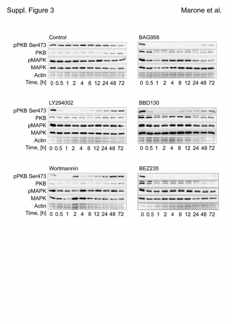
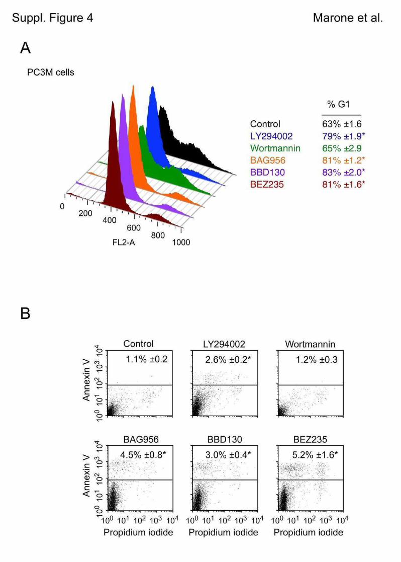
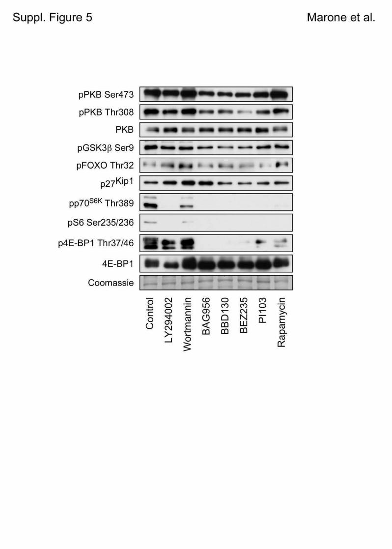
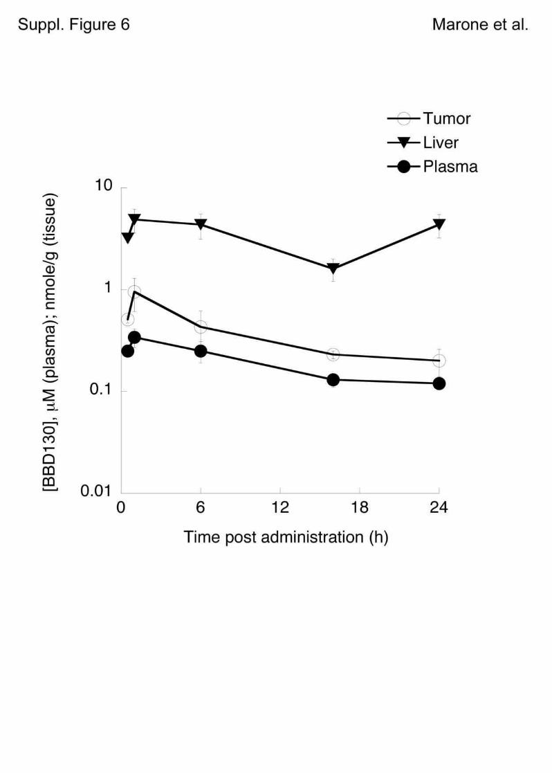
Page 1 of 8
Marone et al.
Supplementary Information for
Targeting Melanoma with Dual PI3K/mTOR Inhibitors
Romina Marone1, Dominik Erhart
1, Thomas Bohnacker
1, Christian Schnell
2, Vladimir
Cmiljanovic3, Frédéric Stauffer
2, Carlos Garcia-Echeverria
2, Bernd Giese
3, Sauveur-Michel
Maira2, Matthias P. Wymann
1
1Inst. Biochemistry and Genetics, Dept. for Biomedicine, Mattenstrasse 28, University of
Basel, CH-4058 Basel, Switzerland; 2Novartis Institutes for Biomedical Research, Oncology
Disease Area, CH-4002 Basel, Switzerland; 3Dept. of Chemistry, University of Basel, CH-
4058 Basel, Switzerland.
To whom correspondence should be addressed:
Matthias P. Wymann
Inst. Biochemistry and Genetics
Dept. of Biomedicine
Mattenstrasse 28
CH-4058 Basel, Switzerland
Tel. +41 61 695 3046; Fax. +41 61 267 3566; [email protected]
Page 2 of 8
Marone et al.
Supplementary Experimental Procedures
Wortmannin-competition assay
Recombinant p110α/p85α protein complex (Millipore) was pre-incubated with the indicated
concentrations of NVP-BBD130, NVP-BAG956 or NVP-BEZ235 at 37°C for 30 min., before
the samples were transferred to ice and wortmannin (200 nM) was added for 15 min. Protein
samples (0.13 µg / lane) were subjected to SDS-PAGE and blotted onto a PVDF membrane.
Immunological detection of wortmannin covalently attached to p110α was achieved using
polyclonal rabbit α-wortmannin antibodies (1). Loading controls for p110α and p85α were
performed using monoclonal anti-p110α (kindly obtained from A. Klippel) and polyclonal anti-
p85a antibodies (1).
Supplementary Figure Legends
Figure S1.
A: Chemical structures of NVP-BAG956, NVP-BBD130 and NVP-BEZ235.
B: Wortmannin competition assay.
Covalent binding of wortmannin to PI3Kα (p85α/p110α complex) was prevented by pre-
incubation of recombinant PI3Kα complex with the depicted PI3K inhibitors. The PI3K
complex was incubated with increasing concentrations of NVP-BAG956, NVP-BBD130 or
NVP-BEZ235, before wortmannin was added, and samples were subsequently subjected to
SDS-PAGE and immunodetection of protein-bound wortmannin, p110α and p85α.
Figure S2. A and B: A2058 cell were treated for 3 days with PI3K inhibitors. Cells were
subsequently trypsinized, washed once with PBS and replated at the same concentration.
Cell number and cell volume were measured for up to 7 days (for NVP-BBD130 treated cells)
in the absence of PI3K inhibitors. Data are means of triplicates ± SEM.
Page 3 of 8
Marone et al.
Figure S3. Changes in the levels of phosphorylated PKB and MAPK in A2058 cells treated
with PI3K inhibitors.
Cells were treated with different PI3K inhibitors and total cell lysates were prepared at
different time intervals. Levels of phosphorylated and total PKB (upper band seen in some
panels is a non-specific interaction) and MAPK were detected by western blotting. Coomassie
blue-stained actin served as loading control.
Figure S4. Effect of PI3K inhibition on cell cycle profile and apoptosis.
A: Determination of cell cycle profile of the prostate cancer cell line PC3M. The cells were
exposed to one dose of PI3K inhibitors for three days. Cell cycle profile was evaluated by
FACS. *p<0.002
B: Evaluation of apoptosis in A2058 cells upon PI3K inhibition. A2058 melanoma cells were
treated for 3 days with PI3K inhibitors. Annexin V positive cells were visualized by FACS.
*p<0.002
Figure S5. Effect of expression of Myr-PKB (constitutively active PKB/Akt) on serum-deprived
and inhibitor-treated HEK293 cells. HEK293 cells expressing Myr-PKB were exposed for 2
hours to the indicated PI3K inhibitors and rapamycin. Phosphoproteins in total cell lysates
were detected by western blotting using the antibodies described in the main text (see
Experimental Procedures).
Figure S6. Pharmacokinetic/pharmacodynamic relationship for BBD130. Mice bearing a
PC3M tumor were treated orally once with a dose of 50 mg/kg BBD130. The animals were
then sacrificed after 0.5, 1, 6, 16 and 24 hours after treatment and tumor, liver and plasma
were collected (n=4). The concentration of the inhibitor in these tissues was quantified using
a reverse-phase high performance liquid chromatography/UV (for a detailed protocol see
Maira et al. (2)).
Page 4 of 8
Marone et al.
Table S1: PTEN, BRaf, NRas and p53 status in melanoma cells.
Protein levels PTEN BRaf NRas p53
Mutation
A2058 -
mut V600E wt
++ V274F
A375 + wt
V600E wt + wt
B16F1 + wt (+)
B16F10 + wt wt (+) wt
C32 -
mut V600E wt
+++ wt
Hs294T - ++
Malme ++ wt
V600E wt (+) wt
Malme3M + wt
wt +
SKMel2 + wt
wt Q61R +++
G245S
SKMel23 -
mut wt wt +
WM35 (+) V600E wt +
1205lu - V600E wt +
Melanocytes ++++
wt wt wt
(+) wt
Status of signaling proteins in human melanocytes, and human and mouse melanoma cells
used here are shown. PTEN protein expression levels and mutations in PTEN, BRaf, NRas
and p53 are depicted. Total cell lysates were analyzed by immuno-blotting for PTEN and p53.
Mutations in PTEN, BRaf, NRas and p53 were identified earlier (public data base of the
Sanger Institute; for B16F1 and F10 see (3); for SKMel23 and Malme see (4, 5); for WM35
and 1205lu see (6)). mut = mutated; wt = wild type.
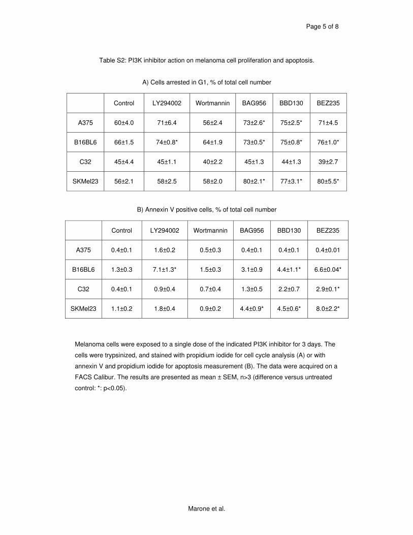
Page 5 of 8
Marone et al.
Table S2: PI3K inhibitor action on melanoma cell proliferation and apoptosis.
A) Cells arrested in G1, % of total cell number
Control LY294002 Wortmannin BAG956 BBD130 BEZ235
A375 60±4.0 71±6.4 56±2.4 73±2.6* 75±2.5* 71±4.5
B16BL6 66±1.5 74±0.8* 64±1.9 73±0.5* 75±0.8* 76±1.0*
C32 45±4.4 45±1.1 40±2.2 45±1.3 44±1.3 39±2.7
SKMel23 56±2.1 58±2.5 58±2.0 80±2.1* 77±3.1* 80±5.5*
B) Annexin V positive cells, % of total cell number
Control LY294002 Wortmannin BAG956 BBD130 BEZ235
A375 0.4±0.1 1.6±0.2 0.5±0.3 0.4±0.1 0.4±0.1 0.4±0.01
B16BL6 1.3±0.3 7.1±1.3* 1.5±0.3 3.1±0.9 4.4±1.1* 6.6±0.04*
C32 0.4±0.1 0.9±0.4 0.7±0.4 1.3±0.5 2.2±0.7 2.9±0.1*
SKMel23 1.1±0.2 1.8±0.4 0.9±0.2 4.4±0.9* 4.5±0.6* 8.0±2.2*
Melanoma cells were exposed to a single dose of the indicated PI3K inhibitor for 3 days. The
cells were trypsinized, and stained with propidium iodide for cell cycle analysis (A) or with
annexin V and propidium iodide for apoptosis measurement (B). The data were acquired on a
FACS Calibur. The results are presented as mean ± SEM, n>3 (difference versus untreated
control: *: p<0.05).
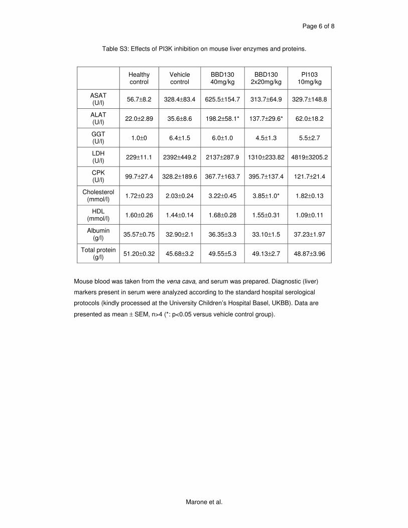
Page 6 of 8
Marone et al.
Table S3: Effects of PI3K inhibition on mouse liver enzymes and proteins.
Healthy control
Vehicle control
BBD130 40mg/kg
BBD130 2x20mg/kg
PI103 10mg/kg
ASAT (U/l)
56.7±8.2 328.4±83.4 625.5±154.7 313.7±64.9 329.7±148.8
ALAT (U/l)
22.0±2.89 35.6±8.6 198.2±58.1* 137.7±29.6* 62.0±18.2
GGT (U/l)
1.0±0 6.4±1.5 6.0±1.0 4.5±1.3 5.5±2.7
LDH (U/l)
229±11.1 2392±449.2 2137±287.9 1310±233.82 4819±3205.2
CPK (U/l)
99.7±27.4 328.2±189.6 367.7±163.7 395.7±137.4 121.7±21.4
Cholesterol (mmol/l)
1.72±0.23 2.03±0.24 3.22±0.45 3.85±1.0* 1.82±0.13
HDL (mmol/l)
1.60±0.26 1.44±0.14 1.68±0.28 1.55±0.31 1.09±0.11
Albumin (g/l)
35.57±0.75 32.90±2.1 36.35±3.3 33.10±1.5 37.23±1.97
Total protein (g/l)
51.20±0.32 45.68±3.2 49.55±5.3 49.13±2.7 48.87±3.96
Mouse blood was taken from the vena cava, and serum was prepared. Diagnostic (liver)
markers present in serum were analyzed according to the standard hospital serological
protocols (kindly processed at the University Children’s Hospital Basel, UKBB). Data are
presented as mean ± SEM, n>4 (*: p<0.05 versus vehicle control group).
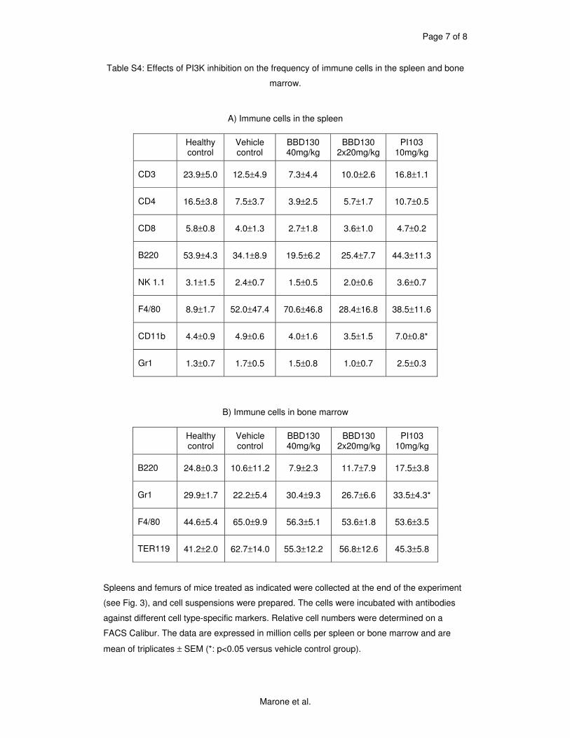
Page 7 of 8
Marone et al.
Table S4: Effects of PI3K inhibition on the frequency of immune cells in the spleen and bone
marrow.
A) Immune cells in the spleen
Healthy control
Vehicle control
BBD130 40mg/kg
BBD130 2x20mg/kg
PI103 10mg/kg
CD3 23.9±5.0 12.5±4.9 7.3±4.4 10.0±2.6 16.8±1.1
CD4 16.5±3.8 7.5±3.7 3.9±2.5 5.7±1.7 10.7±0.5
CD8 5.8±0.8 4.0±1.3 2.7±1.8 3.6±1.0 4.7±0.2
B220 53.9±4.3 34.1±8.9 19.5±6.2 25.4±7.7 44.3±11.3
NK 1.1 3.1±1.5 2.4±0.7 1.5±0.5 2.0±0.6 3.6±0.7
F4/80 8.9±1.7 52.0±47.4 70.6±46.8 28.4±16.8 38.5±11.6
CD11b 4.4±0.9 4.9±0.6 4.0±1.6 3.5±1.5 7.0±0.8*
Gr1 1.3±0.7 1.7±0.5 1.5±0.8 1.0±0.7 2.5±0.3
B) Immune cells in bone marrow
Healthy control
Vehicle control
BBD130 40mg/kg
BBD130 2x20mg/kg
PI103 10mg/kg
B220 24.8±0.3 10.6±11.2 7.9±2.3 11.7±7.9 17.5±3.8
Gr1 29.9±1.7 22.2±5.4 30.4±9.3 26.7±6.6 33.5±4.3*
F4/80 44.6±5.4 65.0±9.9 56.3±5.1 53.6±1.8 53.6±3.5
TER119 41.2±2.0 62.7±14.0 55.3±12.2 56.8±12.6 45.3±5.8
Spleens and femurs of mice treated as indicated were collected at the end of the experiment
(see Fig. 3), and cell suspensions were prepared. The cells were incubated with antibodies
against different cell type-specific markers. Relative cell numbers were determined on a
FACS Calibur. The data are expressed in million cells per spleen or bone marrow and are
mean of triplicates ± SEM (*: p<0.05 versus vehicle control group).
Page 8 of 8
Marone et al.
Supplementary References:
1. Wymann MP, Bulgarelli-Leva G, Zvelebil MJ et al. Wortmannin inactivates
phosphoinositide 3-kinase by covalent modification of Lys-802, a residue involved in the phosphate transfer reaction. Mol Cell Biol 1996;16:1722-33.
2. Maira SM, Stauffer F, Brueggen J et al. Identification and characterization of NVP-BEZ235, a new orally available dual phosphatidylinositol 3-kinase/mammalian target of rapamycin inhibitor with potent in vivo antitumor activity. Mol Cancer Ther 2008
3. Melnikova VO, Bolshakov SV, Walker C, Ananthaswamy HN. Genomic alterations in spontaneous and carcinogen-induced murine melanoma cell lines. Oncogene 2004;23:2347-56.
4. Tsao H, Zhang X, Fowlkes K, Haluska FG. Relative reciprocity of NRAS and PTEN/MMAC1 alterations in cutaneous melanoma cell lines. Cancer Res 2000;60:1800-4.
5. Tsao H, Goel V, Wu H, Yang G, Haluska FG. Genetic interaction between NRAS and BRAF mutations and PTEN/MMAC1 inactivation in melanoma. J Invest Dermatol 2004;122:337-41.
6. Smalley KS, Contractor R, Haass NK et al. Ki67 expression levels are a better marker of reduced melanoma growth following MEK inhibitor treatment than phospho-ERK levels. Br J Cancer 2007;96:445-9.
Author's personal copy
European Journal of Pharmaceutical Sciences 40 (2010) 143–147
Contents lists available at ScienceDirect
European Journal of Pharmaceutical Sciences
journa l homepage: www.e lsev ier .com/ locate /e jps
(E,Z)-3-(3′,5′-Dimethoxy-4′-hydroxy-benzylidene)-2-indolinoneblocks mast cell degranulation
S. Kiefer a, A.C. Mertzb, A. Koryakina a, M. Hamburger a,∗, P. Küenzi a
a Institute of Pharmaceutical Biology, University of Basel, Klingelbergstrasse 50, 4056 Basel, Switzerlandb Institute of Biochemistry and Genetics, Department of Biomedicine, University of Basel, Mattenstrasse 28, 4058 Basel, Switzerland
a r t i c l e i n f o
Article history:
Received 15 January 2010Accepted 20 March 2010Available online 27 March 2010
Keywords:
Isatis tinctoria
WoadDegranulationGranule exocitosisIndolinoneMast cells
a b s t r a c t
(E,Z)-3-(3′,5′-Dimethoxy-4′-hydroxy-benzylidene)-2-indolinone (indolinone) is an alkaloid that has beenidentified as a pharmacologically active compound in extracts of the traditional anti-inflammatory herbIsatis tinctoria. Indolinone has been shown to inhibit compound 48/80-induced mast cell degranulationin vitro. Application of indolinone to bone marrow derived mast cells showed that it was uniformlydistributed in the cytoplasm and that cellular uptake was terminated within minutes. Pre-treatmentof IgE-sensitized mast cells with 100 nM indolinone rendered them insensitive against Fc�RI-receptordependent degranulation. However, upstream signalling induced by antigen such as activation of PI3-Kand MAPK remained unaffected. We conclude that indolinone blocks mast cell degranulation at the levelof granule exocitosis with an IC50 of 54 nm.
© 2010 Elsevier B.V. All rights reserved.
1. Introduction
Isatis tinctoria (woad) is an ancient European dye and medicinalplant. It has traditionally been used as a source of indigo for bluedye and as anti-inflammatory medicine (Danz, 2000). In China, theclosely related Isatis indigotica is monographed in the Chinese Phar-macopoeia where it is indicated as a treatment for inflammation.Natural indigo, obtained by processing of various indigo plants,is used in traditional Chinese medicine to treat inflamed skin,intestines or mucosa (Stöger, 1999–2009). We confirmed the anti-inflammatory and anti-allergic potential of lipophilic I. tinctoria leafextracts some years ago in a pharmacological profiling involvingsome 20 clinically relevant targets (Danz, 2000; Hamburger, 2002),and later in several in vivo models for inflammation and allergy,and in a clinical pilot study for topical application (Heinemann etal., 2004; Recio et al., 2006a,b).
Several pharmacologically active constituents of woad havebeen identified, such as tryptanthrin, indirubin, �-linoleic acid, and(E,Z)-3-(3′,5′-dimethoxy-4′-hydroxy-benzylidene)-2-indolinone
Abbreviations: BMMC, murine bone marrow derived mast cells; DNP, dini-trophenyl; MAPK, mitogen-activated protein kinase; ERK, extracellular-regulatedkinase; PI3-K, phosphatidylinositol 3-kinase; PKB, protein kinase B; SNARE, solubleN-ethylmaleimide-sensitive-factor attachment receptor.
∗ Corresponding author. Tel.: +41 61 2671425; fax: +41 61 2671474.E-mail address: [email protected] (M. Hamburger).
(indolinone). Tryptanthrin is a potent inhibitor of COX-2 (Danzet al., 2001) and 5-LOX (Oberthur et al., 2005), and of nitricoxide (NO) production catalyzed by inducible NO synthase (iNOS)(Ishihara et al., 2000). Indirubin is used in Chinese medicineas an antileukemia drug and has been shown to inhibit cyclin-dependent-kinase 2 (CDK2) (Hoessel et al., 1999). The lipidconstituent �-linolenic acid inhibited 5-LOX (Oberthur et al.,2005) and seems to act as a competitor of arachidonic acid andthus reduces cellular inflammatory responses (Simopoulos, 2002).Indolinone inhibited compound 48/80-induced histamine releasefrom rat peritoneal mast cells but not the related compound(E,Z)-3-(4′-isopropylbenzylidene)-2-indolinone (Ind7) (Ruster etal., 2004).
Indolin-2-one derivatives have been tested in disease mod-els for multiple sclerosis (Bouerat et al., 2005), cancer (Kaur andTalele, 2008), HIV (Boechat et al., 2007), and infectious diseases(Bouchikhi et al., 2008), and an indolin-2-one derivative, sunitinib,was recently approved as tyrosine kinase inhibitor for treatmentof renal cell carcinoma and imatinib-resistant gastrointestinal stro-mal tumours (Atkins et al., 2006). The indolin-2-one scaffold seemsto exhibit some general kinase-inhibitory activity. Substitutedindolin-2-ones occupy the ATP-binding site and act as ATP-mimeticinhibitors (Sun et al., 1998). This suggested that the anti-allergiceffect of indolinone could be related to some kinase-inhibitoryproperties.
Although many cells are involved in allergy and inflamma-tion, mast cells play an important role as initial effectors due
0928-0987/$ – see front matter © 2010 Elsevier B.V. All rights reserved.
doi:10.1016/j.ejps.2010.03.016
Author's personal copy
144 S. Kiefer et al. / European Journal of Pharmaceutical Sciences 40 (2010) 143–147
to their tissular localisation. By release of pro-inflammatorymolecules, mast cells initialise processes that result in earlyand late phase allergic reactions. They are implicated in manydiseases, such as allergy and asthma, gastrointestinal disor-ders, rheumatic diseases and multiple sclerosis (Hofmann andAbraham, 2009). Degranulation of mast cells requires bindingof IgE antibodies to the high affinity IgE receptor (Fc�RI) foractivation and crosslinking of these antibodies by subsequentbinding of an antigen (Rivera et al., 2008; Rivera and Olivera,2008). The crosslinking induces intracellular signalling eventsthat lead to activation of the protein kinase Lyn that in turnphosphorylates immunoreceptor tyrosine-based activation motifs(ITAMs) and Syk kinases. Class IA PI3-K recognise phosphory-lated ITAMs as docking sites, which leads to the productionof PtdIns(3,4,5)P3 that recruits PKB (also known as Akt), lead-ing to its activation and initiates a signalling cascade involvingBruton’s tyrosine kinase (Btk) and phospholipase C� and theRaf-MEK-MAPK pathway. This eventually induces opening ofplasma membrane calcium channels leading to degranulation(Burgoyne and Morgan, 2003). However, the Fc�RI-mediated mastcell response is limited by subsequent de-phosphorylation ofPtdIns(3,4,5)P3 to PtdIns(3,4)P2 by SH2-containing inositol 5′-phosphatase (SHIP).
Subsequent granule exocitosis occurs in a highly regu-lated and conserved way depending on Ca2+ and is related tosynaptic vesicle exocytosis and most likely takes advantageof the same protein components such as SNARE’s (solu-ble N-ethylmaleimide-sensitive-factor attachment receptors)(Burgoyne and Morgan, 2003). Anti-inflammatory, anti-allergic or immunosuppressive drugs such as corticosteroidsor calcineurin-antagonists inhibit degranulation but addi-tionally have many side-effects (Ludowyke and Lagunoff,1985).
The aim of this study was to elucidate whether indolinoneinteracts with the pathways that lead to mast cell activa-tion and degranulation, and at what level this interactionoccurs.
2. Materials and methods
2.1. Chemicals and cell culture
Murine bone marrow derived mast cells (BMMCs) werecultured in Iscove’s modified Dulbecco’s medium (IMDM;Sigma–Aldrich, Buchs, Switzerland) supplemented with 10%heat-inactivated fetal calf serum (FCS; Amimed, Basel, Switzer-land), 100 U/ml penicillin/streptomycin, and 2 mM l-glutamine(both from Invitrogen, Basel, Switzerland). Cells were grownin humified atmosphere containing 5% CO2 and maintainedwith 2 ng/ml recombinant murine interleukin-3 (IL3; Pepro-Tech EC Ltd., London, UK). Chemicals used were: stem cellfactor (SCF; Biosource, Invitrogen, Basel, Switzerland), adeno-sine (Ade; Sigma–Aldrich, Buchs, Switzerland), LY294002 (AlexisCorporation, Lausen, Switzerland), and wortmannin (Wort;Sigma–Aldrich). All solvents used were from Scharlau (Barcelona,Spain).
(E,Z)-3-(3′,5′-Dimethoxy-4′-hydroxy-benzylidene)-2-indolinone (indolinone) and (E,Z)-3-(4′-isopropylbenzylidene)-2-indolinone (Ind7) were synthesised according to a general protocolfor indolinones (Sun et al., 1998). For indolinone, the ratio of slowlyinterconverting E and Z isomers was determined as 81:19 by HPLC(Ruster et al., 2004). Indolinone showed fluorescence when excitedat 488 nm or 405 nm, with an emission maximum at 499 nm. Theabsorption maxima are at 256 and 380 nm.
2.2. Western blot
Immunoblot analysis was performed according to standard pro-cedures. Equal amounts of cellular protein were separated onSDS-PAGE and transferred to a nitrocellulose membrane. Mem-branes were blocked and incubated overnight at 4 ◦C with specificprimary antibody diluted in blocking buffer (5% bovine serumalbumin in TBS-Tween): anti-phospho-PKB (Thr308) 1:1000; anti-phospho-ERK1/2 (Thr202/Tyr204) 1:1000. Specific bands weretagged with HRP conjugated secondary antibodies (Cell SignalingTechnology, Beverly, MA) and detected using enhanced chemilu-minescence (ECL Plus System, GE Healthcare, Little Chalfont, UK).
2.3. Degranulation assays
Release of histamine-containing granules was quantified bythe determination of �-hexosaminidase in cell supernatants asdescribed (Laffargue et al., 2002). Alternatively, Annexin-V stainingof exposed membrane phosphatidylserine was carried out usingthe Annexin-V assay kit (Roche Diagnostics, Rotkreuz, Switzerland)following the manufacturer’s protocol. The cells were then left toadhere to glass coverslips, fixed and assayed as described in Section2.4.
2.4. Fluorescence microscopy
BMMC cells were suspended in PBS and left to adhere to glasscoverslips and fixed in 4% formaldehyde in PBS for a minimumof 15 min at 4 ◦C. Nuclei were stained with DRAQ5 (Alexis Cor-poration, Lausen, Switzerland) according to the manufacturer’sprotocol. Cells plated on coverslips were mounted on glass slideswith Fluorescent Mounting Medium (DakoCytomation, Glostrup,Denmark) and visualised by confocal microscopy (Leica DM RXEscanning confocal microscope) using Leica confocal software, ver-sion 2.5 (Leica Microsystems, Heidelberg, Germany).
2.5. Kinase assay
Appropriate concentrations of PI3-K isoforms p110 �, �, � GST, �tr, and � (fused to interSH2 domains, a kind gift from Novartis, Basel,Switzerland) were mixed with 10 �g/ml l-�-phosphatidylinositol(Sigma–Aldrich, Buchs, Switzerland) dissolved in 0.3% octyl-glucoside kinase buffer (10 mM Tris–HCl, 3 mM MgCl2, 50 mMNaCl, 0.8 mM CHAPS, 1 mM DTT). After 5 min pre-treatment withindolinone or DMSO, 2 �M ATP (Roche, Basel, Switzerland) dis-solved in kinase buffer were added to start the reaction. Reactionswere incubated for 30–90 min, depending on the kinase isoform.Kinase Glo (Promega, Madison, WI) was added to each well tostart the luciferase reaction. After 15 min incubation at room tem-perature, plates (white 96 well luciferase plates (Berthold, BadWildbad, Germany)) were read out in a luminometer (Centro LB960, Berthold) with an integration time of 0.5 s. Samples were runin duplicate.
3. Results
3.1. Localisation of indolinone in BMMC
Due to the fluorescent properties of indolinone (A), we wereable to visualise high concentrations (≥50 �M) to directly deter-mine its localisation in cells and monitor cellular uptake by flowcytometry. Analysis of indolinone-stained cells (50 �M) by con-focal microscopy (Fig. 1B) showed equal distribution throughoutthe cytoplasm, but no staining of the nucleus, suggesting discrete
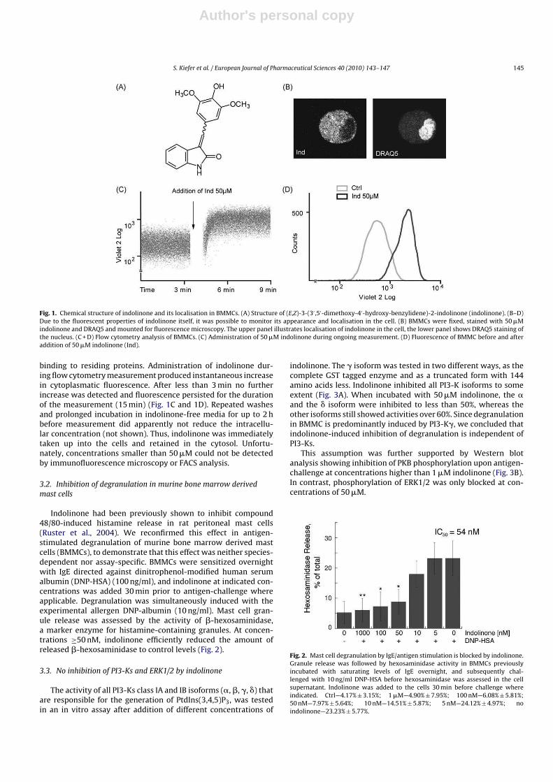
Author's personal copy
S. Kiefer et al. / European Journal of Pharmaceutical Sciences 40 (2010) 143–147 145
Fig. 1. Chemical structure of indolinone and its localisation in BMMCs. (A) Structure of (E,Z)-3-(3′ ,5′-dimethoxy-4′-hydroxy-benzylidene)-2-indolinone (indolinone). (B–D)Due to the fluorescent properties of indolinone itself, it was possible to monitor its appearance and localisation in the cell. (B) BMMCs were fixed, stained with 50 �Mindolinone and DRAQ5 and mounted for fluorescence microscopy. The upper panel illustrates localisation of indolinone in the cell, the lower panel shows DRAQ5 staining ofthe nucleus. (C + D) Flow cytometry analysis of BMMCs. (C) Administration of 50 �M indolinone during ongoing measurement. (D) Fluorescence of BMMC before and afteraddition of 50 �M indolinone (Ind).
binding to residing proteins. Administration of indolinone dur-ing flow cytometry measurement produced instantaneous increasein cytoplasmatic fluorescence. After less than 3 min no furtherincrease was detected and fluorescence persisted for the durationof the measurement (15 min) (Fig. 1C and 1D). Repeated washesand prolonged incubation in indolinone-free media for up to 2 hbefore measurement did apparently not reduce the intracellu-lar concentration (not shown). Thus, indolinone was immediatelytaken up into the cells and retained in the cytosol. Unfortu-nately, concentrations smaller than 50 �M could not be detectedby immunofluorescence microscopy or FACS analysis.
3.2. Inhibition of degranulation in murine bone marrow derived
mast cells
Indolinone had been previously shown to inhibit compound48/80-induced histamine release in rat peritoneal mast cells(Ruster et al., 2004). We reconfirmed this effect in antigen-stimulated degranulation of murine bone marrow derived mastcells (BMMCs), to demonstrate that this effect was neither species-dependent nor assay-specific. BMMCs were sensitized overnightwith IgE directed against dinitrophenol-modified human serumalbumin (DNP-HSA) (100 ng/ml), and indolinone at indicated con-centrations was added 30 min prior to antigen-challenge whereapplicable. Degranulation was simultaneously induced with theexperimental allergen DNP-albumin (10 ng/ml). Mast cell gran-ule release was assessed by the activity of �-hexosaminidase,a marker enzyme for histamine-containing granules. At concen-trations ≥50 nM, indolinone efficiently reduced the amount ofreleased �-hexosaminidase to control levels (Fig. 2).
3.3. No inhibition of PI3-Ks and ERK1/2 by indolinone
The activity of all PI3-Ks class IA and IB isoforms (�, �, �, �) thatare responsible for the generation of PtdIns(3,4,5)P3, was testedin an in vitro assay after addition of different concentrations of
indolinone. The � isoform was tested in two different ways, as thecomplete GST tagged enzyme and as a truncated form with 144amino acids less. Indolinone inhibited all PI3-K isoforms to someextent (Fig. 3A). When incubated with 50 �M indolinone, the �and the � isoform were inhibited to less than 50%, whereas theother isoforms still showed activities over 60%. Since degranulationin BMMC is predominantly induced by PI3-K�, we concluded thatindolinone-induced inhibition of degranulation is independent ofPI3-Ks.
This assumption was further supported by Western blotanalysis showing inhibition of PKB phosphorylation upon antigen-challenge at concentrations higher than 1 �M indolinone (Fig. 3B).In contrast, phosphorylation of ERK1/2 was only blocked at con-centrations of 50 �M.
Fig. 2. Mast cell degranulation by IgE/antigen stimulation is blocked by indolinone.Granule release was followed by hexosaminidase activity in BMMCs previouslyincubated with saturating levels of IgE overnight, and subsequently chal-lenged with 10 ng/ml DNP-HSA before hexosaminidase was assessed in the cellsupernatant. Indolinone was added to the cells 30 min before challenge whereindicated. Ctrl—4.17% ± 3.15%; 1 �M—4.90% ± 7.95%; 100 nM—6.08% ± 5.81%;50 nM—7.97% ± 5.64%; 10 nM—14.51% ± 5.87%; 5 nM—24.12% ± 4.97%; noindolinone—23.23% ± 5.77%.

Author's personal copy
146 S. Kiefer et al. / European Journal of Pharmaceutical Sciences 40 (2010) 143–147
Fig. 3. Indolinone blocks degranulation at the level of granule release. (A) PI3-Kswere co-incubated with ATP and indolinone at indicated concentrations or withDMSO as a control. Kinase activity was determined as consumption of ATP, andresidual ATP was measured by luciferase reaction. Samples were measured as dupli-cates in three independent experiments. The kinase activity of the control wasdefined as 100%. (B) Western blot analysis of BMMC cells (0.5 × 10⁶) previouslyincubated with saturating levels of IgE. The cells were starved in 2% serum for4 h, pre-treated with indolinone or DMSO for 30 min as indicated, and then chal-lenged with 10 ng/ml DNP-HSA for 2 min. Untreated cells were used as control.Shown are typical results from three independent experiments. (C) Mast cells weresensitized with IgE overnight and challenged with 10 ng/ml DNP-HSA (b–f). The fol-lowing substances were added to the cells 30 min before challenge: (a + b) DMSO;(c) 100 nM indolinone; (d) 100 nM Ind7; (e) 1 �M indolinone; (f) 1 �M Ind7. Cellswere fixed with 4% p-formaldehyde and stained with Annexin-V-FITC and analysedby immunofluorescence. Shown is a typical experiment. (D) Western blot analysis ofBMMC cells (0.5 × 10⁶) previously incubated with saturating levels of IgE. The cellswere starved in 2% serum for 4 h, pre-treated with 50 �M indolinone, Ind7 or DMSOfor 30 min as indicated, and then challenged with 10 ng/ml DNP-HSA for 2 min. (E)Chemical structure of Ind7 ((E,Z)-3-(4′-isopropylbenzylidene)-2-indolinone).
Granule exocitosis was efficiently blocked at concentrationsas low as 1 �M (Fig. 3C), as was already suggested by the �-hexosaminidase-release assay (Fig. 2). However, the indolinonederivative Ind7 (Fig. 3E) (Ruster et al., 2004) was completelyineffective at any of the concentrations tested. This finding wasconfirmed by Western blot analysis of IgE-sensitized BMMCs thatwere challenged with antigen for 2 min in absence or presence of50 �M indolinone or Ind7 (Fig. 3D).
4. Discussion
The only drugs in clinical use that directly inhibit mastcell degranulation are disodium cromoglycate and its deriva-tive nedocromil (Corin, 2000). Cromoglycate was developed fromkhellin, a furanochromone from the anti-allergic plant Ammi vis-
naga. It is a mast cell stabilising agent that has been shownto block ion-channels. However, cromoglycate likely has addi-tional effects and its mode of action is still not very clear despitedecades of clinical use (Kay et al., 1987). Various other agents havebeen used as experimental inhibitors of degranulation (Ludowykeand Lagunoff, 1985) but were not suitable as drugs. The mostrecent approaches include Syk kinase inhibitors (Masuda andSchmitz, 2008; Matsubara et al., 2006) and anti-IgE antibodies.One such anti-IgE antibody, omalizumab (Xolair), has recentlyreached the market (Chang and Shiung, 2006; Gomez et al.,2007).
Extracts of I. tinctoria have traditionally been used in the treat-ment of anti-inflammatory complaints. Previous studies assignedspecific facets of the in vitro pharmacological spectrum of theextract to specific compounds (Danz et al., 2001; Oberthur et al.,2005) in this complex multi-component mixture. In the model ofcompound 48/80-stimulated mast cell degranulation, indolinonewas significantly more potent than disodium cromoglycate (IC50 of15 �M (0.0045 �g/ml) vs 1.5 mM). In an earlier study Isatis extracthad an IC50 of 2.3 �g/ml in this assay (Hamburger, 2002). Given thatindolinone is a minor constituent in the extract (typical concentra-tions of 0.04%) (Mohn et al., 2007), it is likely that other componentsof the extract contribute to its mast cell stabilising activity.
Indolinone fulfils Lipinski’s Rule of Five (Lipinski et al., 2001)and, hence, possesses drug-like properties. The compound has acalculated distribution coefficient (clogD) of 2.28 at pH 7, and apolar surface area (PSA) of 67.8 Å2. These physico-chemical prop-erties explain its rapid cellular uptake and distribution in thecytoplasm and, hence, its persistence within the cell and prolongedinhibitory activity.
In this study we have shown that indolinone blocks antigen-induced mast cell degranulation with an IC50 of 54 nM that doesnot involve inhibition of kinases directly downstream of Fc�RI. Itdoes, however, interrupt granule exocytosis by an yet unknownmechanism, possibly by binding to proteins on the surface of gran-ules such as SNARE that play a vital role in regulated exocytosisin mast cells (Puri et al., 2003). Within a series of structurallyrelated benzylidene-2-indolinones (Ruster et al., 2004), inhibitionof mast cell degranulation was only observed with indolinone withno apparent toxicity, as BMMCs and other primary cell lines such aslymph node cells remained unaffected by treatment with ≥50 �Mindolinone for more than a week.
The apparent selectivity of the compound, its potency andfavourable physico-chemical properties render this molecule asan interesting lead structure for development of new anti-allergicagents.
Acknowledgment
Financial support from the Swiss National Science Foundation(Project 205321-116157/1) is gratefully acknowledged.
References
Atkins, M., Jones, C.A., Kirkpatrick, P., 2006. Sunitinib maleate. Nat. Rev. Drug Discov.5, 279–280.
Boechat, N., Kover, W.B., Bongertz, V., Bastos, M.M., Romeiro, N.C., Azevedo, M.L.,Wollinger, W., 2007. Design, synthesis and pharmacological evaluation of HIV-1reverse transcriptase inhibition of new indolin-2-ones. Med. Chem. 3, 533–542.

Author's personal copy
S. Kiefer et al. / European Journal of Pharmaceutical Sciences 40 (2010) 143–147 147
Bouchikhi, F., Rossignol, E., Sancelme, M., Aboab, B., Anizon, F., Fabbro, D., Prud-homme, M., Moreau, P., 2008. Synthesis and biological evaluation of diverselysubstituted indolin-2-ones. Eur. J. Med. Chem. 43, 2316–2322.
Bouerat, L., Fensholdt, J., Liang, X., Havez, S., Nielsen, S.F., Hansen, J.R., Bolvig, S.,Andersson, C., 2005. Indolin-2-ones with high in vivo efficacy in a model formultiple sclerosis. J. Med. Chem. 48, 5412–5414.
Burgoyne, R.D., Morgan, A., 2003. Secretory granule exocitosis. Physiol. Rev. 83,581–632.
Chang, T.W., Shiung, Y.Y., 2006. Anti-IgE as a mast cell-stabilizing therapeutic agent.J. Allergy Clin. Immunol. 117, 1203–1212, quiz 1213.
Corin, R.E., 2000. Nedocromil sodium: a review of the evidence for a dual mechanismof action. Clin. Exp. Allergy 30, 461–468.
Danz, H., 2000. Untersuchungen zur antiinflammatorischen Wirkung und zurAnalytik von Tryptantrin in Isatis tinctoria L. Dr. rer. nat. Thesis. Biologisch-Pharmazeutische Fakultät, Friedrich-Schiller University, Jena.
Danz, H., Stoyanova, S., Wippich, P., Brattstrom, A., Hamburger, M., 2001. Identifica-tion and isolation of the cyclooxygenase-2 inhibitory principle in Isatis tinctoria.Planta Med. 67, 411–416.
Gomez, G., Jogie-Brahim, S., Shima, M., Schwartz, L.B., 2007. Omalizumab reversesthe phenotypic and functional effects of IgE-enhanced Fc epsilonRI on humanskin mast cells. J. Immunol. 179, 1353–1361.
Hamburger, M., 2002. Isatis tinctoria—from the rediscovery of an ancient medici-nal plant towards a novel anti-inflammatory phytopharmaceutical. Phytochem.Rev. 1, 333–344.
Heinemann, C., Schliemann-Willers, S., Oberthur, C., Hamburger, M., Elsner, P., 2004.Prevention of experimentally induced irritant contact dermatitis by extracts ofIsatis tinctoria compared to pure tryptanthrin and its impact on UVB-inducederythema. Planta Med. 70, 385–390.
Hoessel, R., Leclerc, S., Endicott, J.A., Nobel, M.E., Lawrie, A., Tunnah, P., Leost, M.,Damiens, E., Marie, D., Marko, D., Niederberger, E., Tang, W., Eisenbrand, G.,Meijer, L., 1999. Indirubin, the active constituent of a Chinese antileukaemiamedicine, inhibits cyclin-dependent kinases. Nat. Cell Biol. 1, 60–67.
Hofmann, A.M., Abraham, S.N., 2009. New roles for mast cells in modulatingallergic reactions and immunity against pathogens. Curr. Opin. Immunol. 21,679–686.
Ishihara, T., Kohno, K., Ushio, S., Iwaki, K., Ikeda, M., Kurimoto, M., 2000. Tryptanthrininhibits nitric oxide and prostaglandin E(2) synthesis by murine macrophages.Eur. J. Pharmacol. 407, 197–204.
Kaur, K., Talele, T.T., 2008. 3D QSAR studies of 1,3,4-benzotriazepine derivatives asCCK(2) receptor antagonists. J. Mol. Graph Model. 27, 409–420.
Kay, A.B., Walsh, G.M., Moqbel, R., MacDonald, A.J., Nagakura, T., Carroll, M.P.,Richerson, H.B., 1987. Disodium cromoglycate inhibits activation of humaninflammatory cells in vitro. J. Allergy Clin. Immunol. 80, 1–8.
Laffargue, M., Calvez, R., Finan, P., Trifilieff, A., Barbier, M., Altruda, F., Hirsch, E.,Wymann, M.P., 2002. Phosphoinositide 3-kinase gamma is an essential amplifierof mast cell function. Immunity 16, 441–451.
Lipinski, C.A., Lombardo, F., Daminy, B.W., Feeney, P.J., 2001. Experimental andcomputational approchaches to estimate solubility and permeability in drugdiscovery and development settings. Adv. Drug Deliv. Rev. 46, 3–26.
Ludowyke, R., Lagunoff, D., 1985. Drug inhibition of mast cell secretion. Prog. DrugRes. 29, 277–301.
Masuda, E.S., Schmitz, J., 2008. Syk inhibitors as treatment for allergic rhinitis. Pulm.Pharmacol. Ther. 21, 461–467.
Matsubara, S., Li, G., Takeda, K., Loader, J.E., Pine, P., Masuda, E.S., Miyahara, N., Miya-hara, S., Lucas, J.J., Dakhama, A., Gelfand, E.W., 2006. Inhibition of spleen tyrosinekinase prevents mast cell activation and airway hyperresponsiveness. Am. J.Respir. Crit. Care Med. 173, 56–63.
Mohn, T., Potterat, O., Hamburger, M., 2007. Quantification of active principles andpigments in leaf extracts of Isatis tinctoria by HPLC/UV/MS. Planta Med. 73,151–156.
Oberthur, C., Jaggi, R., Hamburger, M., 2005. HPLC based activity profiling for 5-lipoxygenase inhibitory activity in Isatis tinctoria leaf extracts. Fitoterapia 76,324–332.
Puri, N., Kruhlak, M.J., Whiteheart, S.W., Roche, P.A., 2003. Mast cell degranula-tion requires N-ethylmaleimide-sensitive factor-mediated SNARE disassembly.J. Immunol. 171, 5345–5352.
Recio, M.C., Cerda-Nicolas, M., Hamburger, M., Rios, J.L., 2006a. Anti-arthritic activityof a lipophilic woad (Isatis tinctoria) extract. Planta Med. 72, 715–720.
Recio, M.C., Cerda-Nicolas, M., Potterat, O., Hamburger, M., Rios, J.L., 2006b. Anti-inflammatory and antiallergic activity in vivo of lipophilic Isatis tinctoria extractsand tryptanthrin. Planta Med. 72, 539–546.
Rivera, J., Fierro, N.A., Olivera, A., Suzuki, R., 2008. New insights on mast cell activa-tion via the high affinity receptor for IgE. Adv. Immunol. 98, 85–120.
Rivera, J., Olivera, A., 2008. A current understanding of Fc epsilon RI-dependent mastcell activation. Curr. Allergy Asthma Rep. 8, 14–20.
Ruster, G.U., Hoffmann, B., Hamburger, M., 2004. Inhibitory activity of indolin-2-one derivatives on compound 48/80-induced histamine release from mast cells.Pharmazie 59, 236–237.
Simopoulos, A.P., 2002. Omega-3 fatty acids in inflammation and autoimmune dis-eases. J. Am. Coll. Nutr. 21, 495–505.
Stöger, E., 2009. Arzneibuch der chinesischen Medizin, 2nd edition. DeutscherApotheker-Verlag, Stuttgart.
Sun, L., Tran, N., Tang, F., App, H., Hirth, P., McMahon, G., Tang, C., 1998. Synthesisand biological evaluations of 3-substituted indolin-2-ones: a novel class of tyro-sine kinase inhibitors that exhibit selectivity toward particular receptor tyrosinekinases. J. Med. Chem. 41, 2588–2603.
Please cite this article in press as: S. Kiefer, et al., Separation and detection of all phosphoinositide isomers by ESI-MS, J. Pharm. Biomed. Anal.(2010), doi:10.1016/j.jpba.2010.03.029
ARTICLE IN PRESSG Model
PBA-7648; No. of Pages 7
Journal of Pharmaceutical and Biomedical Analysis xxx (2010) xxx–xxx
Contents lists available at ScienceDirect
Journal of Pharmaceutical and Biomedical Analysis
journa l homepage: www.e lsev ier .com/ locate / jpba
Separation and detection of all phosphoinositide isomers by ESI-MS
Sabine Kiefer a, Johannes Rogger a, Anna Meloneb, Ann C. Mertzb,Anna Koryakina a, Matthias Hamburger a, Peter Küenzi a,∗
a Institute of Pharmaceutical Biology, University of Basel, Klingelbergstrasse 50, 4056 Basel, Switzerlandb Institute of Biochemistry and Genetics, Department of Biomedicine, University of Basel, Mattenstrasse 28, 4058 Basel, Switzerland
a r t i c l e i n f o
Article history:
Received 10 December 2009Received in revised form 22 March 2010Accepted 23 March 2010Available online xxx
Keywords:
PhosphoinositidesPhosphatidylinositol 3,4,5 triphosphateLC–MSESIReversed phase ion-pair chromatography
a b s t r a c t
Phosphoinositides (PIs) play fundamental roles as signalling molecules in numerous cellular processes.Direct analysis of PIs is typically accomplished by metabolic labelling with 3H-inositol or inorganic 32Pfollowed by deacylation, ion-exchange chromatography and flow scintillation detection. This analysis islaborious, time-consuming, and involves massive amounts of radioactivity. To overcome these limitationswe established a robust, non-radioactive LC–ESI–MS assay for the separation and analysis of deacylatedPIs that allows discrimination of all isomers without the need for radioactive labelling. We applied themethod to various cell types to study the PI levels upon specific stimulation.
© 2010 Elsevier B.V. All rights reserved.
1. Introduction
The inositol containing glycerophospholipids, collectivelyknown as phosphoinositides (PIs), play a fundamental role indiverse cellular functions such as cell growth and differentiation,motility, calcium mobilisation and oncogenesis [1,2]. The familyof the phosphoinositides consists of the non-phosphorylated pre-cursor phosphatidylinositol (PtdIns) and seven derivatives withdifferent phosphorylation patterns on the myo-inositol ring, wherethe 3-, 4- and 5-positions can be phosphorylated by specific kinases(Fig. 1A).
PIs derived from PtdIns and its phosphorylation pro-ducts phosphatidylinositol-4-phosphate (PtdIns4P) and phosphatidylinositol-4,5-bisphosphate (PtdIns(4,5)P2) form the so calledcanonical pathway [3] and are believed to be kept at constantlevels at the plasma membrane. The other PIs are considered tobe low-abundant signalling molecules that transiently appearupon stimulation. Stimulation with growth factors or insulinleads to increased PtdIns(3,4,5)P3 levels, which in turn producesspecific cellular responses. The bisphosphorylated PIs containingPtdIns(3,4)P2, PtdIns(4,5)P2, and PtdIns(3,5)P2 moieties play dis-tinct roles in signal prolongation after PtdIns(3,4,5)P3 inducingstimuli, regulation of the actin cytoskeleton and vesicle transport,
∗ Corresponding author. Present address: CSL Behring, Wankdorfstrasse 10, 3022Bern, Switzerland. Tel.: +41 31 344 40 48.
E-mail addresses: [email protected], [email protected](P. Küenzi).
respectively [4–8]. PIs containing phosphoinositide monophos-phates were long thought to be mere intermediates in the pathwaybut are now recognised to possess specific functions by themselvesin protein sorting, vesicular trafficking and in osmotic stressresponse [9–12].
Analysis of PIs has been achieved in several ways. Most fre-quently, metabolic radioisotope labelling with inorganic 32P or3H-inositol, lipid extraction, hydrolysis followed by chromato-graphic separation and radiographic analysis of phosphoinositideshas been used [13–15]. Metabolic labelling involves very high dosesof radioactivity (GBq), long labelling times and only detects theturnover of PIs, whereas dormant pools of PIs remain unlabelled.More recent approaches include fluorescent-labelled binding pro-teins for specific PIs, and antibodies directed against PIs [16,17].However, differentiation of all the mono- and bisphosphorylatedpositional isomer PIs, has not been achieved yet.
Two fundamentally different approaches have been pursued inPI analysis: (i) a comprehensive profiling of intact PIs [18–21] and(ii) head group analysis after cleavage of the lipid moieties [22,23].The first approach used in lipidomics leads to a highly complexpicture due to a plethora of closely related molecules that onlyvary in their lipid moieties. This approach including quantificationhas been successfully applied to cells producing phophoinositideswith limited complexity in their lipid residues, such as platelets[24–26]. Even though the analysis of intact PIs would be the pre-ferred approach, it is currently unsuitable for most cells due tothe overwhelming complexity of their PI patterns. Therefore, sep-aration and detection of the head groups following deacylation(Fig. 1B) is a more suitable approach if the focus lies on the detec-
0731-7085/$ – see front matter © 2010 Elsevier B.V. All rights reserved.
doi:10.1016/j.jpba.2010.03.029
Please cite this article in press as: S. Kiefer, et al., Separation and detection of all phosphoinositide isomers by ESI-MS, J. Pharm. Biomed. Anal.(2010), doi:10.1016/j.jpba.2010.03.029
ARTICLE IN PRESSG Model
PBA-7648; No. of Pages 7
2 S. Kiefer et al. / Journal of Pharmaceutical and Biomedical Analysis xxx (2010) xxx–xxx
Fig. 1. (A) Structure of a typical PtdIns, sn-1-stearoyl-2-arachidonyl-phosphatidylinositol and the performed deacylation step, resulting in cleavage of the lipid moiety. (B)Structure of phosphatidylinositol-3,4,5-trisphosphate (PtdInsP3); numbering of the myo-inositol ring is indicated.
tion of the change in phosphorylation pattern that exerts the majorinfluence on the subsequently elicited signalling. Given the lowabundance of certain PIs, even the analysis of deacylated lipids ishighly challenging.
As alternatives to radioisotope labelling, analysis by massspectrometry [27], suppressed conductivity detection [28] andevaporative light scattering detection (ELSD) [29] have been used.Unfortunately, none of these allowed a discrimination of all iso-mers. An LC–ESI–MS method for separation of deacylated PtdInsP2
isomers has been recently published [14]. However, chromatogra-phy suffered from poor peak shape and co-elution of PtdIns(3,5)P2
and PtdIns(3,4,5)P3.Since the PtdIns moieties of PIs can be interconverted by specific
kinases and phosphatases, inhibition, stimulation, modification, ordeletion of one of these enzymes may have profound implicationson the biological response. Therefore, separation and simultane-ous detection of all headgroups of PIs is of major importance fora better understanding of their biological roles, and a robust andsensitive method is of general interest to researchers involved incell signalling.
2. Experimental
2.1. LC–MS instrumentation
HPLC separation was carried out on a series 1100 systemequipped with degasser, binary high pressure mixing pump, andcolumn thermostat (Agilent Technologies). A liquid handler 215(Gilson) was used as autosampler. The HPLC was coupled to anEsquire 3000 ion trap mass spectrometer equipped with an elec-trospray (ESI) interface (Bruker Daltonics). Data acquisition andprocessing was performed using HyStar 3.0 software from BrukerDaltonics.
2.2. LC–MS method
2.2.1. Ion-pair chromatography
N,N-dimethylhexylamine (DMHA; Acros, Thermo Fisher) wasused as ion-pair reagent. Mobile phase A consisted of water con-taining 5 mM DMHA and 4 mM glacial acetic acid (Sigma–Aldrich)and mobile phase B of acetonitrile or methanol with 5 mM DMHAand 4 mM glacial acetic acid. All solvents were from Scharlau(Scharlau, Barcelona, Spain).
2.2.2. Columns
Various columns were tested for suitability in phosphoinosi-tide analysis, including Atlantis C18 (150 mm × 4.6 mm, 5 �m)and T3 (150 mm × 3.5 mm, 3 �m; Waters, Baden, Switzerland),
Nucleosil C100 (250 mm × 4.6 mm, 5 �m; Macherey-Nagel, Düren,Germany), liChrospher diol (125mm × 4.0 mm, 5 �m; Merck,Darmstadt, Germany) and Aqua C18 (250 mm × 4.6 mm, 5 �m and75 mm × 2.0 mm, 3 �m; Phenomenex, Torrance, CA).
2.2.3. Separation of phosphoinositides
2.2.3.1. Method 1. Separation of deacylated PIs with different num-bers of phosphorylations; PtdIns, PtdInsP, PtdInsP2 and PtdInsP3,was achieved by ion-pair chromatography on a Aqua C18 column(3 �m, 125 Å, 75 mm × 2.0 mm). A gradient from mobile phase B(acetonitrile) 0.1 to 50% in 25 min and a wash step (50% B to 100%B in 3 min, 100% B for 12 min, 100% B to 0.1% B in 5 min, 0.1% B for5 min) was applied.
2.2.3.2. Method 2. Separation of deacylated PIs with differentnumbers of phosphorylation plus additional separation of phos-phoinositides bisphosphate isomers PtdIns(4,5)P2, PtdIns(3,5)P2,PtdIns(3,4)P2, was achieved by ion-pair chromatography on anAqua C18 column (125 Å, 250 mm × 4.6 mm, 5 �m). A gradient frommobile phase B (acetonitrile) 15 to 35% in 40 min followed by a washsequence (35% B to 100% B in 2 min, 100% B for 15 min, 100% B to15% B in 3 min, 15% B for 5 min) was applied.
2.2.3.3. Method 3. Separation of PtdIns(3)P, PtdIns(4)P andPtdIns(5)P, additionally to separation of all other PIs, was per-formed with methanol as mobile phase B and a gradient from 15 to50% in 60 min, followed by a wash step (50% B to 100% B in 2 min,100% B for 15 min, 100% B to 15% B in 3 min, 15% B for 5 min).
2.3. Mass spectrometry
Negative ion LC–MS spectra on the ion trap instrument wererecorded after optimization of settings, under ion charge controlconditions (ICC 20000) at a scan speed of 13,000 m/z/s, using a gaussfilter width of 0.2 m/z. Nitrogen was used as a drying gas at a flowrate of 10 l/min and as a nebulizing gas at a pressure of 30 psi. Thenebulizer temperature was set to 300 ◦C. Spectra were recorded inthe range of m/z 200–600 in negative mode. Capillary voltage was at4500 V, endplate offset at −500 V, capillary end voltage at −115.0 V,skimmer voltage −40.0 V and trap drive at 53.4.
2.4. Flow scintillation analysis
Levels of radioactively labelled intracellular phosphatidylinosi-tides were determined essentially as described [30]. Briefly, 4million cells were incubated with 500 �Ci 32Pi for 60 min at 37 ◦C.After removal of non-incorporated 32Pi, cells were extracted asdescribed below. The column effluent was splitted and exam-
Please cite this article in press as: S. Kiefer, et al., Separation and detection of all phosphoinositide isomers by ESI-MS, J. Pharm. Biomed. Anal.(2010), doi:10.1016/j.jpba.2010.03.029
ARTICLE IN PRESSG Model
PBA-7648; No. of Pages 7
S. Kiefer et al. / Journal of Pharmaceutical and Biomedical Analysis xxx (2010) xxx–xxx 3
ined online with a FLO-ONE A500 �-detector (Packard–PerkinElmer).
2.5. Chemicals and cell culture
Murine bone marrow cells were cultured in Iscove’s ModifiedDulbecco’s medium (IMDM; Sigma–Aldrich) supplemented with10% heat-inactivated fetal calf serum (FCS; Amimed, Basel, Switzer-land), 100 U/ml penicillin/streptomycin and 2 mM l-glutamine(both from Invitrogen, Basel, Switzerland). Cells were grown inhumified atmosphere containing 5% CO2 and maintained with2 ng/ml recombinant murine interleukin-3 (IL3; PeproTech EC Ltd.,London, UK).
Human embryonic kidney cells HEK 293 were cultured inDulbecco’s Modified Eagle’s Medium (DMEM; Sigma–Aldrich) sup-plemented with 10% FCS, 100 U/ml penicillin/streptomycin and2 mM l-glutamine. Cells were transfected with jetPEI cationic poly-mer transfection reagent (Polyplus-Transfection, Illkirch, France)according to the manufacturers’ instructions. Twenty-four hourbefore transfection, cells were plated at 106 cells/25 cm2 flask, thentransfected with 2.6 �g GST-Vps34 and 0.4 �g Myc-S6K. Thirty hourafter transfection, cells were starved over night and experimentswere performed the following day.
Platelets were isolated from blood of healthy donors. Blood sam-ples were mixed with acid citrate dextrose ACD (10.1 mM glucose,30 �M citric acid, pH 6.5 in 0.9% NaCl, all from Sigma–Aldrich) andcentrifuged for 5 min at 1000 × g [31]. Platelet rich plasma wascollected and washed in PBS.
Chemicals used for experiments were: adenosine (Ade),N-formyl-Met-Leu-Phe (fMLP), wortmannin (wort) (all fromSigma–Aldrich).
Phosphoinositide standards used were: Phosphoinositidessodium salt from bovine brain (Sigma–Aldrich), PtdIns(3,4)P2,PtdIns(3,5)P2 and PtdIns(3,4,5)P2 as 1,2-dioctanoyl-sn-glycero-3-phosphoinositolphosphates ammonium salt and PtdIns3P andPtdIns5P as 1-heptadecanoyl-2-(5Z,8Z,11Z,14Z-eicosatetraenoyl)-sn-glycero-3-phosphoinositolphosphates ammonium salt fromAvanti Polar Lipids (Avanti Polar Lipids, Alabaster, AL).
2.6. Extraction, deacylation and sample preparation
Extraction of PIs was adapted from Ogiso et al. [27], whodescribed a modified acidic Bligh-Dyer extraction [32] with addi-tion of NaCl to the aqueous phase to help reducing loss of PIs.Briefly, ca. 106 cells were extracted with 2 ml methanol, 2 ml 1 MHCl, 0.15 ml 2 M NaCl and 2 ml chloroform (solvents from Scharlau,other reagents from Sigma–Aldrich). Methanol was supplementedwith PhosSTOP (Roche, Basel, Switzerland), 1 mM NaF, 3 mM BHTand 0.5 mM phosphatidic acid (all from Sigma–Aldrich). The twophases were mixed well and centrifuged shortly for separation.The lower organic phase was removed, evaporated by nitrogenstream and transferred to deacylation. Dried samples were incu-bated with methylamine solution in water/methanol/n-butanol(43:46:11) at 53 ◦C for 50 min, all solvent was evaporated undervacuum, and then extracted with a mixture of n-butanol/petrolether 40–60◦/ethyl formiate (20:4:1) and water [33]. The water-phase was dried in vacuum and the samples were dissolved in 40 �lof solvent A for LC–MS analysis.
3. Results and discussion
3.1. Separation of phosphoinositides in order of increasing
phosphorylation
Separation of anionic or phosphorylated compounds is typi-cally achieved by ion-exchange chromatography. However, typical
ion-pairing reagents are not volatile and, hence, not compatiblewith LC–MS. We tested several volatile and MS-compatible ion-pairing reagents, such as formic acid, ammonium formiate andN,N-dimethyl-hexylamine (DMHA) and applied them on variouscolumns (Nucleosil C100, LiChrospher Diol and Phenomenex AquaC18). The only acceptable separation of a phosphoinositide refer-ence mixture was achieved on a short (75 mm) Phenomenex AquaC18 column with the addition of DMHA. Subsequently, we testeddifferent gradient profiles, column temperatures, pH and concen-trations of DMHA to optimize separation. Column temperature hada slight impact, and the best separation was obtained at 15 ◦C. Incontrast, pH of the mobile phase was critical. Best results wereobtained around pH 7, while lower pH values lead to peak tailingand split peaks and higher pH resulted in shorter retention times.Increase of DMHA concentration from 5 to 10 mM and 20 mM didnot enhance the quality of the separation. A water-acetonitrile gra-dient was applied and the final gradient program was 0.1–50% ACN(containing 5 mM DMHA) in 25 min, leading to the separation of aPI standard mixture shown in Fig. 2A. Peaks shown resulted from0.1 �g of deacylated PtdIns(3,4,5)P3 standard mixed with 4 �g ofdeacylated phosphoinositde extract (mixture of PtdIns, PtdIns4P
and PtdIns(4,5)P2).Separation of a mixture of PIs standards was also achieved under
isocratic conditions (27% ACN and 73% water) but separation ofbiological samples, however, could not be achieved under theseconditions, probably due to interference with the biological matrix.
To test the applicability of our method to biological samples,we analysed mast cell extracts. Mast cells are known to producelarge amounts of PtdIns(3,4,5)P3 upon activation that can be pro-voked in vitro by stimulation with adenosine [30]. Murine bonemarrow derived mast cells (BMMCs) were stimulated with 5 �Madenosine for 30 s, the lipids were extracted, deacylated and anal-ysed with method 1. Our method clearly succeeded in reproducingthe increased amounts of PtdIns(3,4,5)P3 upon stimulation of mastcells with adenosine, whereas peaks of PtdIns and PtdInsP remainedconstant (Fig. 2B and C).
3.2. Positional isomer separation of phosphatidylinositol
bisphosphates
To achieve separation of PtdInsP2 positional isomers vari-ous columns were tested, including Nucleosil C100, Atlantis C18,Atlantis T3 and Phenomenex Aqua C18. The separation was onlyachieved on a Phenomenex Aqua column (column length 250 mm)and a water–acetonitrile gradient (containing 5 mM DMHA as ion-pair reagent) (method 2). A mixture of standards of all PtdInsP2
isomers was separated in the elution order of PtdIns(3,4)P2,PtdIns(4,5)P2 and PtdIns(3,5)P2 (Fig. 3A and B). Peaks shownresulted from 0.1 �g of deacylated PtdIns(3,5)P2 and PtdIns(3,4)P2
standard mixed with 4 �g of deacylated phosphoinositde extract(mixture of PtdIns, PtdIns4P and PtdIns(4,5)P2). Several other sol-vent mixtures and addition of modifiers were tested. A separationwith a different elution order (PtdIns(3,4)P2, PtdIns(3,5)P2 andPtdIns(4,5)P2) was obtained with a water–methanol gradient (con-taining 5 mM DMHA) (method 3) (Fig. 3C and D).
Vps34 transfected HEK 293 cells under hyperosmolar stresswere used as a model to test the analysis of PtdIns(3,5)P2 frombiological samples. The PI3-kinase Vps34 is known to stimulateosmotic stress related production of PtdIns(3,5)P2 in yeast [22,28].HEK Vps34 were incubated for 10 min in a medium supplementedwith 1 M NaCl solution to induce stimulation of Vps34, and gen-eration of PtdIns3P leading to production of PtdIns(3,5)P2. As canbe seen in Fig. 4A and B, the transfection with Vps34 alreadyinduced some production of PtdIns(3,5)P2, which was then furtherincreased upon NaCl hyperosmotic stimulation.
Please cite this article in press as: S. Kiefer, et al., Separation and detection of all phosphoinositide isomers by ESI-MS, J. Pharm. Biomed. Anal.(2010), doi:10.1016/j.jpba.2010.03.029
ARTICLE IN PRESSG Model
PBA-7648; No. of Pages 7
4 S. Kiefer et al. / Journal of Pharmaceutical and Biomedical Analysis xxx (2010) xxx–xxx
Fig. 2. (A) Extracted ion chromatogram (EIC) of deacylated phosphoinositide stan-dards (PtdIns, PtdInsP, PtdInsP2 and PtdInsP3) mixture. (B) EIC of cell samples from 4mio BMMC and standards. Cells were stimulated with adenosine (Ade) 5 �M for 30 sto induce production of PtdInsP3 . (C) Peaks of PtdInsP2 and PtdInsP3 from controlcells in relation to stimulated cells, levels of PtdInsP3 increased after stimulationwith adenosine. Column: Phenomenex Aqua C18 (75 mm × 2 mm, 3 �m). Solvent A:H2O (+5 mM DMHA), solvent B: acetonitrile (+5 mM DMHA). Gradient: 0.1% B to 50%B in 25 min.
Analysis of PtdIns(3,4)P2 in biological samples was tested withhuman blood platelets. In comparison to many other cell mod-els, platelets produce PtdIns(3,4)P2 in relatively large amountsupon activation. PtdIns(3,4,5)P3 is degraded to PtdIns(3,4)P2 bythe 5-phosphatase SHIP1 [34]. PtdIns(3,4)P2 is responsible for thepersistence of the signal induced by PtdIns(3,4,5)P3 [8,35]. Fordetection of PtdIns(3,4)P2, platelets were stimulated for 90 s withfMLP. This resulted in elevated amounts of PtdIns(3,4)P2 whichwere not present in control cells (Fig. 4C and D).
3.3. Positional isomer separation of phosphatidylinositol
monophosphates
Separation of the mono-phosphorylated isomers was onlyachieved with methanol–water mixtures as mobile phase (Fig. 5Aand B), whereas the use of acetonitrile resulted in co-elution ofPtdIns3P and PtdIns5P.
The method was applied to Vps34 transfected HEK cells asmodel. Vps34 is stimulated by amino acid addition through a yetunknown mechanism [36]. Starvation and subsequent amino acidsupplementation stimulates Vps34 and induces the generation ofPtdIns3P [37,38]. Thus, HEK Vps34 cells were serum and aminoacid starved for 12 and 2 h, respectively. By addition of serum andamino acids, cells were stimulated for 30 min prior to extractionof lipids, deacylation and analysis. The stimulation with serum andamino acids induced production of PtdIns3P, which, in contrast, wasinhibited by incubation with the PI3-kinase inhibitor wortmannin15 min prior to and during stimulation with amino acids and serum(Fig. 5C and D).
3.4. Comparison with radiolabelling method
For comparison with the standard detection method of scin-tillation analysis, we applied radiolabelled samples to the newlydeveloped HPLC method combined with subsequent scintillationanalysis. This also gave a reconfirmation of the peaks measuredwith MS. The large loop size of 1 ml within the flow scintilla-tion analyser and a flow rate of only 0.5 ml/min resulted in verybroad and asymmetric peaks. Nevertheless, peaks of the major PIscould be detected (Fig. 6A and B). A radioactive labelled cell sampleshowed all major PI peaks (Fig. 6C) at the same retention times aswhen detected with MS. So did a radioactive labelled standard ofPtdIns3P (Fig. 6C) that was clearly different to the retention time ofthe cellular PtdIns4P peak.
3.5. Discussion
The separation of PIs differing in number of phosphorylationswas successfully achieved with method 1 and applied to analysisof phosphoinositides in cell samples. For analysis of PtdIns(3,4,5)P3
this method offers a good alternative to the assay involving radi-olabelling and anion-exchange HPLC. The short analysis timefacilitates handling of large sample numbers.
Separation of isomers was achieved on the same column type,but of longer size and hence increased separation capacity (method2). This assay offers new perspectives for research on phosphoinosi-tide signalling. The low-abundant PtdInsP2 isomers can now beseparated and analysed without the need for radioactive labelling.
Replacing acetonitrile by methanol enabled the separation ofPtdIns3P, PtdIns4P and PtdIns5P (method 3). This isomer sepa-ration has not been possible before. Also the PtdInsP2 isomerscould be separated with method 3, albeit in a different elutionorder when compared to method 2. This hampered a completeseparation of the highly abundant PtdIns(4,5)P2 from the trace iso-mer PtdIns(3,5)P2. Therefore, analysis of the biologically importantPtdIns(3,5)P2 should be performed with method 2.
Separation of PtdInsP2 isomers has been shown before on acyclodextrin column [14], but co-eluting peaks of PtdIns(3,4,5)P3
and PtdIns(3,5)P2 limited the usefulness of the method. Also, sep-aration of PtdInsP isomers was neither shown nor discussed inthat publication. Compared to a cyclodextrin column the polarendcapped RP-column used here exhibits a more predictablechromatographic behaviour, offers more options for method refine-ment, and is widely applicable. Furthermore, robustness of thecolumn and reproducibility and stability of separations are veryhigh.
The methods presented here were successfully applied to rele-vant biological samples. The extraction procedure of PIs remains amajor concern. As extensively discussed by Ogiso et al. [27], recov-ery rates of PIs are generally poor and decrease with increasingphosphorylation. Due to the amphiphilic properties these lipidsare difficult to extract. The ionic headgroup adsorbs easily to glasssurfaces, whereas the lipid moiety adsorbs to plastic. However,
Please cite this article in press as: S. Kiefer, et al., Separation and detection of all phosphoinositide isomers by ESI-MS, J. Pharm. Biomed. Anal.(2010), doi:10.1016/j.jpba.2010.03.029
ARTICLE IN PRESSG Model
PBA-7648; No. of Pages 7
S. Kiefer et al. / Journal of Pharmaceutical and Biomedical Analysis xxx (2010) xxx–xxx 5
Fig. 3. (A and B) Separation of a standard mixture of PIs containing all PtdInsP2 regioisomers. (B) Elution of the isomers of PtdInsP2 in following order: PtdIns(3,4)P2 ,PtdIns(4,5)P2 and PtdIns(3,5)P2 . Column: Phenomenex Aqua C18 (250 mm × 4.6 mm, 5 �m). A: H2O (+5 mM DMHA), B: acetonitrile (+5 mM DMHA). Gradient: 15% B to 35% Bin 40 min. (C and D) Separation of a standard mixture of PIs containing all PtdInsP2 regioisomers. (D) Separation of PtdInsP2 isomers in sequence of PtdIns(3,4)P2 , PtdIns(3,5)P2
and PtdIns(4,5)P2 . Column: Phenomenex Aqua C18 (250 mm × 4.6 mm, 5 �m). A: Methanol (+5 mM DMHA), B: acetonitrile (+5 mM DMHA). Gradient: 15% B to 50% B in 60 min.
measures can be taken to reduce loss of analytes, such as use ofsilanized glassware to prevent adsorption at glass surfaces [26],and lipid pre-treatment of plastic surfaces with lipids as adsorptionprotectants. We used plastic tubes, added lipids as adsorption pro-tectants, and phosphatase inhibitors to prevent hydrolysis. Thesecombined measures noticably increased extraction yields. How-ever, further optimization is needed towards a robust and fullyvalidated quantitative analysis of PI headgroups. This could be
achieved by spiking with suitable internal standards at the begin-ning of the extraction procedure which ideally should be a PI witha stable-isotope labelled headgroup. However, such standards arenot commercially available. An issue is the efficiency of extrac-tion of phospholipids with different degrees of phosphorylation.There are clear indications that replacing the widely used extrac-tion with chloroform/methanol by butanol increases the yield ofhighly phosphorylated PtdIns isoforms ([26], Traynor-Kaplan and
Fig. 4. Analysis of phosphoinositide cell samples. (A and B) Extracted ion chromatograms (EIC) of HEK Vps34 cell samples showed increased amounts of PtdIns(3,5)P2 ,that were further increased by stimulation with 1 M NaCl for 10 min. (C and D) EIC of lipids extracted from platelets, control sample and stimulated with fMLP for 1 min.Stimulation induced generation of PtdIns(3,4)P2 that was not present in the control sample. Column: Phenomenex Aqua C18 (250 mm × 4.6 mm, 5 �m). A: H2O (+5 mMDMHA), B: acetonitrile (+5 mM DMHA). Gradient: 15% B to 35% B in 40 min.
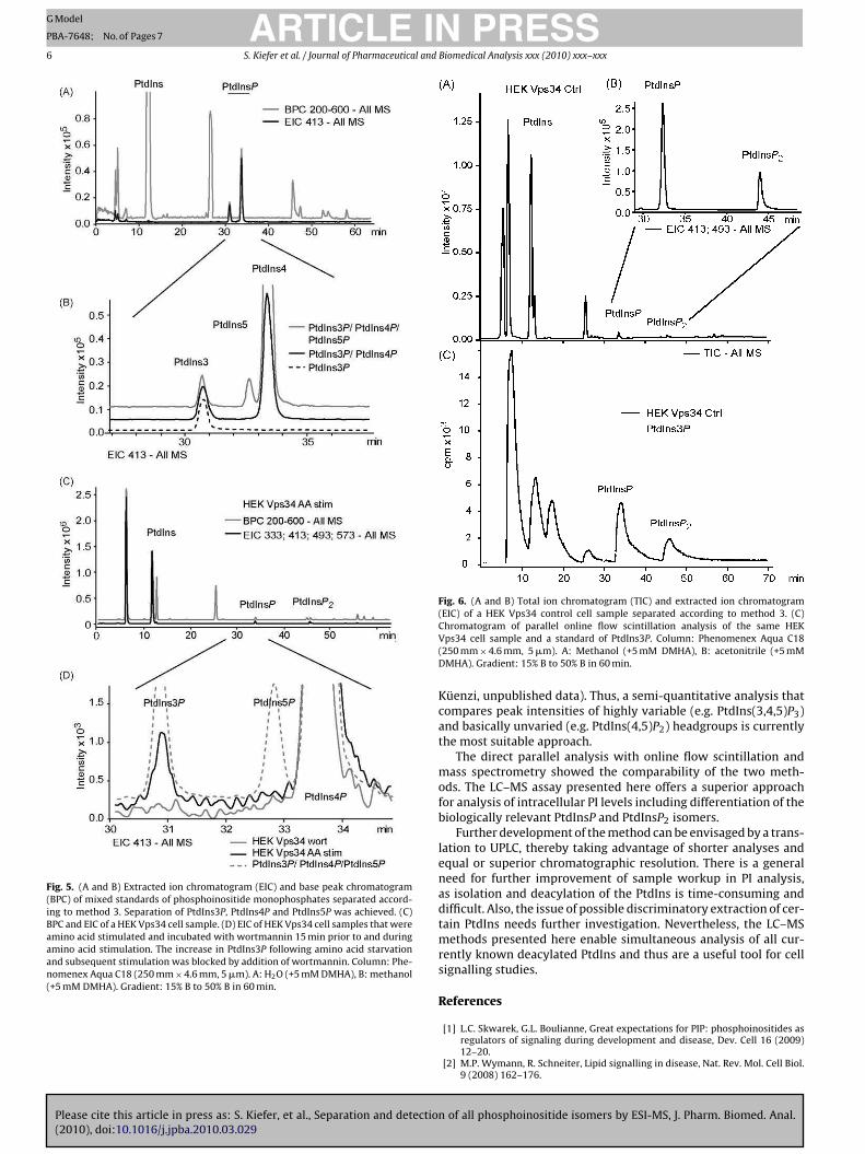
Please cite this article in press as: S. Kiefer, et al., Separation and detection of all phosphoinositide isomers by ESI-MS, J. Pharm. Biomed. Anal.(2010), doi:10.1016/j.jpba.2010.03.029
ARTICLE IN PRESSG Model
PBA-7648; No. of Pages 7
6 S. Kiefer et al. / Journal of Pharmaceutical and Biomedical Analysis xxx (2010) xxx–xxx
Fig. 5. (A and B) Extracted ion chromatogram (EIC) and base peak chromatogram(BPC) of mixed standards of phosphoinositide monophosphates separated accord-ing to method 3. Separation of PtdIns3P, PtdIns4P and PtdIns5P was achieved. (C)BPC and EIC of a HEK Vps34 cell sample. (D) EIC of HEK Vps34 cell samples that wereamino acid stimulated and incubated with wortmannin 15 min prior to and duringamino acid stimulation. The increase in PtdIns3P following amino acid starvationand subsequent stimulation was blocked by addition of wortmannin. Column: Phe-nomenex Aqua C18 (250 mm × 4.6 mm, 5 �m). A: H2O (+5 mM DMHA), B: methanol(+5 mM DMHA). Gradient: 15% B to 50% B in 60 min.
Fig. 6. (A and B) Total ion chromatogram (TIC) and extracted ion chromatogram(EIC) of a HEK Vps34 control cell sample separated according to method 3. (C)Chromatogram of parallel online flow scintillation analysis of the same HEKVps34 cell sample and a standard of PtdIns3P. Column: Phenomenex Aqua C18(250 mm × 4.6 mm, 5 �m). A: Methanol (+5 mM DMHA), B: acetonitrile (+5 mMDMHA). Gradient: 15% B to 50% B in 60 min.
Küenzi, unpublished data). Thus, a semi-quantitative analysis thatcompares peak intensities of highly variable (e.g. PtdIns(3,4,5)P3)and basically unvaried (e.g. PtdIns(4,5)P2) headgroups is currentlythe most suitable approach.
The direct parallel analysis with online flow scintillation andmass spectrometry showed the comparability of the two meth-ods. The LC–MS assay presented here offers a superior approachfor analysis of intracellular PI levels including differentiation of thebiologically relevant PtdInsP and PtdInsP2 isomers.
Further development of the method can be envisaged by a trans-lation to UPLC, thereby taking advantage of shorter analyses andequal or superior chromatographic resolution. There is a generalneed for further improvement of sample workup in PI analysis,as isolation and deacylation of the PtdIns is time-consuming anddifficult. Also, the issue of possible discriminatory extraction of cer-tain PtdIns needs further investigation. Nevertheless, the LC–MSmethods presented here enable simultaneous analysis of all cur-rently known deacylated PtdIns and thus are a useful tool for cellsignalling studies.
References
[1] L.C. Skwarek, G.L. Boulianne, Great expectations for PIP: phosphoinositides asregulators of signaling during development and disease, Dev. Cell 16 (2009)12–20.
[2] M.P. Wymann, R. Schneiter, Lipid signalling in disease, Nat. Rev. Mol. Cell Biol.9 (2008) 162–176.
Please cite this article in press as: S. Kiefer, et al., Separation and detection of all phosphoinositide isomers by ESI-MS, J. Pharm. Biomed. Anal.(2010), doi:10.1016/j.jpba.2010.03.029
ARTICLE IN PRESSG Model
PBA-7648; No. of Pages 7
S. Kiefer et al. / Journal of Pharmaceutical and Biomedical Analysis xxx (2010) xxx–xxx 7
[3] B. Payrastre, K. Missy, S. Giuriato, S. Bodin, M. Plantavid, M. Gratacap, Phos-phoinositides: key players in cell signalling, in time and space, Cell. Signal. 13(2001) 377–387.
[4] I.H. Batty, J. van der Kaay, A. Gray, J.F. Telfer, M.J. Dixon, C.P. Downes, The controlof phosphatidylinositol 3,4-bisphosphate concentrations by activation of theSrc homology 2 domain containing inositol polyphosphate 5-phosphatase 2,SHIP2, Biochem. J. 407 (2007) 255–266.
[5] M.J. Clague, S. Urbe, J. de Lartigue, Phosphoinositides and the endocytic path-way, Exp. Cell Res. 315 (2008) 1627–1631.
[6] D. Manna, A. Albanese, W.S. Park, W. Cho, Mechanistic basis of differentialcellular responses of phosphatidylinositol 3,4-bisphosphate and phos-phatidylinositol 3,4,5-trisphosphate-binding pleckstrin homology domains, J.Biol. Chem. 282 (2007) 32093–32105.
[7] R.H. Michell, V.L. Heath, M.A. Lemmon, S.K. Dove, Phosphatidylinositol 3,5-bisphosphate: metabolism and cellular functions, Trends Biochem. Sci. 31(2006) 52–63.
[8] G. Di Paolo, P. De Camilli, Phosphoinositides in cell regulation and membranedynamics, Nature 443 (2006) 651–657.
[9] J.B. Morris, K.A. Hinchliffe, A. Ciruela, A.J. Letcher, R.F. Irvine, Thrombin stim-ulation of platelets causes an increase in phosphatidylinositol 5-phosphaterevealed by mass assay, FEBS Lett. 475 (2000) 57–60.
[10] C. Pendaries, H. Tronchere, C. Racaud-Sultan, F. Gaits-Iacovoni, S. Coronas, S.Manenti, M.P. Gratacap, M. Plantavid, B. Payrastre, Emerging roles of phos-phatidylinositol monophosphates in cellular signaling and trafficking, Adv.Enzyme Regul. 45 (2005) 201–214.
[11] D. Sbrissa, O.C. Ikonomov, R. Deeb, A. Shisheva, Phosphatidylinositol 5-phosphate biosynthesis is linked to PIKfyve and is involved in osmotic responsepathway in mammalian cells, J. Biol. Chem. 277 (2002) 47276–47284.
[12] A.E. Wurmser, J.D. Gary, S.D. Emr, Phosphoinositide 3-kinases and their FYVEdomain-containing effectors as regulators of vacuolar/lysosomal membranetrafficking pathways, J. Biol. Chem. 274 (1999) 9129–9132.
[13] H. Guillou, L.R. Stephens, P.T. Hawkins, Quantitative measurement of phos-phatidylinositol 3,4,5-trisphosphate, Methods Enzymol. 434 (2007) 117–130.
[14] P.T. Ivanova, S.B. Milne, M.O. Byrne, Y. Xiang, H.A. Brown, Glycerophospholipididentification and quantitation by electrospray ionization mass spectrometry,Methods Enzymol. 432 (2007) 21–57.
[15] K.R. Auger, L.A. Serunian, S.P. Soltoff, P. Libby, L.C. Cantley, PDGF-dependenttyrosine phosphorylation stimulates production of novel polyphosphoinosi-tides in intact cells, Cell 57 (1989) 167–175.
[16] H. Hama, J. Torabinejad, G.D. Prestwich, D.B. DeWald, Measurement andimmunofluorescence of cellular phosphoinositides, Methods Mol. Biol. 284(2004) 243–258.
[17] T.E. Rusten, H. Stenmark, Analyzing phosphoinositides and their interactingproteins, Nat. Methods 3 (2006) 251–258.
[18] S.B. Milne, P.T. Ivanova, D. DeCamp, R.C. Hsueh, H.A. Brown, A targeted massspectrometric analysis of phosphatidylinositol phosphate species, J. Lipid Res.46 (2005) 1796–1802.
[19] M. Pulfer, R.C. Murphy, Electrospray mass spectrometry of phospholipids, MassSpectrom. Rev. 22 (2003) 332–364.
[20] M.R. Wenk, L. Lucast, G. Di Paolo, A.J. Romanelli, S.F. Suchy, R.L. Nussbaum, G.W.Cline, G.I. Shulman, W. McMurray, P. De Camilli, Phosphoinositide profiling incomplex lipid mixtures using electrospray ionization mass spectrometry, Nat.Biotechnol. 21 (2003) 813–817.
[21] A.D. Postle, D.C. Wilton, A.N. Hunt, G.S. Attard, Probing phospholipid dynam-ics by electrospray ionisation mass spectrometry, Prog. Lipid Res. 46 (2007)200–224.
[22] S.K. Dove, F.T. Cooke, M.R. Douglas, L.G. Sayers, P.J. Parker, R.H. Michell, Osmoticstress activates phosphatidylinositol-3,5-bisphosphate synthesis, Nature 390(1997) 187–192.
[23] M. Laffargue, J.M. Ragab-Thomas, A. Ragab, J. Tuech, K. Missy, L. Monnereau, U.Blank, M. Plantavid, B. Payrastre, P. Raynal, H. Chap, Phosphoinositide 3-kinaseand integrin signalling are involved in activation of bruton tyrosine kinase inthrombin-stimulated platelets, FEBS Lett. 443 (1999) 66–70.
[24] R.D. Byrne, M. Garnier-Lhomme, K. Han, M. Dowicki, N. Michael, N. Totty, V.Zhendre, A. Cho, T.R. Pettitt, M.J. Wakelam, D.L. Poccia, B. Larijani, PLCgammais enriched on poly-phosphoinositide-rich vesicles to control nuclear envelopeassembly, Cell Signal. 19 (2007) 913–922.
[25] M. Garnier-Lhomme, R.D. Byrne, T.M.C. Hobday, S. Gschmeissner, R. Woschol-ski, D.L. Poccia, E.J. Dufourc, B. Larijani, Nuclear envelope remnants: fluidmembanes enriched in STEROLS and ployphosphoinositides, PLOS One 4 (2009)1–11.
[26] T.R. Pettitt, S.K. Dove, A. Lubben, S.D. Calaminus, M.J. Wakelam, Analysisof intact phosphoinositides in biological samples, J. Lipid Res. 47 (2006)1588–1596.
[27] H. Ogiso, R. Taguchi, Reversed-phase LC/MS method for polyphosphoinositideanalyses: changes in molecular species levels during epidermal growth factoractivation in A431 cells, Anal. Chem. 80 (2008) 9226–9232.
[28] C. Nasuhoglu, S. Feng, J. Mao, M. Yamamoto, H.L. Yin, S. Earnest, B. Barylko, J.P.Albanesi, D.W. Hilgemann, Nonradioactive analysis of phosphatidylinositidesand other anionic phospholipids by anion-exchange high-performance liquidchromatography with suppressed conductivity detection, Anal. Biochem. 301(2002) 243–254.
[29] T. Gunnarsson, L. Ekblad, A. Karlsson, P. Michelsen, G. Odham, B. Jergil, Sepa-ration of polyphosphoinositides using normal-phase high-performance liquidchromatography and evaporative light scattering detection or electrospraymass spectrometry, Anal. Biochem. 254 (1997) 293–296.
[30] M. Laffargue, R. Calvez, P. Finan, A. Trifilieff, M. Barbier, F. Altruda, E. Hirsch,M.P. Wymann, Phosphoinositide 3-kinase gamma is an essential amplifier ofmast cell function, Immunity 16 (2002) 441–451.
[31] B. Dewald, M. Baggiolini, Platelet-activating factor as a stimulus of exocytosisin human neutrophils, Biochim. Biophys. Acta 888 (1986) 42–48.
[32] E.G. Bligh, W.J. Dyer, A rapid method of total lipid extraction and purification,Can. J. Biochem. Physiol. 37 (1959) 911–917.
[33] N.G. Clarke, R.M. Dawson, Alkaline O leads to N-transacylation. A new methodfor the quantitative deacylation of phospholipids, Biochem. J. 195 (1981)301–306.
[34] M.P. Gratacap, S. Severin, G. Chicanne, M. Plantavid, B. Payrastre, Different rolesof SHIP1 according to the cell context: the example of blood platelets, Adv.Enzyme Regul. 48 (2008) 240–252.
[35] A. Sorisky, W.G. King, S.E. Rittenhouse, Accumulation of PtdIns(3,4)P2 andPtdIns(3,4,5)P3 in thrombin-stimulated platelets. Different sensitivities to Ca2+
or functional integrin, Biochem. J. 286 (Pt 2) (1992) 581–584.[36] P. Gulati, G. Thomas, Nutrient sensing in the mTOR/S6K1 signalling pathway,
Biochem. Soc. Trans. 35 (2007) 236–238.[37] T. Nobukuni, M. Joaquin, M. Roccio, S.G. Dann, S.Y. Kim, P. Gulati, M.P. Byfield,
J.M. Backer, F. Natt, J.L. Bos, F.J. Zwartkruis, G. Thomas, Amino acids mediatemTOR/raptor signaling through activation of class 3 phosphatidylinositol 3OH-kinase, Proc. Natl. Acad. Sci. U.S.A. 102 (2005) 14238–14243.
[38] M.P. Byfield, J.T. Murray, J.M. Backer, hVps34 is a nutrient-regulated lipidkinase required for activation of p70 S6 kinase, J. Biol. Chem. 280 (2005)33076–33082.

������������������ �����������������������������������������������
�����������
�������������� ��������������������������������� ������������
����������������������������
���������������������������������������������� ��������������������������������������������������!∀���������������������#��������������������������������������������������������������∀��������������∃�
����� ���������������������%������������������������������������&∀�������∀������∀������
������������������%���������������������������������������������������� ������∀������
� ∋��(����������)�������������������������������∗����������������������∀������������������ �����������������#����������������������∗���������������������������
� +��������������������,��������������������������∀��������������������#�∀����������� ���������������������������
� %����∗������ �������−������.�����������������������������
/�����0����.0�����������������������������12��2���3�4������ %����0�5�6�3778
!���������������������� �������∀�������������������������#��������������
%��9�������%�����������&�������������������∗�������������������������� ���������.�����0����.0�����������������������������12��2���3�4����������������
:�����������������0%��9�������%�����������∗����������������������������� ���������;����������������������������∗����������������������������������������� ���������.������∀���������%��%��������������������������<������������������������������������������∗�����������������9������9��������������� �������∀��������������������������������� ���������������%����������������)�������������9�������%��������������������=��������� �����